


Acta Veterinaria et Zootechnica Sinica ›› 2025, Vol. 56 ›› Issue (8): 4101-4111.doi: 10.11843/j.issn.0366-6964.2025.08.047
• Research Notes • Previous Articles Next Articles
TIAN Ru( ), FU Xingwei, HU Leyu, ZHU Mingjun*(), TONG Dewen*()
Received:2024-10-28
Online:2025-08-23
Published:2025-08-28
Contact:
ZHU Mingjun, TONG Dewen
E-mail:2775744728@qq.com;nabtchina@126.com;dwtong@nwsuaf.edu.cn
CLC Number:
TIAN Ru, FU Xingwei, HU Leyu, ZHU Mingjun, TONG Dewen. Isolation and Pathogenicity Analysis of a GⅡa Porcine Epidemic Diarrhea Virus[J]. Acta Veterinaria et Zootechnica Sinica, 2025, 56(8): 4101-4111.
Table 1
Primer information"
| 引物名称 Primer name | 引物序列(5′→3′) Sequence | 退火温度/℃ Annealing temperature | 片段长度/bp Product length |
| PEDV | F:CTTTGGTGGTAATGTGGCTG | 56 | 273 |
| R:TCAACAGCTGTGTCCCATTC | |||
| TGEV | F:GGGCCAACGTAAAGAGCTTCC | 52 | 820 |
| R:GCTCTGACCTTTCTGCAG | |||
| PDCoV | F:CCCCAAACCAACTAAGGACAAG | 56 | 125 |
| R:AAGCATCATCCCACTCCCAATC | |||
| PoRV | F:GAAACGGAATAGCTCCACA | 54 | 271 |
| R:GAATAATCAAATCCAGCCAC | |||
| PCV2 | F:ACCAGCGCACTTCGGCAGCGGC | 53 | 537 |
| R:GCTTCTGCATTTTCCCGCTCAC | |||
| PRV | F:CACGGAGGAGGAGCTGGGGCT | 58 | 217 |
| R:GTCCACGCCCCGCTTGAAGCT | |||
| PPV | F:AATACTTGGGGGAGGGCTTG | 57 | 415 |
| R:CTAGGTGCTGCTGGTGTGTA |
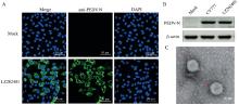
Fig. 1
Identification results of PEDV LZ202401 strain A. IFA analysis results of virus infected Vero E6 cells (Mock. Control group; LZ202401. Treatment group; PEDV N protein anti-fluorescence); B. Western blot verification results (PEDV-N. Target protein 57 ku; β-actin. Reference protein 43 ku; CV777. PEDV classical strain); C. Transmission electron microscope image of LZ202401 strain"
Fig. 2
S gene sequence analysis of LZ202401 strain A. Phylogenetic tree of S gene sequence of LZ202401 strain; B. Analysis of neutralizing epitope mutation sites in S protein"
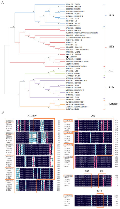
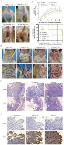
Fig. 3
Pathogenicity analysis of LZ202401 strain A. Piglet diarrhea; B. Piglet diarrhea score (Solid score 0, semi-solid score 1, liquid score 2);C. Piglet survival curve; D. Anatomy of abdominal pathological changes; E. Tissue section observation (10×); F. Immunohistochemistry detection results (20×) (lesion characteristics marked by red arrows)"

| 1 | 刘政龙,王金良,沈志强,等.猪流行性腹泻病毒研究进展[J].中国畜牧兽医,2015,42(9):2506-2511. |
| LIUZ L,WANGJ L,SHENZ Q,et al.Research progress of porcine epidemic diarrhea virus[J].China Animal Husbandry and Veterinary Medicine,2015,42(9):2506-2511. | |
| 2 |
PENSAERTM B,DEBOUCKP.A new coronavirus-like particle associated with diarrhea in swine[J].Arch Virol,1978,58(3):243-247.
doi: 10.1007/BF01317606 |
| 3 | 宣华,邢德坤,王殿瀛,等.应用猪胎肠单层细胞培养猪流行性腹泻病毒的研究[J].兽医大学学报,1984(3):202-208. |
| XUANH,XINGD K,WANGD Y,et al.Study on the culture of porcine epidemic diarrhea virus adapted to fetal porcine intestine primary cell monolayer[J].Journal of Veterinary Science and Technology,1984(3):202-208. | |
| 4 | 王颢然,高利,高翔,等.2008—2018年我国猪流行性腹泻病毒混合感染分析[J].中国动物传染病学报,2020,28(3):97-103. |
| WANGH R,GAOL,GAOX,et al.Analysis of mixed infection of porcine epidemic diarrhea virus in China from 2008 to 2018[J].Chinese Journal of Animal Infectious Diseases,2020,28(3):97-103. | |
| 5 | 朱子健,闫丽辉,鞠妍,等.2015—2016年我国东北地区猪病毒性腹泻流行病学调查[J].中国预防兽医学报,2017,39(5):356-360. |
| ZHUZ J,YANL H,JUY,et al.Epidemiological investigation of porcine viral diarrhea in Northeast China from 2015 to 2016[J].Chinese Journal of Preventive Veterinary Medicine,2017,39(5):356-360. | |
| 6 | 徐丽华,李军星,苏菲,等.2011—2017年浙江省猪病毒性腹泻病的流行病学调查[J].中国兽医科学,2018,48(5):625-630. |
| XUL H,LIJ X,SUNF,et al.Epidemiological investigation of porcine viral diarrhea in Zhejiang Province from 2011 to 2017[J].Chinese Veterinary Science,2018,48(5):625-630. | |
| 7 |
CUIJ T,QIANH,HOUC Y,et al.Characteristics of the spike and ORF3 genes of porcine epidemic diarrhea virus in Henan and Shanxi provinces of China[J].Arch Virol,2020,165(10):2323-2333.
doi: 10.1007/s00705-020-04744-x |
| 8 |
李良华,宋忠旭,董斌科,等.猪流行性腹泻防控关键技术[J].畜牧与饲料科学,2019,40(11):109-112.
doi: 10.12160/j.issn.1672-5190.2019.11.027 |
|
LIL H,SONGZ X,DONGB K,et al.Key technologies for prevention and control of porcine epidemic diarrhea[J].Animal Husbandry and Feed Science,2019,40(11):109-112.
doi: 10.12160/j.issn.1672-5190.2019.11.027 |
|
| 9 |
JUNGK,HUH,SAIFL J.Porcine deltacoronavirus infection: Etiology, cell culture for virus isolation and propagation, molecular epidemiology and pathogenesis[J].Virus Res,2016,226,50-59.
doi: 10.1016/j.virusres.2016.04.009 |
| 10 |
GERBERP F,GONGQ,HUANGY W,et al.Detection of antibodies against porcine epidemic diarrhea virus in serum and colostrum by indirect ELISA[J].Vet J,2014,202(1):33-36.
doi: 10.1016/j.tvjl.2014.07.018 |
| 11 |
LIZ L,CHENF,YUANY,et al.Sequence and phylogenetic analysis of nucleocapsid genes of porcine epidemic diarrhea virus (PEDV) strains in China[J].Arch Virol,2013,158(6):1267-1273.
doi: 10.1007/s00705-012-1592-4 |
| 12 |
SIF S,HUX X,WANGC Y,et al.Porcine epidemic diarrhea virus (PEDV) ORF3 enhances viral proliferation by inhibiting apoptosis of infected cells[J].Viruses,2020,12(2):214.
doi: 10.3390/v12020214 |
| 13 |
KIRCHDOERFERR N,BHANDARIM,MARTINIO,et al.Structure and immune recognition of the porcine epidemic diarrhea virus spike protein[J].Structure,2021,29(4):385-392.
doi: 10.1016/j.str.2020.12.003 |
| 14 |
SATOT,TAKEYAMAN,KATSUMATAA,et al.Mutations in the spike gene of porcine epidemic diarrhea virus associated with growth adaptation in vitro and attenuation of virulence in vivo[J].Virus Genes,2011,43(1):72-78.
doi: 10.1007/s11262-011-0617-5 |
| 15 | HUANGY W,DICKERMANA W,PIÑEYROP,et al.Origin, evolution, and genotyping of emergent porcine epidemic diarrhea virus strains in the United States[J].mBio,2013,4(5):e00737-00713. |
| 16 | 尤永君,刘思桐,王幸,等.猪流行性腹泻病毒流行病学调查及毒株致病力研究[J].天津科技,2023,50(S1):81-87. |
| YOUY J,LIUS T,WANGX,et al.Epidemiological investigation and pathogenicity study of porcine epidemic diarrhea virus[J].Tianjin Science and Technology,2023,50(S1):81-87. | |
| 17 |
JUNGK,SAIFL J,WANGQ H.Porcine epidemic diarrhea virus (PEDV): An update on etiology, transmission, pathogenesis, and prevention and control[J].Virus Res,2020,286,198045.
doi: 10.1016/j.virusres.2020.198045 |
| 18 |
LIUH X,YINX R,TIANH L,et al.The S protein of a novel recombinant PEDV strain promotes the infectivity and pathogenicity of PEDV in Mid-west China[J].Transbound Emerg Dis,2022,69(6):3704-3723.
doi: 10.1111/tbed.14740 |
| 19 | 沈海燕,黄俊,张琬玓,等.一株猪流行性腹泻病毒的分离鉴定及其ORF3和S基因序列分析[J].黑龙江畜牧兽医,2024(2):79-87. |
| SHENH Y,HUANGJ,ZHANGW D,et al.Isolation and identification of a porcine epidemic diarrhea virus and its ORF3 and S gene sequence analysis[J].Heilongjiang Animal Science and Veterinary Medicine,2024(2):79-87. | |
| 20 |
SIG B,NIUJ W,ZHOUX,et al.Use of dual priming oligonucleotide system-based multiplex RT-PCR assay to detect five diarrhea viruses in pig herds in South China[J].AMB Express,2021,11(1):99.
doi: 10.1186/s13568-021-01255-z |
| 21 |
莫红芳,何东贤,熊晓妍,等.广西某猪场猪伪狂犬病病毒的分离鉴定[J].动物医学进展,2023,44(10):123-128.
doi: 10.3969/j.issn.1007-5038.2023.10.023 |
|
MOH F,HED X,XIONGX Y,et al.Isolation and identification of porcine pseudorabies virus from a pig farm in Guangxi[J].Progress in Veterinary Medicine,2023,44(10):123-128.
doi: 10.3969/j.issn.1007-5038.2023.10.023 |
|
| 22 |
HOFMANNM,WYLERR.Propagation of the virus of porcine epidemic diarrhea in cell culture[J].J Clin Microbiol,1988,26(11):2235-2239.
doi: 10.1128/jcm.26.11.2235-2239.1988 |
| 23 |
SUNY S,GONGT,WUD D,et al.Isolation, identification, and pathogenicity of porcine epidemic diarrhea virus[J].Front Microbiol,2023,14,1273589.
doi: 10.3389/fmicb.2023.1273589 |
| 24 |
LEIJ L,MIAOY Q,BIW R,et al.Porcine epidemic diarrhea virus: Etiology, epidemiology, antigenicity, and control strategies in China[J].Animals,2024,14(2):294.
doi: 10.3390/ani14020294 |
| 25 | 刘秋燕,涂玉蓉,江梦雅,等.猪流行性腹泻疾病的诊断与防控[J].广东畜牧兽医科技,2024,49(04):91-96. |
| LIUQ Y,TUY R,JIANGM Y,et al.Diagnosis and control of porcine epidemic diarrhea disease[J].Guangdong Journal of Animal and Veterinary Science,2024,49(04):91-96. | |
| 26 | 耿超,陆泓宇,梁婉,等.猪流行性腹泻病毒HB2018的分离鉴定及致病性[J].中国兽医学报,2021,41(09):1704-1709. |
| GENGC,LUH Y,LIANGW,et al.Isolation, identification and pathogenicity of porcine epidemic diarrhea virus HB2018[J].Chinese Journal of Veterinary Science,2021,41(09):1704-1709. | |
| 27 |
GUOY L,SUIL,KONGD M,et al.Porcine epidemic diarrhea virus strain CH/HLJ/18 isolated in China: characterization and phylogenetic analysis[J].Virol J,2024,21(1):28.
doi: 10.1186/s12985-023-02233-6 |
| 28 | 刘维哲,罗成刚,袁蓉,等.一株猪流行性腹泻病毒强毒株的分离与鉴定[J].畜牧兽医学报,2024,55(7):3049-3063. |
| LIUW Z,LUOC G,YUANR,et al.Isolation and identification of a highly pathogenic strain of porcine epidemic diarrhea virus[J].Acta Veterinaria et Zootechnica Sinica,2024,55(7):3049-3063. | |
| 29 | 王金坡,崔亮亮,孙秀秀,等.哺乳仔猪自然感染猪流行性腹泻病毒病例的病理形态学观察[J].中国兽医学报,2020,40(12):2362-2367. |
| WANGJ P,CUIL L,SUNX X,et al.Pathomorphological observation of naturally infected suckling piglets with porcine epidemic diarrhea virus[J].Chinese Journal of Veterinary Science,2020,40(12):2362-2367. | |
| 30 |
LIW T,VAN KUPPEVELDF J M,HEQ G,et al.Cellular entry of the porcine epidemic diarrhea virus[J].Virus Res,2016,226,117-127.
doi: 10.1016/j.virusres.2016.05.031 |
| 31 |
LUOH J,LIANGZ P,LINJ J,et al.Research progress of porcine epidemic diarrhea virus S protein[J].Front Microbiol,2024,15,1396894.
doi: 10.3389/fmicb.2024.1396894 |
| [1] | ZENG Shengxin, SONG Chengqi, SHEN Kaiyuan, HAO Guoxin, WANG Yakun, WANG Xin, WANG Xiaoxu, LIU Zhijie, LIU Yongbo, LIU Yongsheng, YANG Shunli, FU Zhixin. Isolation, Identification of a Deer Clostridium perfringens and Pathogenicity Analysis in Mouse [J]. Acta Veterinaria et Zootechnica Sinica, 2025, 56(8): 3976-3984. |
| [2] | LI Zhiqiang, CHEN Xueqing, ZHANG Yuanshu. Detection of Angiotensin Converting Enzyme 2 in Intestinal Tissues of Clinically Infected Porcine Epidemic Diarrhea Virus Piglets and Analysis of Its Relationship with Intestinal Pathological Changes [J]. Acta Veterinaria et Zootechnica Sinica, 2025, 56(7): 3463-3473. |
| [3] | WANG Yunke, WANG Na, YUE Ke, HE Kunmiao, ZHANG Xing, LIU Yao, ZHANG Gaiping. Substances with Inhibitory Effects on Porcine Epidemic Diarrhea Virus Replication in vitro [J]. Acta Veterinaria et Zootechnica Sinica, 2025, 56(6): 2577-2589. |
| [4] | WU Chao, MING Wenhan, LU Shuwan, YANG Caimei, LIU Jinsong, MA Xiang, ZHANG Ruiqiang. Innate Immune Evasion Mechanisms of Porcine Epidemic Diarrhea Virus and Advances in Prevention and Control Strategies [J]. Acta Veterinaria et Zootechnica Sinica, 2025, 56(6): 2590-2599. |
| [5] | ZHOU Min, TANG Deyuan, ZENG Zhiyong, WANG Bin, HUANG Tao, HU Wenwen, MAO Yinming, ZHOU Piao, HE Song. Research Progress on the Interaction between Porcine Epidemic Diarrhea Virus Proteins and Host Proteins [J]. Acta Veterinaria et Zootechnica Sinica, 2025, 56(6): 2600-2612. |
| [6] | LI Chengcheng, ZHAO Yongxiang, CAO Qiuxia, SONG Xu, LI Yupeng, FAN Baochao, GUO Rongli, XU Yefen, LI Bin. Tight Junction Protein CLDN4 Promotes Porcine Epidemic Diarrhea Virus Infection [J]. Acta Veterinaria et Zootechnica Sinica, 2025, 56(6): 2826-2835. |
| [7] | WANG Yan, HUANG Xinyu, WU Guiying, WU Wanping, Lü Qizhuang. Genetic Variation Analysis of Porcine Circovirus Type 3 in China from 2017 to 2023 [J]. Acta Veterinaria et Zootechnica Sinica, 2025, 56(6): 2857-2867. |
| [8] | CHEN Yunlong, FAN Gang, FAN Xinyi, GUO Yongchao, ZHANG Xinmiao, WANG Yan, ZHANG Shiqiang. Isolation and Identification of a Novel Porcine Circovirus Type 2d Strain with Multiple Nucleotide Substitutions [J]. Acta Veterinaria et Zootechnica Sinica, 2025, 56(6): 3032-3040. |
| [9] | WANG Shujuan, WANG Dongfang, MA Zhenyuan, ZHAO Xueli, LIU Ying, YANG Haibo, CHAI Mao, WANG Cui, YAN Ruoqian. Isolation, Identification and Evolutionary Analysis of a Strain of Senecavirus A [J]. Acta Veterinaria et Zootechnica Sinica, 2025, 56(6): 3041-3046. |
| [10] | WANG Yanan, GUO Yaru, JIANG Yanping, CUI Wen, LI Jiaxuan, LI Yijing, WANG Li. Isolation, Identification and Pathogenicity Analysis of Porcine Rotavirus [J]. Acta Veterinaria et Zootechnica Sinica, 2025, 56(5): 2259-2269. |
| [11] | LI Ting, ZHANG Chengcheng, WANG Xiuling, ZHANG Xiaorong, WU Yantao. Isolation and Identification of a Novel Duck Reovirus Strain and the Sequence Analysis of Its σC Gene [J]. Acta Veterinaria et Zootechnica Sinica, 2025, 56(5): 2520-2524. |
| [12] | WU Peiling, LI Yixuan, WANG Haojie, LI Yafei, LIU Shaomeng, LIU Qingyun, WANG Xiangru. Research Progress of Porcine Epidemic Diarrhea Vaccine for Pigs [J]. Acta Veterinaria et Zootechnica Sinica, 2025, 56(3): 1042-1058. |
| [13] | YU Xinya, HE Haijian, WANG Lei, NI Yuchen, DU Jing, ZHOU Yingshan, DONG Wanyu, WANG Xiaodu. Effect of lncRNA 18850 on Porcine Epidemic Diarrhea Virus Replication [J]. Acta Veterinaria et Zootechnica Sinica, 2025, 56(3): 1366-1375. |
| [14] | LIU Jian, YU Zehai, ZHANG Meiyu, LI Dan, WANG Jun, LIU Fangqin, ZHANG Qun, XU Shouzhen. Full-genome Analysis of a Bovine Enterovirus Type F and the Establishment of an Indirect ELISA Method for Antibody Detection [J]. Acta Veterinaria et Zootechnica Sinica, 2025, 56(2): 814-825. |
| [15] | ZHANG Dongxuan, WANG Zhihao, QIAO Yan, ZHAO Xiaoxiao, FAN Songjie, ZHANG Chao. Prokaryotic Expression of S1 Protein in Porcine Epidemic Diarrhea Virus and Screening of Its Aptamers [J]. Acta Veterinaria et Zootechnica Sinica, 2025, 56(2): 839-850. |
| Viewed | ||||||
|
Full text |
|
|||||
|
Abstract |
|
|||||