


Acta Veterinaria et Zootechnica Sinica ›› 2025, Vol. 56 ›› Issue (5): 2259-2269.doi: 10.11843/j.issn.0366-6964.2025.05.024
• Preventive Veterinary Medicine • Previous Articles Next Articles
WANG Yanan1( ), GUO Yaru1, JIANG Yanping1,2, CUI Wen1,2, LI Jiaxuan1,2, LI Yijing1,2,*(), WANG Li1,2,*()
Received:2024-05-10
Online:2025-05-23
Published:2025-05-27
Contact:
LI Yijing, WANG Li
E-mail:wyn19971224@163.com;yijingli@163.com;wanglicau@163.com
CLC Number:
WANG Yanan, GUO Yaru, JIANG Yanping, CUI Wen, LI Jiaxuan, LI Yijing, WANG Li. Isolation, Identification and Pathogenicity Analysis of Porcine Rotavirus[J]. Acta Veterinaria et Zootechnica Sinica, 2025, 56(5): 2259-2269.
Table 1
Sequence information of specific primers"
| 序号 Number | 引物名称 Primer name | 引物序列(5′→3′) Primer sequence | 扩增长度/bp Fragment length |
| 1 | PoRV-VP1-F1 PoRV-VP1-R1 | ATGGGGAAGTACAATCTAATCTT CTCGTTCTGACGGAAATAATC | 2 012 |
| 2 | PoRV-VP1-F2 PoRV-VP1-R2 | AGATTATTTCCGTCAGAACG TTTGCGTATATATGTGATCGTAG | 941 |
| 3 | PoRV-VP2-F1 PoRV-VP2-R1 | ACGCGTCGACCATGGCGTACA GCATAGTTGGAAACTGTTGTC | 1 574 |
| 4 | PoRV-VP2-F2 PoRV-VP2-R2 | GGATGCATTATAGAAATGGTG CCGCTCGAGAATTACAGTTCG | 1 112 |
| 5 | PoRV-VP3-F PoRV-VP3-R | GGCTWTTAAAGCARTATTAGTAGTG GGT CAC ATC ATG ACT AGT GTG | 2 508 |
| 6 | PoRV-VP4-F PoRV-VP4-R | ATGGCTTCGCTCATTTATAGACAACTA ACTACTTACAGTCTACATTGCATGATT | 2 337 |
| 7 | PoRV-VP6-F PoRV-VP6-R | GGCTTTWAAACGAAGTCTTC GGTCACATAATCTCACT | 1 356 |
| 8 | PoRV-VP7-F PoRV-VP7-R | GCCTTTAAAAGCGAGAATT GGTCACATCATACAACTCTA | 1 062 |
| 9 | PoRV-NSP1-F PoRV-NSP1-R | CCATGGCTACTTTTAAGGATGCT CTAGGCGCTACTCTAGTGCAGGG | 1 517 |
| 10 | PoRV-NSP2-F PoRV-NSP2-R | GGCTTTTAAAGCGTCTCAG GGTCACATAAGCGCTTTC | 1 059 |
| 11 | PoRV-NSP3-F PoRV-NSP3-R | GGCTTTTAATGCTTTTCAGTG GGTCACATAACGCCCCTATAG | 1 073 |
| 12 | PoRV-NSP4-F PoRV-NSP4-R | GGWYACRYTAAGACCRTTCC GGCTTTTAAAGCGCTACAG | 747 |
| 13 | PoRV-NSP5-F PoRV-NSP5-R | GGCTTTTAAAGCGCTACAG GGTCACAAAACGGGAGT | 664 |
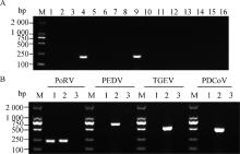
Fig. 1
PCR results of small intestinal tissue samples A. Detection of PoRV in small intestinal tissue disease materials: M. DL2000 DNA Marker; 1-15. Small intestinal tissue disease materials; 16. Negative control. B. Detection of PEDV, TGEV and PDCoV in PoRV-positive small intestinal tissue samples: M. DL2000 Marker; 1. Small intestinal tissue disease material; 2. Positive sample; 3. Negative sample"

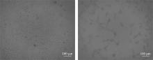
Fig. 2
Virus isolation and culture results A. MA104 cells; B. MA104 cells infected with PoRV"
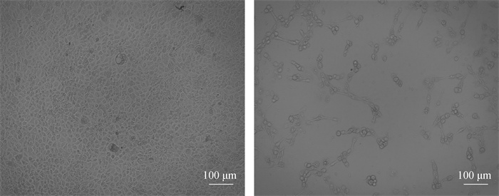
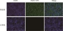
Fig. 3
Immunofluorescence identification result"
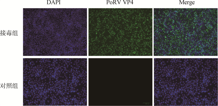
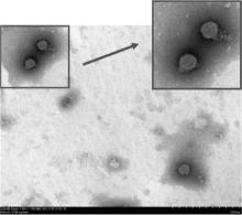
Fig. 4
Electron microscopic observation results of PoRV isolate"

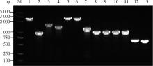
Fig. 5
PoRV whole genome sequence amplification"

Table 2
Genotyping of each segment of PoRV"
| 基因 Gene | 长度/bp Length | 基因型 Genotype | 参考序列名称 Reference sequence name | 参考序列序列号 Reference strains accession numbers | 同源性/% Homology |
| VP1 | 3 295 | R1 | RVA/Pig-tc/USA/OSU/1975/G5P9[7] | GU199514.1 | 99.76 |
| VP2 | 2 686 | C1 | RVA/Pig-tc/USA/LS00006_OSU/1975/G5P[X] | KR052758.1 | 99.85 |
| VP3 | 2 563 | M1 | RVA/Pig-tc/USA/LS00006_OSU/1975/G5P[X] | KR052759.1 | 99.88 |
| VP4 | 2 337 | P7 | RVA/Pig-tc/ESP/OSU-C5111/2010/G5P[7] | KJ450845.1 | 99.75 |
| VP6 | 1 356 | I5 | RVA/Pig-tc/ESP/OSU-C5111/2010/G5P[7] | KJ450847.1 | 99.85 |
| VP7 | 1 062 | G5 | RVA/porcine/CH/2020/OSU/serotype 5 | OR091159.1 | 99.81 |
| NSP1 | 1 569 | A1 | RVA/Pig-tc/USA/LS00006_OSU/1975/G5P[X] | KR052752.1 | 99.61 |
| NSP2 | 1 059 | N1 | RV/KOR/C-1 | PP100168.1 | 99.72 |
| NSP3 | 1 075 | T1 | RVA/KOR/OSU/G5P7 | JX971586.1 | 100.00 |
| NSP4 | 744 | E1 | RVA/KOR/174-1/G8P7 | KF500216.1 | 99.73 |
| NSP5 | 664 | H1 | RVA/Porcine-tc/KOR/K71/2006/G5P[7] | MF940468.1 | 99.55 |
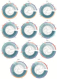
Fig. 6
Genetic Evolution Analysis of HLJ/2021 Strain"
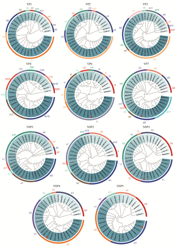
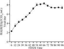
Fig. 7
Growth curve of PoRV"
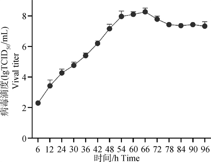

Fig. 8
Clinical symptoms after PoRV infection A. Clinical symptoms of PoRV-infected piglets; B. Changes in body weight of PoRV-infected piglets; C. Changes in body temperature of PoRV-infected piglets. *P < 0.05, **P < 0.01, ****P < 0.000 1"
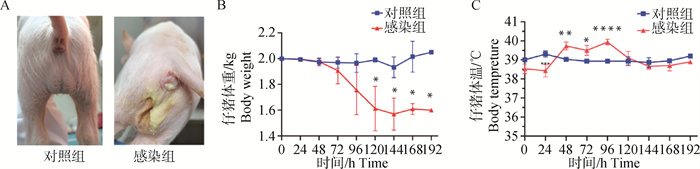
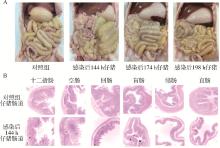
Fig. 9
Pathological changes in PoRV-infected piglets A. Necropsy changes of PoRV-infected piglets; B. Histopathological changes of PoRV-infected piglets (4×)"

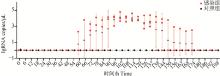
Fig. 10
Virus shedding results of piglets"
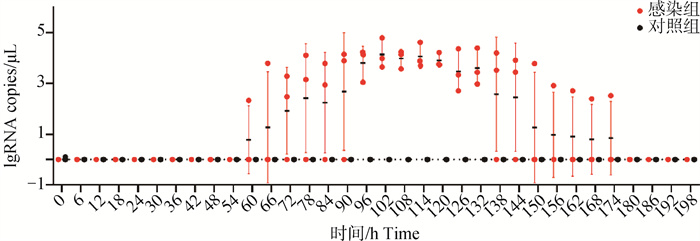
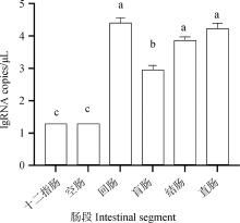
Fig. 11
Viral load in intestine of piglets Identical letters indicate no significant difference between groups, while different letters indicate a significant difference between groups (P < 0.05)"
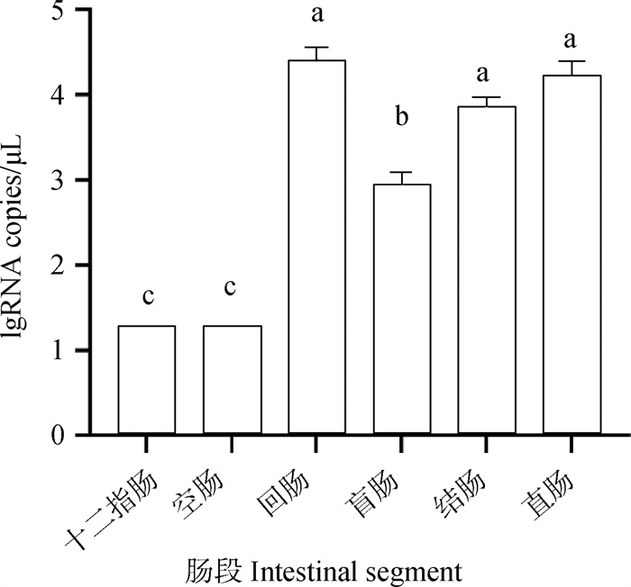
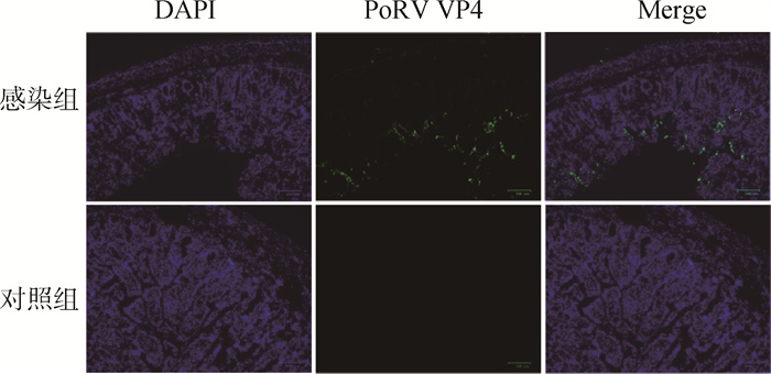
Fig. 12
IFA detection results of virus in intestinal tissue"
| 1 |
FUKUSHO A , SHIMIZU Y , ITO Y . Isolation of cytopathic porcine rotavirus in cell roller culture in the presence of trypsin[J]. Arch Virol, 1981, 69 (1): 49- 60.
doi: 10.1007/BF01315265 |
| 2 |
VIASOVA A N , AMIMO J O , SAIF L J . Porcine rotaviruses: epidemiology, immune responses and control strategies[J]. Viruses, 2017, 9 (3): 48.
doi: 10.3390/v9030048 |
| 3 |
THEUNS S , DESMARETS L M , HEYLEN E , et al. Porcine group A rotaviruses with heterogeneous VP7 and VP4 genotype combinations can be found together with enteric bacteria on Belgian swine farms[J]. Vet Microbiol, 2014, 172 (1-2): 23- 34.
doi: 10.1016/j.vetmic.2014.04.002 |
| 4 |
TAO R , CHANG X , ZHOU J , et al. Molecular epidemiological investigation of group A porcine rotavirus in East China[J]. Front Vet Sci, 2023, 10, 1138419.
doi: 10.3389/fvets.2023.1138419 |
| 5 |
QIAO M , LI M , LI Y , et al. Recent molecular characterization of porcine rotaviruses detected in China and their phylogenetic relationships with human rotaviruses[J]. Viruses, 2024, 16 (3): 453.
doi: 10.3390/v16030453 |
| 6 |
DOERKSEN T , CHRISTENSEN T , LU A , et al. Assessment of porcine Rotavirus-associated virome variations in pigs with enteric disease[J]. Vet Microbiol, 2022, 270, 109447.
doi: 10.1016/j.vetmic.2022.109447 |
| 7 |
MATTHIJNSSENS J , CIARLET M , RAHMAN M , et al. Recommendations for the classification of group A rotaviruses using all 11 genomic RNA segments[J]. Arch Virol, 2008, 153 (8): 1621- 1629.
doi: 10.1007/s00705-008-0155-1 |
| 8 |
NURDIN JA , KOTAKI T , KAWAGISHI T , et al. N-Glycosylation of Rotavirus NSP4 protein affects viral replication and pathogenesis[J]. J Virol, 2023, 97 (1): e0186122.
doi: 10.1128/jvi.01861-22 |
| 9 |
ANGEL J , TANG B , FENG N , et al. Studies of the role for NSP4 in the pathogenesis of homologous murine rotavirus diarrhea[J]. J Infect Dis, 1998, 177 (2): 455- 458.
doi: 10.1086/517374 |
| 10 |
WANG J , ZHOU J , ZHU X , et al. Isolation and characterization of a G9P[23] porcine rotavirus strain AHFY2022 in China[J]. Microb Pathog, 2024, 190, 106612.
doi: 10.1016/j.micpath.2024.106612 |
| 11 |
ZHANG F , LUO Y , LIN C , et al. Epidemiological monitoring and genetic variation analysis of pathogens associated with porcine viral diarrhea in southern China from 2021 to 2023[J]. Front Microbiol, 2024, 15, 1303915.
doi: 10.3389/fmicb.2024.1303915 |
| 12 | 谷拉强, 陶然, 程曦, 等. 2022—2023年我国部分地区猪A群轮状病毒分子流行病学调查分析[J]. 南方农业学报, 2023, 54 (10): 3083- 3091. |
| GU L Q , TAO R , CHENG X , et al. Molecular epidemiological survey of group A porcine rotavirus in selected areas of China during 2022-2023[J]. Journal of Southern Agriculture, 2023, 54 (10): 3083- 3091. | |
| 13 | 甸子芩, 樊茂, 蒋红君, 等. 2015-2017年云南省轮状病毒感染情况及基因分型分析[J]. 中国病原生物学杂志, 2019, 14 (01): 78-82, 87. |
| DIAN Z Q , FAN M , JIANG H J , et al. Analysis of the prevalence of rotavirus infection and genotyping of rotavirus isolated in Yunnan Province from 2015 to 2017[J]. Journal of Pathogen Biology, 2019, 14 (01): 78-82, 87. | |
| 14 | MEBUS C A . Reovirus and rotavirus infections[J]. Proc Annu Meet U S Anim Health Assoc, 1975 (79): 345- 349. |
| 15 | WOODE G N . Pathogenic rotaviruses isolated from pigs and calves[J]. Ciba Found Symp, 1976 (42): 251- 271. |
| 16 | 黄小波, 徐璐, 曹三杰, 等. 猪轮状病毒OSU株的培养特性与致病性研究[J]. 中国人兽共患病学报, 2012, 28 (02): 120-123, 147. |
| HUANG X B , XU L , CAO S J , et al. Culture characteristics and pathogenicity of porcine rotavirus OSU strain[J]. Chinese Journal of Zoonoses, 2012, 28 (02): 120-123, 147. | |
| 17 | 宋菲菲, 郝洪吉, 张艳红, 等. Vero细胞培养轮状病毒关键条件的优化[J]. 中国生物制品学杂志, 2014, 27 (10): 1345- 1347. |
| SONG F F , HAO H J , ZHANG Y H , et al. Optimization of key condition for culture of rotavirus in Vero cells[J]. Chin J Biologicals, 2014, 27 (10): 1345- 1347. | |
| 18 |
MIAO Q , PAN Y , GONG L , et al. Full genome characterization of a human-porcine reassortment G12P[7] rotavirus and its pathogenicity in piglets[J]. Transbound Emerg Dis, 2022, 69 (6): 3506- 3517.
doi: 10.1111/tbed.14712 |
| 19 |
LAGAN P , MOONEY M H , LEMON K . Genome analyses of species A rotavirus isolated from various mammalian hosts in Northern Ireland during 2013-2016[J]. Virus Evol, 2023, 9 (2): vead039.
doi: 10.1093/ve/vead039 |
| 20 |
GAO L , SHEN H , ZHAO S , et al. Isolation and pathogenicity analysis of a G5P[23] porcine rotavirus strain[J]. Viruses, 2023, 16 (1): 21.
doi: 10.3390/v16010021 |
| 21 |
LI Q , WANG Z , JIANG J , et al. Outbreak of piglet diarrhea associated with a new reassortant porcine rotavirus B[J]. Vet Microbiol, 2024, 288, 109947.
doi: 10.1016/j.vetmic.2023.109947 |
| 22 | 樊高, 申翰钦, 王连想, 等. 猪轮状病毒的分离鉴定和全基因组序列分析[J]. 中国兽医杂志, 2023, 59 (6): 47- 54. |
| FAN G , SHEN H Q , WANG L X , et al. Isolation, identification and whole genome sequence analysis of porcine rotavirus[J]. Chinese Journal of Veterinary Medicine, 2023, 59 (06): 47- 54. | |
| 23 | 刘小兰, 刘昌锦, 余文洋, 等. 猪轮状病毒江西株AY01的分离鉴定[J]. 中国畜牧兽医, 2022, 49 (8): 3151- 3162. |
| LIU X L , LIU C J , YU W Y , et al. Isolation and identification of porcine rotavirus Jiangxi strain AY01[J]. China Animal Husbandry & Veterinary Medicine, 2022, 49 (08): 3151- 3162. | |
| 24 | 乔成鹏. 2015~2016年中国部分地区猪轮状病毒感染检测及病毒分离与鉴定[D]. 大庆: 黑龙江八一农垦大学, 2019. |
| QIAO C P. Detection, Isolation and identification of porcine rotavirus infection in some areas of China from 2015 to 2016[D]. Daqing: Heilongiiang Bayi Agricultural University, 2019. (in Chinese) | |
| 25 |
WANG Z , LV C , XU X , et al. The dynamics of a Chinese porcine G9P[23] rotavirus production in MA-104 cells and intestines of 3-day-old piglets[J]. J Vet Med Sci, 2018, 80 (5): 790- 797.
doi: 10.1292/jvms.17-0657 |
| 26 |
KIM H H , PARK J G , MATTHIJNSSENS J , et al. Pathogenicity of porcine G9P[23] and G9P[7] rotaviruses in piglets[J]. Vet Microbiol, 2013, 166 (1-2): 123- 137.
doi: 10.1016/j.vetmic.2013.05.024 |
| 27 | 何晓明, 田小艳, 王东东, 等. 2021—2022年我国部分地区猪轮状病毒分子流行病学调查[J]. 畜牧与兽医, 2024, 56 (3): 77- 85. |
| HE X M , TIAN X Y , WANG D D , et al. Molecular epidemiology investigation of porcine rotavirus in some regions of China in 2021-2022[J]. Animal Husbandry & Veterinary Medicine, 2024, 56 (3): 77- 85. | |
| 28 | 陈小飞, 张斌, 张春红, 等. 猪A群轮状病毒SCJY-13株的分离鉴定及致病性分析[J]. 中国畜牧兽医, 2022, 49 (2): 660- 668. |
| CHEN X F , ZHANG B , ZHANG C H , et al. Isolation, identification and pathogenicity analysis of porcine rotavirus group A strain SCJY-13[J]. China Animal Husbandry &, Veterinary Medicine, 2022, 49 (2): 660- 668. |
| [1] | LI Ting, ZHANG Chengcheng, WANG Xiuling, ZHANG Xiaorong, WU Yantao. Isolation and Identification of a Novel Duck Reovirus Strain and the Sequence Analysis of Its σC Gene [J]. Acta Veterinaria et Zootechnica Sinica, 2025, 56(5): 2520-2524. |
| [2] | LIU Jian, YU Zehai, ZHANG Meiyu, LI Dan, WANG Jun, LIU Fangqin, ZHANG Qun, XU Shouzhen. Full-genome Analysis of a Bovine Enterovirus Type F and the Establishment of an Indirect ELISA Method for Antibody Detection [J]. Acta Veterinaria et Zootechnica Sinica, 2025, 56(2): 814-825. |
| [3] | DU Qingjie, WU Liping, ZHANG Fan, DAI Pengxiu, FENG Xiancheng, ZHANG Xinke. Difference Analysis of Oral Flora in Dogs with Periodontitis and Drug Resistance of Oral Porphyromonas [J]. Acta Veterinaria et Zootechnica Sinica, 2025, 56(2): 934-942. |
| [4] | Liguo GAO, Hanqin SHEN, Yiquan CHEN, Sheng CHEN, Wencheng LIN, Feng CHEN. Prokaryotic Expression of Recombinant VP6* Protein of Porcine Rotavirus and Establishment of Indirect ELISA Detection Method [J]. Acta Veterinaria et Zootechnica Sinica, 2024, 55(9): 4021-4028. |
| [5] | Shan ZHANG, Dahu LIU, Baojing LIU, Lin LIANG, Ruiying LIANG, Xinming TANG, Xusheng QIU, Chan DING, Jiabo DING, Shaohua HOU. Isolation, Identification and Pathogenicity Analysis of a Pigeon Paramyxovirus-1 Strain [J]. Acta Veterinaria et Zootechnica Sinica, 2024, 55(9): 4051-4060. |
| [6] | Bilin XIE, Zhimin LIN, Binbin LIN, Yijuan XU, Fengqiang LIN, Lu YAN, Huini WU, Cuiting LI, Haiou ZHOU, Zhaolong LI. Isolation, Identification and Pathogenicity Analysis of Riemerella anatipestifer Strain LC1 and CX1 [J]. Acta Veterinaria et Zootechnica Sinica, 2024, 55(9): 4196-4203. |
| [7] | Ning PENG, Yaxu LIANG, Fei LONG, Dongming YU, Xiang ZHONG. Inhibitory Effect of Resveratrol on Rotavirus-infected Porcine Intestinal Epithelial Cells IPEC-J2 [J]. Acta Veterinaria et Zootechnica Sinica, 2024, 55(9): 4213-4225. |
| [8] | Cheng YANG, Ye LIU, Ning CHENG, Kaiyue WANG, Xinlei LI, Jiuying SUN, Junping HAN, Wenjun LI, Huanhuan WANG, Xiao SHAO, Xuejiao CHENG, Yingfeng SUN. Genomic Characterization of a Recombinant Strain of PRRSV-2 between Lineages 1.8 and 1.5 [J]. Acta Veterinaria et Zootechnica Sinica, 2024, 55(8): 3570-3578. |
| [9] | Yue LI, Changchun ZHANG, Guangyu LIU, Mengyuan GAO, Chaojun FU, Jiabao XING, Sijia XU, Qiyuan KUANG, Jing LIU, Xiaopeng GAO, Heng WANG, Lang GONG, Guihong ZHANG, Yankuo SUN. Application and Analysis of Meta-transcriptomics Sequencing Technology in the Diagnosis of Viral Diarrhea Diseases in Piglets [J]. Acta Veterinaria et Zootechnica Sinica, 2024, 55(8): 3579-3589. |
| [10] | Huanqin ZHENG, Xiaomin JIANG, Hong YUE, Baoyan WANG, Yang LIU, Xingxiao ZHANG, Jianlong ZHANG, Hongwei ZHU. Isolation, Identification and Partial Biological Characteristics Analysis of Feline Herpesvirus-1 [J]. Acta Veterinaria et Zootechnica Sinica, 2024, 55(7): 3040-3048. |
| [11] | Jitong LI, Tong ZHU, Junfeng LÜ, Yuehua GAO, Feng HU, Kexiang YU, Minxun SONG, Jianlin WANG, Yufeng LI. Isolation and Identification of Novel Picornavirus from Ducks and Whole Genome Sequence Analysis [J]. Acta Veterinaria et Zootechnica Sinica, 2024, 55(7): 3075-3084. |
| [12] | Bohua LIU, Hanyu FU, Yuheng WANG, Suolangsizhu, Jiaqiang NIU, Yuhua BAO, Jiakui LI, Yefen XU. Isolation, Identification and Genome Analysis of Type B Pasteurella multocida Isolated from Yak in Tibetan Nakchu City [J]. Acta Veterinaria et Zootechnica Sinica, 2024, 55(7): 3105-3118. |
| [13] | ZHENG Rui, LIU Zishi, ZHANG Kangyou, YAN Yong, WEI Ling, ZEREN Wengmu, DINGZE Demi, HUANG Jianjun, WANG Li, WEI Yong. Isolation, Identification and Biological Characterization of Colletotrichum jasminigenum in Stems of Peanuts [J]. Acta Veterinaria et Zootechnica Sinica, 2024, 55(5): 2206-2213. |
| [14] | HU Zeqi, LI Runcheng, TAN Zuming, XIE Xiuyan, WANG Jiangping, QIN Lejuan, LI Rong, GE Meng. Establishment and Preliminary Application of PEDV, PoRVA and PDCoV TaqMan Triple RT-qPCR Assay [J]. Acta Veterinaria et Zootechnica Sinica, 2024, 55(5): 2267-2272. |
| [15] | TIAN Rui, XU Sixiang, XIE Feng, LIU Guangjin, WANG Gang, LI Qingxia, DAI Lei, XIE Guoxin, ZHANG Qiongwen, LU Yajing, WANG Guangwen, WANG Jinxiu, ZHANG Wei. Bioinformatics Analysis of the Genome of Clostridium perfringens Isolated from Cattle [J]. Acta Veterinaria et Zootechnica Sinica, 2024, 55(4): 1707-1715. |
| Viewed | ||||||
|
Full text |
|
|||||
|
Abstract |
|
|||||