


Acta Veterinaria et Zootechnica Sinica ›› 2025, Vol. 56 ›› Issue (7): 3463-3473.doi: 10.11843/j.issn.0366-6964.2025.07.038
• Basic Veterinary Medicine • Previous Articles Next Articles
LI Zhiqiang( ), CHEN Xueqing, ZHANG Yuanshu*()
Received:2024-09-03
Online:2025-07-23
Published:2025-07-25
Contact:
ZHANG Yuanshu
E-mail:2080627737@qq.com;zhangyuanshu_njau@163.com
CLC Number:
LI Zhiqiang, CHEN Xueqing, ZHANG Yuanshu. Detection of Angiotensin Converting Enzyme 2 in Intestinal Tissues of Clinically Infected Porcine Epidemic Diarrhea Virus Piglets and Analysis of Its Relationship with Intestinal Pathological Changes[J]. Acta Veterinaria et Zootechnica Sinica, 2025, 56(7): 3463-3473.
Table 1
Prime sequences of PEDV-M gene"
| 基因 | 引物序列(5′→3′) | 扩增产物大小/bp |
| Gene | Primer sequence (5′→3′) | PCR product size (bp) |
| PEDV-M-F | TTCCCGTTGATGAGGTGAT | 552 |
| PEDV-M-R | AAGCATTGACTGAACGACC |
Table 2
Prime sequences of the targeted gene and GAPDH"
| 基因 | 引物序列(5′→3′) | 扩增产物大小/bp |
| Gene | Primer sequence (5′→3′) | PCR product size (bp) |
| PEDV-N-F | CGGGTGCCATTATCTCTCTATG | 117 |
| PEDV-N-R | CCCAATTTGCTGGTCCTTATTC | |
| ISG15-F | GGGACCTGACGGTGAAGATA | 105 |
| ISG15-R | CGCCGATCTTCTGGGTGAT | |
| ISG54-F | CACCTCTGGACTGGCAATAGC | 100 |
| ISG54-R | GTCAGGATTCAGCCGAATGG | |
| ISG56-F | GCTTTCAAATCCCTTCCGCTAT | 105 |
| ISG56-R | GCCTTGGCCCGTTCATAAT | |
| GAPDH-F | AAGGTGGTGAAGCAGGCGTC | 152 |
| GAPDH-R | AGCTTGACGAAGTGGTCGTTGAG |

Fig. 1
Identification of PEDV and its expression in the small intestine A. Detection of PEDV-M gene in intestinal tissue samples of piglets; B. Detection of PEDV-N protein; C. Detection of PEDV-N gene; M. DL1500 DNA marker; 1. Negative control; 2. Positive control; 3. The sample to be tested; **.P < 0.01, very significantly different compared with the control group"
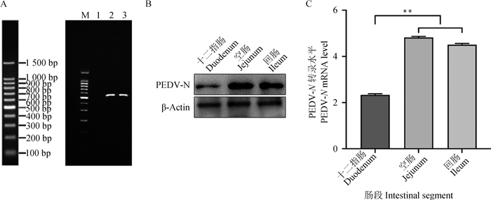
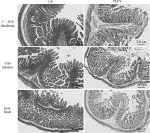
Fig. 2
PED piglets small intestinal histopathological changes (HE staining) Ctrl. Healthy piglet; PEDV. Piglets infected with PEDV"


Fig. 3
Changes of inflammatory factors in different sections of intestinal tissues of piglets (n=3) A. Changes in TNF-α content in piglet small intestine; B. Changes in IL-6 content in piglet small intestine; C. Changes in IL-8 content in piglet small intestine; D. Changes in IL-1β content in piglet small intestine; E. Changes in IL-10 content in piglet small intestine; F. Changes in TGF-β1 content in piglet small intestine; **P < 0.01, very significantly different compared with the control group (Ctrl)"
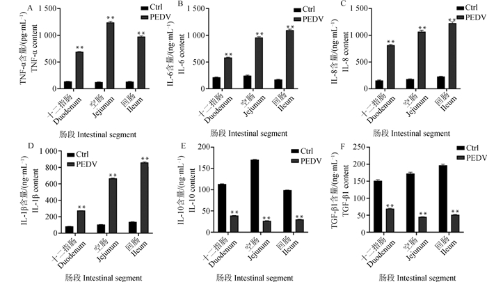
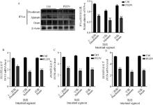
Fig. 4
Expression changes of IFN-α protein and related ISGs in different intestinal tissues of piglets (n=3) A. Expression of IFN-α protein in small intestine; B-D. Expression of ISG15, ISG54 and ISG56 genes in the small intestine; *P < 0.05, significantly different compared with the control group (Ctrl); **P < 0.01, very significantly different compared with the control group (Ctrl)"


Fig. 5
Changes of Ang Ⅱ and Ang (1-7) contents in different intestinal tissues of piglets (n=3) A. Changes in Ang Ⅱ content in piglet small intestine; B. Changes in Ang (1-7) content in piglet small intestine; **P < 0.01, very significantly different compared with the control group (Ctrl)"
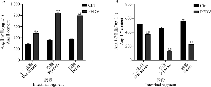
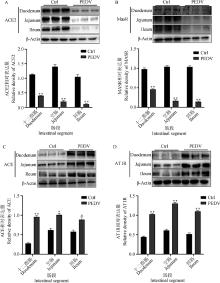
Fig. 6
Expression changes of ACE2, ACE, MasR and AT1R proteins in various intestinal tissues of piglets (n=3) A. Expression of ACE2 protein in small intestine; B. Expression of MasR protein in small intestine; C. Expression of ACE protein in small intestine; D. Expression of AT1R protein in small intestine; *P < 0.05, significantly different compared with the control group (Ctrl); ** P < 0.01, very significantly different compared with the control group (Ctrl)"
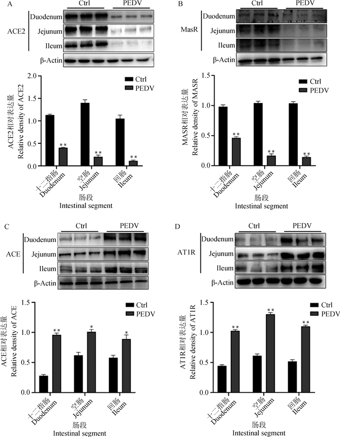
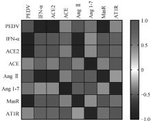
Fig. 7
Heatmap of PEDV and IFN-α and RAS components Blue. Negative correlation; Red. Positive correlation"

| 1 | VICKERS C , HALES P , KAUSHIK V , et al. Hydrolysis of biological peptides by human angiotensin-converting enzyme-related carboxypeptidase[J]. J Biol Chem, 2002, 277 (17): 14838- 14843. |
| 2 | LAMBERT D W , CLARKE N E , TURNER A J . Not just angiotensinases: new roles for the angiotensin-converting enzymes[J]. Cell Mol Life Sci, 2010, 67 (1): 89- 98. |
| 3 | 王珊珊, 黄瑜, 张树坤, 等. 血管紧张素转化酶2(ACE2)的基因克隆及在仔猪体内的表达分布[J]. 中国兽医学报, 2013, 33 (10): 1579- 1583. |
| WANG S S , HUANG Y , ZHANG S K , et al. Cloning of angiotensin converting enzyme 2(ACE2) gene and its expression distribution in piglets[J]. Chinese Journal of Veterinary Science, 2013, 33 (10): 1579- 1583. | |
| 4 | 权素玉, 王睿杰, 孙俊杰, 等. 血管紧张素转化酶2和中性氨基酸转运载体B0AT1在不同日龄猪肠道中的表达差异[J]. 畜牧与兽医, 2015, 47 (4): 36- 39. |
| QUAN S Y , WANG R J , SUN J J , et al. Different expression of angiotensin converting enzyme 2 and the neutral amino acid transporter B0AT1 in the intestinal tissues of different growth stage pigs[J]. Animal Husbandry & Veterinary Medicine, 2015, 47 (4): 36- 39. | |
| 5 | KEMP J R , UNAL H , DESNOYER R , et al. Angiotensin Ⅱ-regulated microRNA 483-3p directly targets multiple components of the renin-angiotensin system[J]. J Mol Cell Cardiol, 2014, 75 (7): 25- 39. |
| 6 | KIHARA M , SATO K , HASHIMOTO T , et al. Expression of endothelial nitric oxide synthase is suppressed in the renal vasculature of angiotensinogen-gene knockout mice[J]. Cell Tissue Res, 2006, 323 (2): 313- 320. |
| 7 | HASHIMOTO T , PERLOT T , REHMAN A , et al. ACE2 links amino acid malnutrition to microbial ecology and intestinal inflammation[J]. Nature, 2012, 487 (7408): 477. |
| 8 | PERLOT T , PENNINGER J M . ACE2-From the renine angiotensin system to gut microbiota and Malnutrition[J]. Microb Infect, 2013, 15 (13): 866- 873. |
| 9 | KOCHERHANS R , BRIDGEN A , ACKERMANN M , et al. Completion of the porcine epidemic diarrhoea coronavirus (PEDV) genome sequence[J]. Virus Genes, 2001, 23 (2): 137- 144. |
| 10 | SHIBATA I , TSUDA T , MORI M , et al. Isolation of porcine epidemic diarrhea virus in porcine cell cultures and experimental infection of pigs of different ages[J]. Vet Microbiol, 2000, 72 (3-4): 173- 182. |
| 11 | LI Y , WU Q , HUANG L , et al. An alternative pathway of enteric PEDV dissemination from nasal cavity to intestinal mucosa in swine[J]. Nat Commun, 2018, 9 (1): 3811. |
| 12 | BEAM A , GOEDE D , FOX A , et al. A porcine epidemic diarrhea virus outbreak in one geographic region of the United States: descriptive epidemiology and investigation of the possibility of airborne virus spread[J]. PLoS One, 2015, 10 (12): e0144818. |
| 13 | KIM Y , LEE C . Porcine epidemic diarrhea virus induces caspase-independent apoptosis through activation of mitochondrial apoptosis-inducing factor[J]. Virology, 2014 (460-461): 180- 193. |
| 14 | DING Z , FANG L , JING H , et al. Porcine epidemic diarrhea virus nucleocapsid protein antagonizes beta interferon production by sequestering the interaction between IRF3 and TBK1[J]. J Virol, 2014, 88 (16): 8936- 8945. |
| 15 | KOYAMA S , ISHII K J , COBAN C , et al. Innate immune response to viral infection[J]. Cytokine, 2008, 43 (3): 336- 341. |
| 16 | 李莎莎. 猪流行性腹泻病毒M蛋白抑制Ⅰ型干抚素产生的分子机制[D]. 北京: 中国农业科学院, 2022. |
| LI S S. Molecular Mechanism of porcine epidemic diarrhea virus membrane protein to inhibit type I interferon production[D]. Beijing: Chinese Academy of Agricultural Sciences, 2022. (in Chinese) | |
| 17 | PENNINGER J M , GRANT M B , SUNG J J Y . The role of angiotensin converting enzyme 2 in modulating gut microbiota, intestinal inflammation, and coronavirus infection[J]. Gastroenterology, 2021, 160 (1): 39- 46. |
| 18 | YISIREYILI M , UCHIDA Y , YAMAMOTO K , et al. Angiotensin receptor blocker irbesartan reduces stress-induced intestinal inflammation via AT1a signaling and ACE2-dependent mechanism in mice[J]. Brain Behav Immun, 2018, 69, 167- 179. |
| [1] | LI Qingyun, CAO Fengfeng, XING Zhou, LI Zhuoying, TAO Jinzhong. Progress in the Study of the Formation and Lysis of Corpus Luteum in Dairy Cows and Its Role in the Maintenance of Pregnancy [J]. Acta Veterinaria et Zootechnica Sinica, 2025, 56(7): 3088-3095. |
| [2] | WANG Yunke, WANG Na, YUE Ke, HE Kunmiao, ZHANG Xing, LIU Yao, ZHANG Gaiping. Substances with Inhibitory Effects on Porcine Epidemic Diarrhea Virus Replication in vitro [J]. Acta Veterinaria et Zootechnica Sinica, 2025, 56(6): 2577-2589. |
| [3] | WU Chao, MING Wenhan, LU Shuwan, YANG Caimei, LIU Jinsong, MA Xiang, ZHANG Ruiqiang. Innate Immune Evasion Mechanisms of Porcine Epidemic Diarrhea Virus and Advances in Prevention and Control Strategies [J]. Acta Veterinaria et Zootechnica Sinica, 2025, 56(6): 2590-2599. |
| [4] | ZHOU Min, TANG Deyuan, ZENG Zhiyong, WANG Bin, HUANG Tao, HU Wenwen, MAO Yinming, ZHOU Piao, HE Song. Research Progress on the Interaction between Porcine Epidemic Diarrhea Virus Proteins and Host Proteins [J]. Acta Veterinaria et Zootechnica Sinica, 2025, 56(6): 2600-2612. |
| [5] | LI Chengcheng, ZHAO Yongxiang, CAO Qiuxia, SONG Xu, LI Yupeng, FAN Baochao, GUO Rongli, XU Yefen, LI Bin. Tight Junction Protein CLDN4 Promotes Porcine Epidemic Diarrhea Virus Infection [J]. Acta Veterinaria et Zootechnica Sinica, 2025, 56(6): 2826-2835. |
| [6] | WU Qiong, LI Lingdan, YUAN Hui, BIN Chen, DENG Ke, LI Wei, YE Shiyi, LI Guopan, SHEN Qingchun, XIONG Tao. Prokaryotic Expression of PoIFN-α 8s and Identification of Its Activity in vitro and in vivo [J]. Acta Veterinaria et Zootechnica Sinica, 2025, 56(5): 2413-2423. |
| [7] | WU Peiling, LI Yixuan, WANG Haojie, LI Yafei, LIU Shaomeng, LIU Qingyun, WANG Xiangru. Research Progress of Porcine Epidemic Diarrhea Vaccine for Pigs [J]. Acta Veterinaria et Zootechnica Sinica, 2025, 56(3): 1042-1058. |
| [8] | YU Xinya, HE Haijian, WANG Lei, NI Yuchen, DU Jing, ZHOU Yingshan, DONG Wanyu, WANG Xiaodu. Effect of lncRNA 18850 on Porcine Epidemic Diarrhea Virus Replication [J]. Acta Veterinaria et Zootechnica Sinica, 2025, 56(3): 1366-1375. |
| [9] | ZHANG Dongxuan, WANG Zhihao, QIAO Yan, ZHAO Xiaoxiao, FAN Songjie, ZHANG Chao. Prokaryotic Expression of S1 Protein in Porcine Epidemic Diarrhea Virus and Screening of Its Aptamers [J]. Acta Veterinaria et Zootechnica Sinica, 2025, 56(2): 839-850. |
| [10] | WANG Yi, HOU Lulu, FANG Fei, GAO Linying, XIE Shumin, WANG Yu. Fluoride Induced Small Intestine Oxidative Damage in Broilers via Autophagy and Ferroptosis [J]. Acta Veterinaria et Zootechnica Sinica, 2025, 56(1): 442-454. |
| [11] | Yiqian FU, Dongge LIANG, Mingyang WANG, Jiajia PAN, Yanbin YANG, Lei ZENG, Xiangtao KANG. Construction of Interferon Regulatory Factor Knockdown Cell Line and Its Effect on Pseudorabies Virus Proliferation [J]. Acta Veterinaria et Zootechnica Sinica, 2024, 55(9): 4100-4109. |
| [12] | Weizhe LIU, Chenggang LUO, Rong YUAN, Yijie LIAO, Yimin WEN, Ying SUN, Enbo YU, Sanjie CAO, Xiaobo HUANG. Isolation and Identification of a Highly Pathogenic Strain of Porcine Epidemic Diarrhea Virus [J]. Acta Veterinaria et Zootechnica Sinica, 2024, 55(7): 3049-3063. |
| [13] | Dongliang LI, Guanmin ZHENG, Shuai LI, Hongsen ZHU, Chao WU. Differential Expression of Transcriptome in Jejunal of Piglets Infected with Porcine Epidemic Diarrhea Virus [J]. Acta Veterinaria et Zootechnica Sinica, 2024, 55(6): 2652-2661. |
| [14] | WANG Jing, ZHANG Shujuan, HU Xia, LIU Xiangyang, ZHANG Xingcui, SONG Zhenhui. CD44 Regulates Na+/H+ Exchanger 3 Activity by Influencing Porcine Epidemic Diarrhea Virus Replication [J]. Acta Veterinaria et Zootechnica Sinica, 2024, 55(5): 2176-2185. |
| [15] | HU Zeqi, LI Runcheng, TAN Zuming, XIE Xiuyan, WANG Jiangping, QIN Lejuan, LI Rong, GE Meng. Establishment and Preliminary Application of PEDV, PoRVA and PDCoV TaqMan Triple RT-qPCR Assay [J]. Acta Veterinaria et Zootechnica Sinica, 2024, 55(5): 2267-2272. |
| Viewed | ||||||
|
Full text |
|
|||||
|
Abstract |
|
|||||