


Acta Veterinaria et Zootechnica Sinica ›› 2025, Vol. 56 ›› Issue (8): 3976-3984.doi: 10.11843/j.issn.0366-6964.2025.08.035
• Preventive Veterinary Medicine • Previous Articles Next Articles
ZENG Shengxin1( ), SONG Chengqi2, SHEN Kaiyuan2, HAO Guoxin2, WANG Yakun2, WANG Xin2, WANG Xiaoxu3, LIU Zhijie3, LIU Yongbo2, LIU Yongsheng2, YANG Shunli2, FU Zhixin2,*()
Received:2024-11-01
Online:2025-08-23
Published:2025-08-28
Contact:
FU Zhixin
E-mail:zengshengxin@126.com;fzxljl@163.com
CLC Number:
ZENG Shengxin, SONG Chengqi, SHEN Kaiyuan, HAO Guoxin, WANG Yakun, WANG Xin, WANG Xiaoxu, LIU Zhijie, LIU Yongbo, LIU Yongsheng, YANG Shunli, FU Zhixin. Isolation, Identification of a Deer Clostridium perfringens and Pathogenicity Analysis in Mouse[J]. Acta Veterinaria et Zootechnica Sinica, 2025, 56(8): 3976-3984.
Table 1
The sequences of primers"
| 基因 Genes | 引物序列(5′→3′) Primer sequences | 片段长度/bp Product length |
| 16S rRNA | F-AGAGTTTGATCCTGGCTCAG | 1 465[ |
| R- GGTTACCTTGTTACGACTT | ||
| cpa | F-GCTAATGTTACTGCCGTTGA | 324 |
| R-CCTCTGATACATCGTGTAAG | ||
| cpb | F-GCGAATATGCTGAATCATCTA | 196 |
| R-GCAGGAACATTAGTATATCTTC | ||
| etx | F-GCGGTGATATCCATCTATTC | 655 |
| R-CCACTTACTTGTCCTACTAAC | ||
| itx | F-ACTACTCTCAGACAAGACAG | 446 |
| R-CTTTCCTTCTATTACTATACG | ||
| NetB | F-GCTGGTGCTGGAATAAATGC | 384 |
| R-TCGCCATTGAGTAGTTTCCC | ||
| cpe | F-GGAGATGGTTGGATATTAGG | 233 |
| R-GGACCAGCAGTTGTAGATA |

Fig. 1
Culture characteristics and staining microscopy of the isolates A. TSC medium; B. 7% Sheep blood agar medium; C. Isolated bacteria observed under microscopy (10×100)"
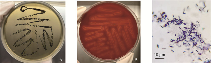

Fig. 2
PCR amplification electrophoresis map of 16S rRNA gene M. DNA marker; 1. Isolate sample; N. Blank control"
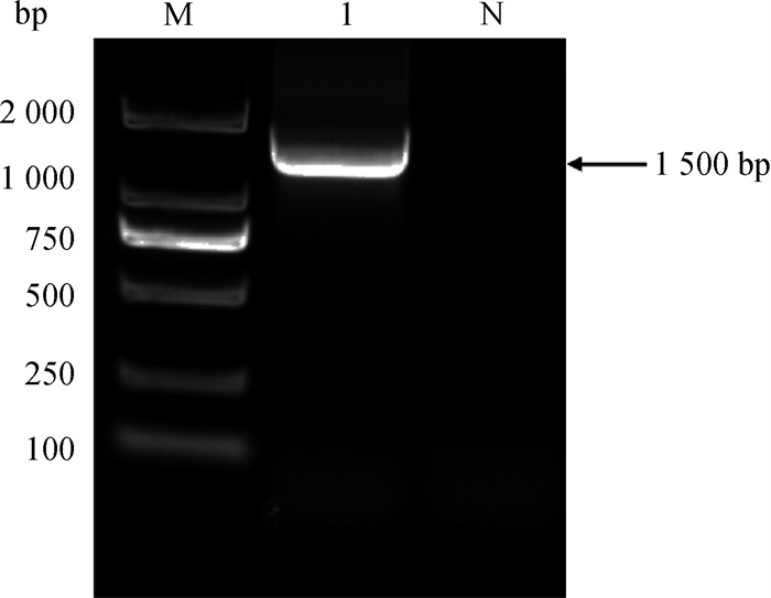
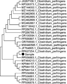
Fig. 3
Phylogenetic tree of 16S rRNA gene Sample. Isolate sample"


Fig. 4
Electropherogram of PCR amplification results of toxin genes M. DNA marker; 1. cpa gene; 2. cpb gene; 3. etx gene; 4. Iota gene; 5. NetB gene; 6. cpe gene"
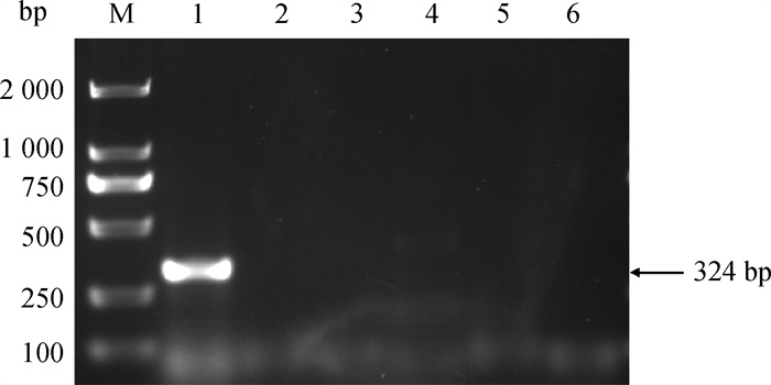
Table 2
Drug sensitivity test results"
| 药物种类 Drug type | 药物名称 Drugs | 缩写 Abbreviations | 药物含量/(μg·片-1) Drug content | 结果 Results |
| 氨基糖苷类 Aminoglycoside | 阿米卡星 Amikacin | AMK | 30 | R |
| 链霉素 Streptomycin | S | 10 | R | |
| 卡那霉素 Kanamycin | KAN | 30 | R | |
| 庆大霉素 Gentamicin | GM | 120 | S | |
| β-内酰胺类 β-lactams | 氨苄西林 Ampicillin | AMP | 10 | S |
| 头孢氨苄 Cefalexin | CN | 30 | S | |
| 苯唑西林 Oxacillin | OX | 1 | R | |
| 头孢唑林 Cefazolin | CZ | 30 | S | |
| 头孢他啶 Ceftazidime | CAZ | 30 | S | |
| 青霉素 Penicillin | PEN | 10 | S | |
| 四环素类 Tetracyclines | 强力霉素 Doxycycline | DO | 30 | S |
| 米诺环素 Minocycline | MI | 30 | S | |
| 四环素 Tetracycline | TET | 30 | S | |
| 多肽类 Polypeptide | 万古霉素 Vancomycin | VAN | 30 | R |
| 多黏菌素B Polymyxin B | PB | 300 IU | S | |
| 氯霉素类 Chloram phenicols | 氟苯尼考 Florfenicol | FFC | 30 | S |
| 喹诺酮类 Quinolones | 环丙沙星 Ciprofloxacin | CIP | 5 | I |
| 林可酰胺类 Lincosamides | 克林霉素 Clindamycin | CC | 2 | R |
| 大环内酯类 Macrolides | 红霉素 Erythromycin | E | 15 | R |
| 碳青霉烯类 Carbapenem | 亚胺培南 Imipenem | IPM | 10 | S |
| 磺胺类 Sulfonamides | 复方新诺明 Compound sulfamethoxazole | SXT | 25 | R |
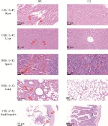
Fig. 5
HE staining results of tissues D. Experimental group; H. Healthy control group"

| 1 | KIUR,HALLL J.An update on the human and animal enteric pathogen Clostridium perfringens[J].Emerg Microbes Infect,2018,7(1):141. |
| 2 | 王晓旭,鲍坤,徐超,等.梅花鹿魏氏梭菌病的诊断与防治[J].特产研究,2023,45(3):13-17. |
| WANGX X,BAOK,XUC,et al.Diagnosis and control of clostridium wegelii disease in deer[J].Specialty Research,2023,45(3):13-17. | |
| 3 | 李浩.梅花鹿仔鹿的饲养管理技术要点[J].山东畜牧兽医,2024,45(8):37-38, 41. |
| LIH.Key technical points of feeding and management of Sika deer[J]. Shandong Animal Husbandry and Veterinary Science,2024,45(8):37-38, 41. | |
| 4 | 赵禹博,王祺伟.探讨梅花鹿魏氏梭菌病的诊断与防治[J].吉林畜牧兽医,2024,45(7):121-123. |
| ZHAOY B,WANGQ W.Study on the diagnosis and prevention of Clostridiosis of sika deer[J].Jilin Animal Husbandry and Veterinary Science,2024,45(7):121-123. | |
| 5 | 闫新华,赵传芳,王长凤.鹿肠毒血症的综合诊断[J].特种经济动植物,2002(10):38-39. |
| YANX H,ZHAOC F,WANGC F.Comprehensive diagnosis of deer enterotoxemia[J].Special Economic Flora and Fauna,2002(10):38-39. | |
| 6 | NGUYENT T,VU-KHACH,NGUYENT D.Isolation and characterization of Clostridium perfringens strains isolated from ostriches (Struthio camelus) in Vietnam[J].Vet World,2020,13(8):1679-1684. |
| 7 | 王冬经,吴金措姆,曾江勇.牦牛源A型产气荚膜梭菌的分离鉴定及生物学特性研究[J].中国畜牧兽医,2024,51(1):229-241. |
| WANGD J,WUJ Z,ZENGJ Y.Isolation, identification and biological characteristics of Clostridium perfringens type A from yaks[J].Chinese Journal of Animal Husbandry and Veterinary Medicine,2024,51(1):229-241. | |
| 8 | BUCHANANR E,GIBBONSN E.伯杰氏鉴定细菌学手册[M].8版北京:中国科学院微生物研究所,1984. |
| BUCHANANR E,GIBBONSN E.Berger's Manual of analytical bacteriology[M].8th editionBeijing:Institute of Microbiology, Chinese Academy of Sciences,1984. | |
| 9 | NCCLS. Performances tandards for antimicrobial disk and dilution susceptibility tests for bacteria isolated from animals[S]. Approved Standard, 2002: M31-A2. |
| 10 |
谭娟娟,杨贝莹,武前悦,等.江西地区野猪菌群多样性分析及其携带产气荚膜梭菌的分离鉴定[J].畜牧兽医学报,2025,56(4):1876-1886.
doi: 10.11843/j.issn.0366-6964.2025.04.035 |
|
TANJ J,YANGB Y,WUQ Y,et al.Diversity analysis of wild boar flora and isolation and identification of the carrier Clostridium perfringens in Jiangxi[J].Acta Veterinaria et Zootechnica Sinica,2025,56(4):1876-1886.
doi: 10.11843/j.issn.0366-6964.2025.04.035 |
|
| 11 | ROODJ I,ADAMSV,LACEYJ,et al.Expansion of the Clostridium perfringens toxin-based typing scheme[J].Anaerobe,2018,53,5-10. |
| 12 |
田睿,徐思翔,谢烽,等.黄牛源产气荚膜梭菌分离株基因组的生物信息学分析[J].畜牧兽医学报,2024,55(4):1707-1715.
doi: 10.11843/j.issn.0366-6964.2024.04.032 |
|
TIANR,XUS X,XIEF,et al.Bioinformatics analysis of genome of Clostridium perfringens isolates from cattle[J].Acta Veterinaria et Zootechnica Sinica,2024,55(4):1707-1715.
doi: 10.11843/j.issn.0366-6964.2024.04.032 |
|
| 13 | 吴钰兴,邹潍力,程子馨,等.1株猪源A型产气荚膜梭菌的分离鉴定及致病性研究[J].中国畜牧兽医,2024,51(6):2697-2706. |
| WUY X,ZOUW L,CHENGZ X,et al.Isolation, identification and pathogenicity of A strain of Clostridium perfringens type A from swine[J].Chinese Journal of Animal Science and Veterinary Medicine,2024,51(6):2697-2706. | |
| 14 |
吴克,冯航,王娟,等.关中奶山羊源D型产气荚膜梭菌全基因组序列测定及其分子特征分析[J].畜牧兽医学报,2022,53(11):3967-3974.
doi: 10.11843/j.issn.0366-6964.2022.11.022 |
|
WUK,FENGH,WANGJ,et al.Genome sequence analysis and molecular characteristics analysis of Clostridium perfringens type D from Guanzhong dairy goats[J]. Acta Veterinaria et Zootechnica Sinica,2022,53(11):3967-3974.
doi: 10.11843/j.issn.0366-6964.2022.11.022 |
|
| 15 |
阚刘刚,刘艳,吴媛媛,等.鸡坏死性肠炎生物性防控研究进展[J].畜牧兽医学报,2019,50(6):1123-1134.
doi: 10.11843/j.issn.0366-6964.2019.06.002 |
|
KANL G,LIUY,WUY Y,et al.Research progress on biological prevention and control of necrotizing enteritis in chickens[J].Acta Veterinaria et Zootechnica Sinica,2019,50(6):1133-1134.
doi: 10.11843/j.issn.0366-6964.2019.06.002 |
|
| 16 | 唐诗. 检测免源A型产气荚膜梭菌α毒素量子点免疫层析试纸条的试制[D]. 成都: 四川农业大学, 2023. |
| TANG S. Preparation of Quantum-dot immunochromatographic strip for detection of α-toxin from Clostridium perfringens type A[D]. Chengdu: Sichuan Agricultural University, 2023. (in Chinese) | |
| 17 | 董秀梅,李秀云,刘念,等.狼源产气荚膜梭菌的分离鉴定及分子分型[J].中国预防兽医学报,2022,44(8):831-836. |
| DONGX M,LIX Y,LIUN,et al.Isolation, identification and molecular typing of Clostridium perfringens from wolf[J].Chinese Journal of Preventive Veterinary Medicine,2022,44(8):831-836. | |
| 18 | 王斌,张娜,冯航,等.朱鹮产气荚膜梭菌的分离鉴定和致病性[J].中国兽医学报,2021,41(9):1770-1773, 1784. |
| WANGB,ZHANGN,FENGH,et al.Isolation, identification and pathogenicity of Clostridium perfringens in crested ibis[J]. Chinese Journal of Veterinary Medicine,2021,41(9):1770-1773, 1784. | |
| 19 | 孙立明,武士训,常柏林,等.梅花鹿肠毒血症的诊断[J].中国畜禽传染病,1992(1):28-29. |
| SUNL M,WUS X,CHANGB L,et al.Diagnosis of enterotoxemia in sika deer[J].Chinese Infectious Diseases of Livestock and Poultry,1992(1):28-29. | |
| 20 | 赵永江,刘丽洁,詹武,等.规模化鹿场爆发鹿魏氏梭菌和巴氏杆菌病[J].畜牧兽医杂志,2008(2):125. |
| ZHAOY J,LIUL J,ZHANW,et al.Outbreaks of Clostridium wegelii and pasteurellosis in deer on large-scale deer farms[J]. Journal of Animal Husbandry and Veterinary Medicine,2008(2):125. | |
| 21 | 阎喜军,闫新华,赵传芳,等.鹿魏氏梭菌分离鉴定及部分生物学特性的研究[J].特产研究,1999(1):22-23, 26. |
| YANX J,YANX H,ZHAOC F,et al.Isolation, identification and some biological characteristics of Clostridium wilsoni of deer[J].Specialty Research,1999(1):22-23, 26. | |
| 22 | 张凤翔,张魁,冯丽华,等.鹿猝死症的病因调查[J].中国兽医杂志,1997(12):16-17. |
| ZHANGF X,ZHANGK,FENGL H,et al.Investigation on etiology of sudden death syndrome in deer[J].Chinese Journal of Veterinary Medicine,1997(12):16-17. | |
| 23 | 俞乃胜,李光宗,韦剑珊,等.梅花鹿肠毒血症的细菌学鉴定[J].中国兽医科技,1994(10):28-30. |
| YUN S,LIG Z,WEIJ S,et al.Bacteriological identification of enterotoxemia in Sika deer[J].Chinese Veterinary Science and Technology,1994(10):28-30. | |
| 24 | 王圆圆,李宁,陈国亮,等.河北省承德市一起鹿魏氏梭菌病的紧急流行病学调查[J].中国动物检疫,2018,35(3):14-16. |
| WANGY Y,LIN,CHENG L,et al.An urgent epidemiological investigation of clostridium weilieri disease in deer in Chengde city, Hebei Province[J]. China Animal Quarantine,2018,35(3):14-16. | |
| 25 | 朱凯宗.山东地区鹿肠毒血症的综合诊断[J].畜牧兽医科技信息,2019(9):169-170. |
| ZHUK Z.Comprehensive diagnosis of deer enterotoxemia in Shandong[J].Science and Technology Information of Animal Husbandry and Veterinary Science,2019(9):169-170. | |
| 26 | 罗润波,吴丹,黄家旗,等.青藏高原地区牦牛源产气荚膜梭菌流行病学调查及耐药性分析[J].中国兽医学报,2023,43(9):1851-1858. |
| LUOR B,WUD,HUANGJ Q,et al.Epidemiological investigation and drug resistance analysis of Clostridium perfringens from yaks in Tibetan Plateau[J].Chinese Journal of Veterinary Medicine,2023,43(9):1851-1858. | |
| 27 | WELLINGTONE M,BOXALLA B,CROSSP,et al.The role of the natural environment in the emergence of antibiotic resistance in gram-negative bacteria[J].Lancet Infect Dis,2013,13(2):155-165. |
| 28 | LIJ,ADAMSV,BANNAMT L,et al.Toxin plasmids of Clostridium perfringens[J].Microbiol Mol Biol Rev,2013,77(2):208-233. |
| 29 | ODAM,TERAOY,SAKURAIJ,et al.Membrane-binding mechanism of Clostridium perfringens alpha-toxin[J].Toxins,2015,7(12):5268-5275. |
| 30 | TITBALLR W,NAYLORC E,BASAKA K.The Clostridium perfringens a-toxin[J].Anaerobe,1999,5(9):51-64. |
| 31 | 王璞,盛巧玲,李鑫,等.A型产气荚膜梭菌小鼠感染模型的建立[J].中国兽医杂志,2013(5):36-38, 100. |
| WANGP,SHENGQ L,LIX,et al.Establishment of A mouse infection model of Clostridium perfringens type A[J].Chinese Journal of Veterinary Medicine,2013(5):36-38, 100. | |
| 32 | 吴洁. A型产气荚膜梭菌家兔感染模型的建立[D]. 泰安: 山东农业大学, 2016. |
| WU J. Establishment of infection model of Clostridium perfringens type A rabbit[D]. Taian: Shandong Agricultural University, 2016. (in Chinese) | |
| 33 | 林家琪,金铎,陈诗涵,等.一例马鹿产气荚膜梭菌感染的诊断与防治[J].黑龙江畜牧兽医,2017(10):191-193. |
| LINJ Q,JIND,CHENS H,et al.Diagnosis and prevention of a case of Clostridium perfringens infection in red deer[J].Heilongjiang Animal Husbandry and Veterinary Science,2017(10):191-193. |
| [1] | TIAN Ru, FU Xingwei, HU Leyu, ZHU Mingjun, TONG Dewen. Isolation and Pathogenicity Analysis of a GⅡa Porcine Epidemic Diarrhea Virus [J]. Acta Veterinaria et Zootechnica Sinica, 2025, 56(8): 4101-4111. |
| [2] | ZHANG Xulin, TANG Yu, LIU Lixiang, FAN Bingfeng, ZHANG Yushi, ZHANG Ying, SHAO Jing, SUN Huimin, CHU Xiaoyu, PENG Feiyu, XU Baozeng. Effect of TLR7/8 Agonist R848 on the Efficiency of Y Sperm Sorting in Sika Deer [J]. Acta Veterinaria et Zootechnica Sinica, 2025, 56(7): 3278-3289. |
| [3] | JIA Qiong, GAO Shuaipeng, XIU Yanyu, REN Hongrui, ZHANG Shuyin, YANG Haoyu, FAN Ruiwen. Establishment of Double-antibody Sandwich ELISA Method Based on the Specifiec Nanobody against Clostridium Perfringens β Toxin [J]. Acta Veterinaria et Zootechnica Sinica, 2025, 56(6): 2906-2916. |
| [4] | CHEN Yunlong, FAN Gang, FAN Xinyi, GUO Yongchao, ZHANG Xinmiao, WANG Yan, ZHANG Shiqiang. Isolation and Identification of a Novel Porcine Circovirus Type 2d Strain with Multiple Nucleotide Substitutions [J]. Acta Veterinaria et Zootechnica Sinica, 2025, 56(6): 3032-3040. |
| [5] | WANG Shujuan, WANG Dongfang, MA Zhenyuan, ZHAO Xueli, LIU Ying, YANG Haibo, CHAI Mao, WANG Cui, YAN Ruoqian. Isolation, Identification and Evolutionary Analysis of a Strain of Senecavirus A [J]. Acta Veterinaria et Zootechnica Sinica, 2025, 56(6): 3041-3046. |
| [6] | WANG Yanan, GUO Yaru, JIANG Yanping, CUI Wen, LI Jiaxuan, LI Yijing, WANG Li. Isolation, Identification and Pathogenicity Analysis of Porcine Rotavirus [J]. Acta Veterinaria et Zootechnica Sinica, 2025, 56(5): 2259-2269. |
| [7] | LI Ting, ZHANG Chengcheng, WANG Xiuling, ZHANG Xiaorong, WU Yantao. Isolation and Identification of a Novel Duck Reovirus Strain and the Sequence Analysis of Its σC Gene [J]. Acta Veterinaria et Zootechnica Sinica, 2025, 56(5): 2520-2524. |
| [8] | TAN Juanjuan, YANG Beiying, WU Qianyue, HUA Huiying, CAO Huabin, YAN Hui, ZHANG Jinhua. Diversity Analysis of Wild Boar Flora in Jiangxi Region and the Isolation and Identification of Clostridium perfringens Carried by Wild Boar [J]. Acta Veterinaria et Zootechnica Sinica, 2025, 56(4): 1876-1886. |
| [9] | ABABAIKERI Buweihailiqiemu, AIHEMAITIJIANG Aihemaitiniyazi, MOHAMMADTURSUN Nabijan, SHAN Wenjuan. Preliminary Study on the Function of PRDX1 Antioxidant Gene in Tarim Red Deer [J]. Acta Veterinaria et Zootechnica Sinica, 2025, 56(3): 1216-1230. |
| [10] | LIU Jian, YU Zehai, ZHANG Meiyu, LI Dan, WANG Jun, LIU Fangqin, ZHANG Qun, XU Shouzhen. Full-genome Analysis of a Bovine Enterovirus Type F and the Establishment of an Indirect ELISA Method for Antibody Detection [J]. Acta Veterinaria et Zootechnica Sinica, 2025, 56(2): 814-825. |
| [11] | DU Qingjie, WU Liping, ZHANG Fan, DAI Pengxiu, FENG Xiancheng, ZHANG Xinke. Difference Analysis of Oral Flora in Dogs with Periodontitis and Drug Resistance of Oral Porphyromonas [J]. Acta Veterinaria et Zootechnica Sinica, 2025, 56(2): 934-942. |
| [12] | Guangxuan FAN, Tianjiao WANG, Yimeng DONG, Hongliang WANG, Ning DING, Xinhao WANG, Xiumei XING. Molecular Genealogy Construction and Population Genetic Structure Analysis of Jilin Sika Deer Based on SNP Loci [J]. Acta Veterinaria et Zootechnica Sinica, 2024, 55(9): 3925-3935. |
| [13] | Shan ZHANG, Dahu LIU, Baojing LIU, Lin LIANG, Ruiying LIANG, Xinming TANG, Xusheng QIU, Chan DING, Jiabo DING, Shaohua HOU. Isolation, Identification and Pathogenicity Analysis of a Pigeon Paramyxovirus-1 Strain [J]. Acta Veterinaria et Zootechnica Sinica, 2024, 55(9): 4051-4060. |
| [14] | Yan WANG, Yadong GAO, Chenghui JIANG, Qiaoying ZENG. Isolation and Pathogenicity of a Goose Derived Fowl Adenovirus Type 4 [J]. Acta Veterinaria et Zootechnica Sinica, 2024, 55(9): 4232-4240. |
| [15] | Zhangrong PENG, Haoran SUN, Qiaoru ZHANG, Ying YANG, Hongying GUO, Tong CHANG, Hui ZHAO, Tietao ZHANG. Study on the Pattern of Intramuscular Fat Deposition and Its Influence in Flavor Quality of Sika Deer at Different Ages [J]. Acta Veterinaria et Zootechnica Sinica, 2024, 55(8): 3541-3551. |
| Viewed | ||||||
|
Full text |
|
|||||
|
Abstract |
|
|||||