


Acta Veterinaria et Zootechnica Sinica ›› 2024, Vol. 55 ›› Issue (8): 3678-3687.doi: 10.11843/j.issn.0366-6964.2024.08.039
• Basic Veterinary Medicine • Previous Articles Next Articles
Zuhua YU1,2( ), Mengru GAO1,2, Lei HE1,2, Ying WEI1,2, Jian CHEN1,2, Songbiao CHEN1,2, Ke DING1,2,3,*()
Received:2023-11-08
Online:2024-08-23
Published:2024-08-28
Contact:
Ke DING
E-mail:yzhd05@163.com;keding19@163.com
CLC Number:
Zuhua YU, Mengru GAO, Lei HE, Ying WEI, Jian CHEN, Songbiao CHEN, Ke DING. Effects of mdv1-miR-M4-5p Encoded by MDV on Proliferation and Apoptosis of MDCC-MSB1 Cells[J]. Acta Veterinaria et Zootechnica Sinica, 2024, 55(8): 3678-3687.
Table 1
Real-time PCR primer sequences"
| 名称 Name | 引物序列(5′→3′) Sequence | 用途 Application |
| U6 RT prime | GTCGTATCCAGTGCAGGGTCCGAGGTATTCGCACTGGATACGACCGATACA | 反转录 |
| U6 forward | CGCTTCGGCAGCACATATAC | Real-time PCR |
| miR-M4-5p RT prime | GTCGTATCCAGTGCAGGGTCCGAGGTATTCGCACTGGATACGACGAAGGGTT | 反转录 |
| miR-M4-5p forward | TGCGCTTAATGCTGTATCGGAAC | Real-time PCR |
| miR-M4-5p/U6 reverse | CCAGTGCAGGGTCCGAGGTATT | Real-time PCR |
| GAPDH forward | CCAGAACATCATCCCAGCGT | Real-time PCR |
| GAPDH reverse | CAGGTCAGGTCAACAACAGA | Real-time PCR |
| TGF-β1 forward | GTATTGGGCCAAAGAGCTGC | Real-time PCR |
| TGF-β1 reverse | ATTGCCGTAACCCTGGTACA | Real-time PCR |
| Smad2 forward | GTGGCAGATGAGGGTGATTT | Real-time PCR |
| Smad2 reverse | TGCAGCCACAGGACGAAAC | Real-time PCR |
| Caspase-3 forward | TGTGGACTCTGGAATTCTGC | Real-time PCR |
| Caspase-3 reverse | TGCCTGAACGAGATGACAGT | Real-time PCR |
| Caspase-9 forward | CCGAAGGGAGCAAGCACG | Real-time PCR |
| Caspase-9 reverse | AGGTTGGACTGGGATGGAC | Real-time PCR |
| Cyt-c forward | AGGCAAGCACAAGACTGGA | Real-time PCR |
| Cyt-c reverse | CTGACTATCACCAAGAACCACC | Real-time PCR |
| Bcl-2 forward | TACCAGAGGGACTTCGC | Real-time PCR |
| Bcl-2 reverse | TTGACCCCATCACGGA | Real-time PCR |
| cyclinD1 forward | AGGTTGGACTGGGATGGAC | Real-time PCR |
| cyclinD1 reverse | TGCAGCCACAGGACGAAACAG | Real-time PCR |

Fig. 1
Real-time quantitative PCR results of TGF-β1, Smad2 and genes related to cell proliferation and apoptosis A-H. The relative transcription of mdv1-miR-M4-5p, TGF-β1, Smad2, caspase-3, caspase-9, cyclinD1, Bcl-2 and cyt-c respectively. **. P < 0.01"


Fig. 2
Effects of mdv1-miR-M4-5p on the proliferation of MDCC-MSB1 cells **. P < 0.01"
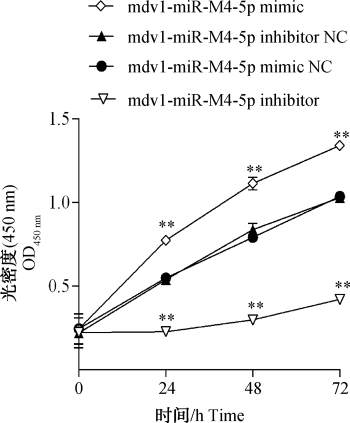

Fig. 3
Effects of mdv1-miR-M4-5p on the cell cycle of MDCC-MSB1 cells A. mdv1-miR-M4-5p mimic NC; B. mdv1-miR-M4-5p mimic; C. The numerical statistics of mdv1-miR-M4-5p mimic and its negative control; D. mdv1-miR-M4-5p inhibitor NC; E. mdv1-miR-M4-5p inhibitor; F. The numerical statistics of mdv1-miR-M4-5p inhibitor and its negative control. *. P < 0.05"
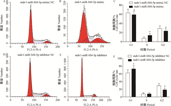

Fig. 4
Effects of mdv1-miR-M4-5p on the apoptosis of MDCC-MSB1 cells A. mdv1-miR-M4-5p mimic NC; B. mdv1-miR-M4-5p mimic; C. mdv1-miR-M4-5p inhibitor NC; D. mdv1-miR-M4-5p inhibitor; E. Percentage of apoptosis in each group. **. P < 0.01"

| 1 |
SZCZEPANEK J , SKORUPA M , TRETYN A . MicroRNA as a potential therapeutic molecule in cancer[J]. Cells, 2022, 11 (6): 1008.
doi: 10.3390/cells11061008 |
| 2 |
LIANG C , YANG J B , LIN X Y , et al. Recent advances in the diagnostic and therapeutic roles of microRNAs in colorectal cancer progression and metastasis[J]. Front Oncol, 2022, 12, 911856.
doi: 10.3389/fonc.2022.911856 |
| 3 |
CHATTERJEE B , SAHA P , BOSE S , et al. MicroRNAs: as critical regulators of tumor-associated macrophages[J]. Int J Mol Sci, 2020, 21 (19): 7117.
doi: 10.3390/ijms21197117 |
| 4 |
HULL R , MARIMA R , ALAOUNA M , et al. Viral encoded miRNAs in tumorigenesis: theranostic opportunities in precision oncology[J]. Microorganisms, 2022, 10 (7): 1448.
doi: 10.3390/microorganisms10071448 |
| 5 |
VOJTECHOVA Z , TACHEZY R . The role of miRNAs in virus-mediated oncogenesis[J]. Int J Mol Sci, 2018, 19 (4): 1217.
doi: 10.3390/ijms19041217 |
| 6 |
MACHADO C B , DA CUNHA L S , DA SILVA MAUÉS J H , et al. Role of miRNAs in human T cell leukemia virus type 1 induced T cell leukemia: a literature review and bioinformatics approach[J]. Int J Mol Sci, 2022, 23 (10): 5486.
doi: 10.3390/ijms23105486 |
| 7 |
TENG M , ZHU Z J , YAO Y X , et al. Critical roles of non-coding RNAs in lifecycle and biology of Marek's disease herpesvirus[J]. Sci China Life Sci, 2023, 66 (2): 251- 268.
doi: 10.1007/s11427-022-2258-4 |
| 8 |
CHEN L , GAO D , SHAO Z Z , et al. miR-155 indicates the fate of CD4+ T cells[J]. Immunol Lett, 2020, 224, 40- 49.
doi: 10.1016/j.imlet.2020.05.003 |
| 9 |
靳睿哲, 王迪娴, 赵乾, 等. miR-155-5p在肿瘤中的表达、功能以及调控作用[J]. 肿瘤防治研究, 2023, 50 (3): 309- 315.
doi: 10.3971/j.issn.1000-8578.2023.22.1026 |
|
JIN R Z , WANG D X , ZHAO Q , et al. miR-155-5p expression, function and regulation in tumors[J]. Cancer Research on Prevention and Treatment, 2023, 50 (3): 309- 315.
doi: 10.3971/j.issn.1000-8578.2023.22.1026 |
|
| 10 |
杨攀, 王萍, 周细武. 循环miR-155作为肿瘤生物标志物的研究进展[J]. 中国细胞生物学学报, 2016, 38 (10): 1281- 1287.
doi: 10.11844/cjcb.2016.10.0138 |
|
YANG P , WANG P , ZHOU X W . Recent advances in circulating miR-155 as tumor biomarker[J]. Chinese Journal of Cell Biology, 2016, 38 (10): 1281- 1287.
doi: 10.11844/cjcb.2016.10.0138 |
|
| 11 |
余祖华, 丁轲, 郁川, 等. gga-miR-155对MDCC-MSB1细胞生物学行为的影响[J]. 畜牧兽医学报, 2018, 49 (11): 2496- 2504.
doi: 10.11843/j.issn.0366-6964.2018.11.022 |
|
YU Z H , DING K , YU C , et al. Effect of gga-miR-155 on the biological behavior of MDCC-MSB1 cells[J]. Acta Veterinaria et Zootechnica Sinica, 2018, 49 (11): 2496- 2504.
doi: 10.11843/j.issn.0366-6964.2018.11.022 |
|
| 12 |
DAVISON A J , EBERLE R , EHLERS B , et al. The order Herpesvirales[J]. Arch Virol, 2009, 154 (1): 171- 177.
doi: 10.1007/s00705-008-0278-4 |
| 13 |
ZHUANG G Q , SUN A J , TENG M , et al. A tiny RNA that packs a big punch: the critical role of a viral miR-155 ortholog in lymphomagenesis in Marek's disease[J]. Front Microbiol, 2017, 8, 1169.
doi: 10.3389/fmicb.2017.01169 |
| 14 | ZHANG Y Y , TANG N , LUO J , et al. Marek's disease virus-encoded microRNA 155 ortholog critical for the induction of lymphomas is not essential for the proliferation of transformed cell lines[J]. J Virol, 2019, 93 (17): e00713- 19. |
| 15 | LUO J , SUN A J , TENG M , et al. Expression profiles of microRNAs encoded by the oncogenic Marek's disease virus reveal two distinct expression patterns in vivo during different phases of disease[J]. J Gen Virol, 2011, 92 (Pt 3): 608- 620. |
| 16 |
ZHAO Y G , XU H T , YAO Y X , et al. Critical role of the virus-encoded microRNA-155 ortholog in the induction of Marek's disease lymphomas[J]. PLoS Pathog, 2011, 7 (2): e1001305.
doi: 10.1371/journal.ppat.1001305 |
| 17 |
YU Z H , TENG M , SUN A J , et al. Virus-encoded miR-155 ortholog is an important potential regulator but not essential for the development of lymphomas induced by very virulent Marek's disease virus[J]. Virology, 2014, 448, 55- 64.
doi: 10.1016/j.virol.2013.09.017 |
| 18 |
ZHAO Y G , YAO Y X , XU H T , et al. A functional microRNA-155 ortholog encoded by the oncogenic Marek's disease virus[J]. J Virol, 2009, 83 (1): 489- 492.
doi: 10.1128/JVI.01166-08 |
| 19 |
CHI J Q , TENG M , YU Z H , et al. Marek's disease virus-encoded analog of microRNA-155 activates the oncogene c-Myc by targeting LTBP1 and suppressing the TGF-β signaling pathway[J]. Virology, 2015, 476, 72- 84.
doi: 10.1016/j.virol.2014.11.027 |
| 20 |
WANG J M , XIANG H J , LU Y F , et al. Role and clinical significance of TGF-β1 and TGF-βR1 in malignant tumors (Review)[J]. Int J Mol Med, 2021, 47 (4): 55.
doi: 10.3892/ijmm.2021.4888 |
| 21 |
KUHIKAR R , KHAN N , PHILIP J , et al. Transforming growth factor β1 accelerates and enhances in vitro red blood cell formation from hematopoietic stem cells by stimulating mitophagy[J]. Stem Cell Res Ther, 2020, 11 (1): 71.
doi: 10.1186/s13287-020-01603-z |
| 22 |
BELKOURCHIA F , DESROSIERS R R . The protein L-Isoaspartyl (D-Aspartyl) methyltransferase regulates glial-to-mesenchymal transition and migration induced by TGF-β1 in human U-87 MG Glioma cells[J]. Int J Mol Sci, 2022, 23 (10): 5698.
doi: 10.3390/ijms23105698 |
| 23 |
DEVAN A R , PAVITHRAN K , NAIR B , et al. Deciphering the role of transforming growth factor-beta 1 as a diagnostic-prognostic-therapeutic candidate against hepatocellular carcinoma[J]. World J Gastroenterol, 2022, 28 (36): 5250- 5264.
doi: 10.3748/wjg.v28.i36.5250 |
| 24 |
KIM N , RYU H , KIM S , et al. CXCR7 promotes migration and invasion in head and neck squamous cell carcinoma by upregulating TGF-β1/Smad2/3 signaling[J]. Sci Rep, 2019, 9 (1): 18100.
doi: 10.1038/s41598-019-54705-x |
| 25 |
DONG Z H , SUN Y Y , WEI G W , et al. Ergosterol ameliorates diabetic nephropathy by attenuating mesangial cell proliferation and extracellular matrix deposition via the TGF-β1/Smad2 signaling pathway[J]. Nutrients, 2019, 11 (2): 483.
doi: 10.3390/nu11020483 |
| 26 | WU S , JI L A , FAN X M , et al. Jieduquyuzishen prescription attenuates renal fibrosis in MRL/lpr mice via inhibiting EMT and TGF-β1/Smad2/3 pathway[J]. Evid Based Complement Alternat Med, 2022, 2022, 4987323. |
| 27 |
ZHANG Y Q , HUA L P , LIN C F , et al. Pien-Tze-Huang alleviates CCl4-induced liver fibrosis through the inhibition of HSC autophagy and the TGF-β1/Smad2 pathway[J]. Front Pharmacol, 2022, 13, 937484.
doi: 10.3389/fphar.2022.937484 |
| 28 |
LOUAFI F , MARTINEZ-NUNEZ R T , SANCHEZ-ELSNER T . MicroRNA-155 targets SMAD2 and modulates the response of macrophages to transforming growth factor-β[J]. J Biol Chem, 2010, 285 (53): 41328- 41336.
doi: 10.1074/jbc.M110.146852 |
| 29 |
MAHESH G , BISWAS R . MicroRNA-155: a master regulator of inflammation[J]. J Interferon Cytokine Res, 2019, 39 (6): 321- 330.
doi: 10.1089/jir.2018.0155 |
| 30 |
XU W D , FENG S Y , HUANG A F . Role of miR-155 in inflammatory autoimmune diseases: a comprehensive review[J]. Inflamm Res, 2022, 71 (12): 1501- 1517.
doi: 10.1007/s00011-022-01643-6 |
| 31 |
COSTINEAN S , ZANESI N , PEKARSKY Y , et al. Pre-B cell proliferation and lymphoblastic leukemia/high-grade lymphoma in Eμ-miR155 transgenic mice[J]. Proc Natl Acad Sci U S A, 2006, 103 (18): 7024- 7029.
doi: 10.1073/pnas.0602266103 |
| 32 |
RILEY K J , RABINOWITZ G S , YARIO T A , et al. EBV and human microRNAs co-target oncogenic and apoptotic viral and human genes during latency[J]. EMBO J, 2012, 31 (9): 2207- 2221.
doi: 10.1038/emboj.2012.63 |
| 33 |
LIU Y , YANG H L , ZHONG F F , et al. Anti-apoptotic function of herpes simplex virus-2 latency-associated transcript RL1 sequence and screening of its encoded microRNAs[J]. Clin Exp Dermatol, 2016, 41 (7): 782- 791.
doi: 10.1111/ced.12671 |
| 34 | LIU X Y , HAPPEL C , ZIEGELBAUER J M . Kaposi's sarcoma-associated herpesvirus microRNAs target GADD45B to protect infected cells from cell cycle arrest and apoptosis[J]. J Virol, 2017, 91 (3): e02045- 16. |
| 35 |
QIE S , DIEHL J A . Cyclin D1, cancer progression, and opportunities in cancer treatment[J]. J Mol Med (Berl), 2016, 94 (12): 1313- 1326.
doi: 10.1007/s00109-016-1475-3 |
| 36 |
MONTALTO F I , DE AMICIS F . Cyclin D1 in cancer: a molecular connection for cell cycle control, adhesion and invasion in tumor and stroma[J]. Cells, 2020, 9 (12): 2648.
doi: 10.3390/cells9122648 |
| 37 |
DADSENA S , ZOLLO C , GARCÍA-SÁEZ A J . Mechanisms of mitochondrial cell death[J]. Biochem Soc Trans, 2021, 49 (2): 663- 674.
doi: 10.1042/BST20200522 |
| 38 |
余祖华, 丁轲, 贾艳艳, 等. 鸡TGFβ1对MDCC-MSB1细胞增殖、凋亡、迁移与侵袭的影响[J]. 畜牧兽医学报, 2020, 51 (10): 2567- 2575.
doi: 10.11843/j.issn.0366-6964.2020.10.025 |
|
YU Z H , DING K , JIA Y Y , et al. Effect of Gallus TGFβ1 on the proliferation, apoptosis, migration and invasion of MDCC-MSB1 cells[J]. Acta Veterinaria et Zootechnica Sinica, 2020, 51 (10): 2567- 2575.
doi: 10.11843/j.issn.0366-6964.2020.10.025 |
| [1] | LU Zenghua, CUI Yan, YU Sijiu, BAI Xuefeng, LU Hongqin, HE Junfeng, LU Kai, ZHAI Guoliang, QI Zhengman. Effect of Erythropoietin on the Expression of Apoptotic Factor in Yak Renal Interstitial Fibroblasts [J]. Acta Veterinaria et Zootechnica Sinica, 2024, 55(8): 3460-3471. |
| [2] | Yudian SUN, Ziyue SONG, Hongliang ZHANG, Zhihua QIN, Hu SHAN, Ruimei YANG. Isolation and ldentification of Duckling Short Beak and Dwarfism Syndrome Virus [J]. Acta Veterinaria et Zootechnica Sinica, 2024, 55(8): 3623-3630. |
| [3] | XU Xiying, WANG Yiheng, OU Qianting, HONG Linyuan, LIU Xujing, LU Xianying, JIA Kun. Effects of Silencing PREX1 Expression on Proliferation and Invasiveness of CHMp [J]. Acta Veterinaria et Zootechnica Sinica, 2024, 55(8): 3706-3713. |
| [4] | Ziyan WANG, Yahui WANG, Tianyi WU, Chen GAO, Zhenwei DU, Fei GE, Xiaobei ZHANG, Wenxuan ZHAO, Lupei ZHANG, Huijiang GAO, Huansheng DONG, Junya LI. INTS11 Promotes the Proliferation of Bovine Myoblasts by Mediating the Transcription of CDK2 and CYCLIND1 [J]. Acta Veterinaria et Zootechnica Sinica, 2024, 55(7): 2927-2939. |
| [5] | Milan MA, Qi WANG, Qiu YAN, Tianan LI, Xingxu ZHAO, Yong ZHANG. Expression of HIG1 Hypoxia Inducible Domain Family Member 1C in Cryptorchidism of Yak and Its Regulatory Mechanism [J]. Acta Veterinaria et Zootechnica Sinica, 2024, 55(7): 2983-2994. |
| [6] | Jinting LUO, Fafang XU, Lei WANG, Xuan LUO, Yuhong MA, Jianbo ZHANG, Weihua HUANG, Yuejun SHANG, Guofang WU. The Effect of RSP on Cell Proliferation and Apoptosis of Porcine Leydig Cells with Hypoxia [J]. Acta Veterinaria et Zootechnica Sinica, 2024, 55(6): 2441-2450. |
| [7] | Huijie REN, Xun MA, Jing WANG, Caixia LIU, Dongdong ZENG, Lijun KOU, Weidi SHI, Shuangfei LÜ, Ruixuan QIAN, Shengjie GAO. Construction and Partial Biological Characteristics Trial of Lm4b_02325/26 Double Gene Deletion Strain of Listeria monocytogenes [J]. Acta Veterinaria et Zootechnica Sinica, 2024, 55(6): 2578-2587. |
| [8] | DONG Shucan, MAO Shuaixiang, WU Cuiying, LI Yaokun, SUN Baoli, GUO Yongqing, DENG Ming, LIU Dewu, LIU Guangbin. The Effect of the Androgen Receptor Inhibitor Enzalutamide on Proliferation and Apoptosis of Goat Ovarian Granulosa Cells [J]. Acta Veterinaria et Zootechnica Sinica, 2024, 55(5): 2022-2031. |
| [9] | WANG Jiying, YIN Ruiru, XIE Xing, WANG Haiyan, LIU Hudong, HU Hui, XIONG Qiyan, FENG Zhixin, SHAO Guoqing, YU Yanfei. Effects of LDH in Mesomycoplasma (Mycoplasma) hyopneumoniae on Apoptosis of Porcine Bronchial Epithelial Cells [J]. Acta Veterinaria et Zootechnica Sinica, 2024, 55(5): 2195-2205. |
| [10] | LI Qiuyun, TIAN Xinyuan, LIAO Wensheng, ZHANG Huanrong, REN Yupeng, YANG Falong, ZHU Jiangjiang, XIANG Hua. Effects of SOCS2 on Proliferation, Cycle and Apoptosis of Turbinate Bone Cells in Goats [J]. Acta Veterinaria et Zootechnica Sinica, 2024, 55(5): 2226-2240. |
| [11] | LAN Xinrui, ZHAO Baobao, ZHANG Bihan, LIN Xiaoyu, MA Huiming, WANG Yongsheng. Effects of β-sitosterol on Porcine Oocyte Maturation and Embryonic Development in Vitro [J]. Acta Veterinaria et Zootechnica Sinica, 2024, 55(4): 1629-1637. |
| [12] | HU Qiaoyan, ZHAI Xiangqin, LI Yidan, HAN Jiale, LEI Chuzhao, DANG Ruihua. Effects of bta-miR-101 on Proliferation, Apoptosis and Secretion of Bovine Testicular Sertoli Cells [J]. Acta Veterinaria et Zootechnica Sinica, 2024, 55(3): 1040-1051. |
| [13] | HUO Yuannan, QIU Meijia, ZHANG Jiaojiao, YANG Weirong, WANG Xianzhong. Arginine and Its Metabolites Attenuate Heat Stress-induced Apoptosis of Immature Boar Sertoli Cells [J]. Acta Veterinaria et Zootechnica Sinica, 2024, 55(2): 587-597. |
| [14] | QIU Wenyue, SU Yiman, YE Jiali, ZHANG Xinting, PANG Xiaoyue, WANG Rongmei, XIE Zimao, ZHANG Hui, TANG Zhaoxin, SU Rongsheng. Study on Asiatic Acid Alleviates LPS-induced Acute Kidney Injury by Regulating Apoptosis and Autophagy of Broilers [J]. Acta Veterinaria et Zootechnica Sinica, 2024, 55(2): 809-821. |
| [15] | CHEN Songbiao, LIU Feifei, SHANG Ke, YU Zuhua, HE Lei, WEI Ying, CHEN Jian, ZHANG Chunjie, CHENG Xiangchao, DING Ke. Molecular Mechanism of the “Battle” between Virus Infection and Host Antiviral Immunity-Apoptosis, Necroptosis and Pyroptosis [J]. Acta Veterinaria et Zootechnica Sinica, 2024, 55(1): 59-70. |
| Viewed | ||||||
|
Full text |
|
|||||
|
Abstract |
|
|||||