


Acta Veterinaria et Zootechnica Sinica ›› 2026, Vol. 57 ›› Issue (1): 443-453.doi: 10.11843/j.issn.0366-6964.2026.01.039
• BASIC VETERINARY MEDICINE • Previous Articles Next Articles
QI Jiahui( ), FU Tingting, ZHENG Minxing, WANG Xuanjing, WU Haiyang, LU Jiayin, LUO Xiaomao, YU Xiuju, WANG Haidong(), YAN Yi()
Received:2025-04-18
Online:2026-01-23
Published:2026-01-26
Contact:
WANG Haidong, YAN Yi
E-mail:547475459@qq.com;sxaudywhd@163.com;sxaudywhd@ 163.com;yanyi@sxau.edu.cn
CLC Number:
QI Jiahui, FU Tingting, ZHENG Minxing, WANG Xuanjing, WU Haiyang, LU Jiayin, LUO Xiaomao, YU Xiuju, WANG Haidong, YAN Yi. Peroxisome Proliferator-activated Receptor α Promotes Skeletal Muscle Development in Mice by Regulating the AMPK/P38 MAPK Pathway[J]. Acta Veterinaria et Zootechnica Sinica, 2026, 57(1): 443-453.
Table 1
Primers sequence used in this study"
引物名称 Primer name | 引物序列(5´→3´) Primer sequences |
|---|---|
| PPARα | F: AGAGCCCCATCTGTCCTCTC; R: ACTGGTAGTCTGCAAAACCAAA |
| MyoD | F: AATGGCTACGACACCGCCTACT; R: GGGTCTGGGTTCCCTGTTCTGT |
| MyoG | F: AACTACCTTCCTGTCCACCTTC; R: CACAGACTTCCTCTTACACACCT |
| 36B4 | F: ACTGAGATTCGGGATATGCTGT; R: CCCACCTTGTCTCCAGTCTTTA |
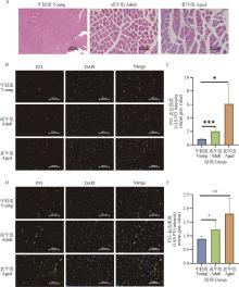
Fig.1
Morphology of gastrocnemius muscle and changes in P21 and P53 expression in young, adult, and aged miceA. HE staining of gastrocnemius muscle in young, adult, and aging mice (bar=100 μm); B, C. Immunofluorescence detection and quantification of P21 expression in mouse gastrocnemius muscle of different ages (in Fig.B, bar=200 μm); D, E. Immunofluorescence detection and quantification of P53 expression in mouse gastrocnemius muscle of different ages (in Fig.D, bar=200 μm). *. P<0.05, **. P < 0.01, ***. P<0.001"

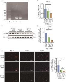
Fig.2
With the increase of age, the expression of PPARα in the gastrocnemius muscle of mice decreased at both mRNA and protein levelsA. PCR detection of PPARα expression in skeletal muscle (M. DL500 DNA marker; 1. Negative control, DEPC water; 2. Positive control, liver tissue cDNA; 3. Target band, gastrocnemius cDNA); B. Real-time quantitative PCR was used to detect the expression of PPARα; C, D. Western blot to detect the expression level and quantitative analysis of PPARα protein; E, F. Immunofluorescence was used to detect the expression and quantification of PPARα in gastrocnemius muscle at different developmental stages (in Fig.E, bar=100 μm). *. P<0.05, **. P<0.01, ***. P<0.001, ****. P<0.000 1"
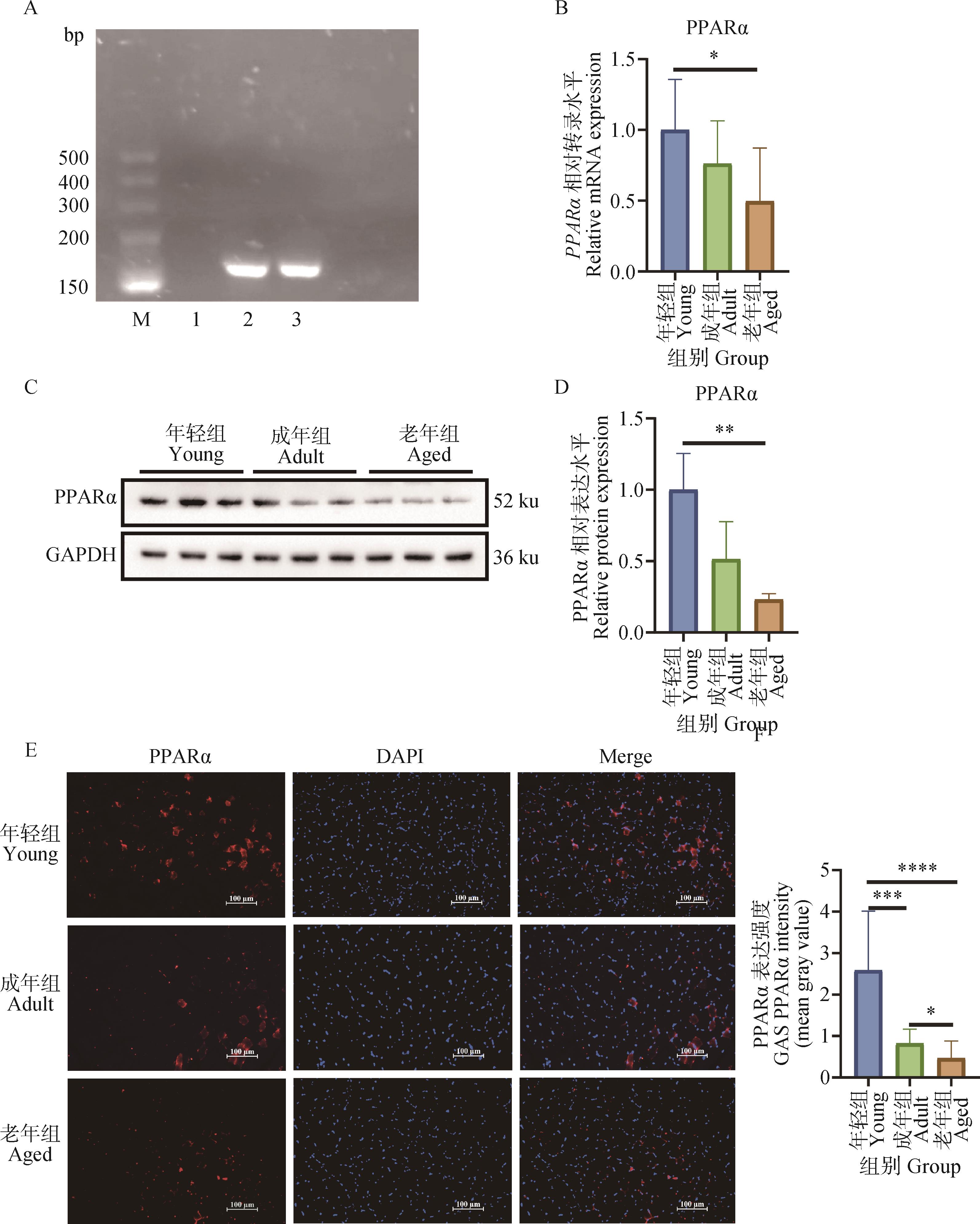
Fig.3
Knockout of PPARα reduces myofiber area and the expression of MyoD and MyoGA. HE staining of WT and PPARα-/- mouse gastrocnemius muscles (bar=100 μm);B. Cross-sectional area analysis of gastrocnemius muscle of WT and PPARα-/- mice;C. Body composition analysis of WT and PPARα-/- mice;D. qRT-PCR was used to detect genes related to skeletal muscle development in WT and PPARα-/- mice; E. Western blot was used to detect the expressions of PPARα, MyoD and MyoG in the gastrocnemius muscle of WT and PPARα-/- mice; F. Relative quantitative statistics of PPARα, MyoD, and MyoG in panel E. *. P<0.05, **. P<0.01, ***. P<0.001"

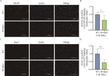
Fig.4
Knockdown of PPARα significantly reduces the positive areas of MyoD and Pax7 in the gastrocnemius muscleA, B. Immunofluorescence detection and quantification of MyoD in the gastrocnemius muscle of WT and PPARα-/- mice (bar=100 μm); C, D. Immunofluorescence detection and quantification of Pax7 in gastrocnemius muscle of WT and PPARα-/- mice (bar=100 μm). *. P<0.05, **. P<0.01"
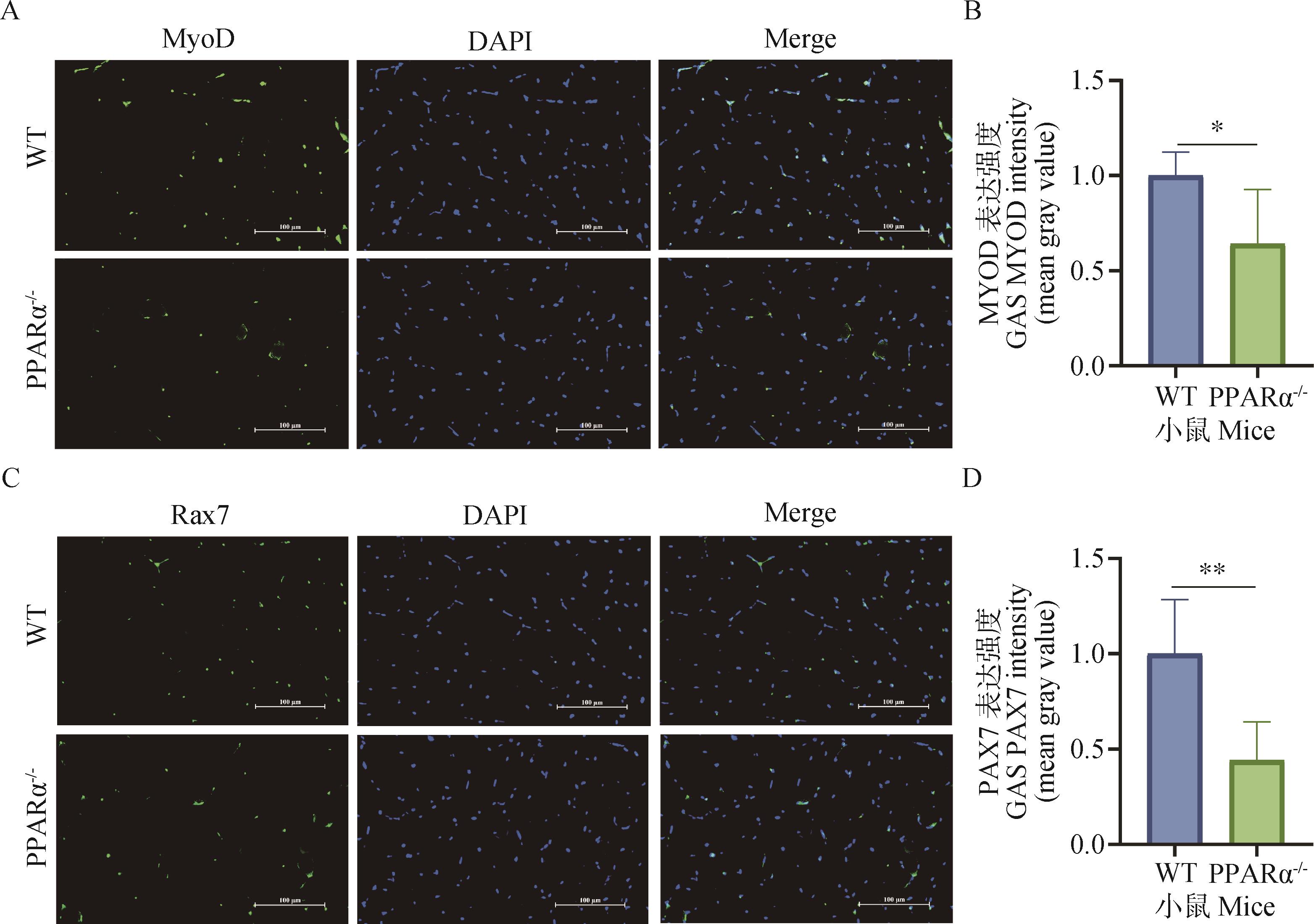
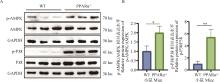
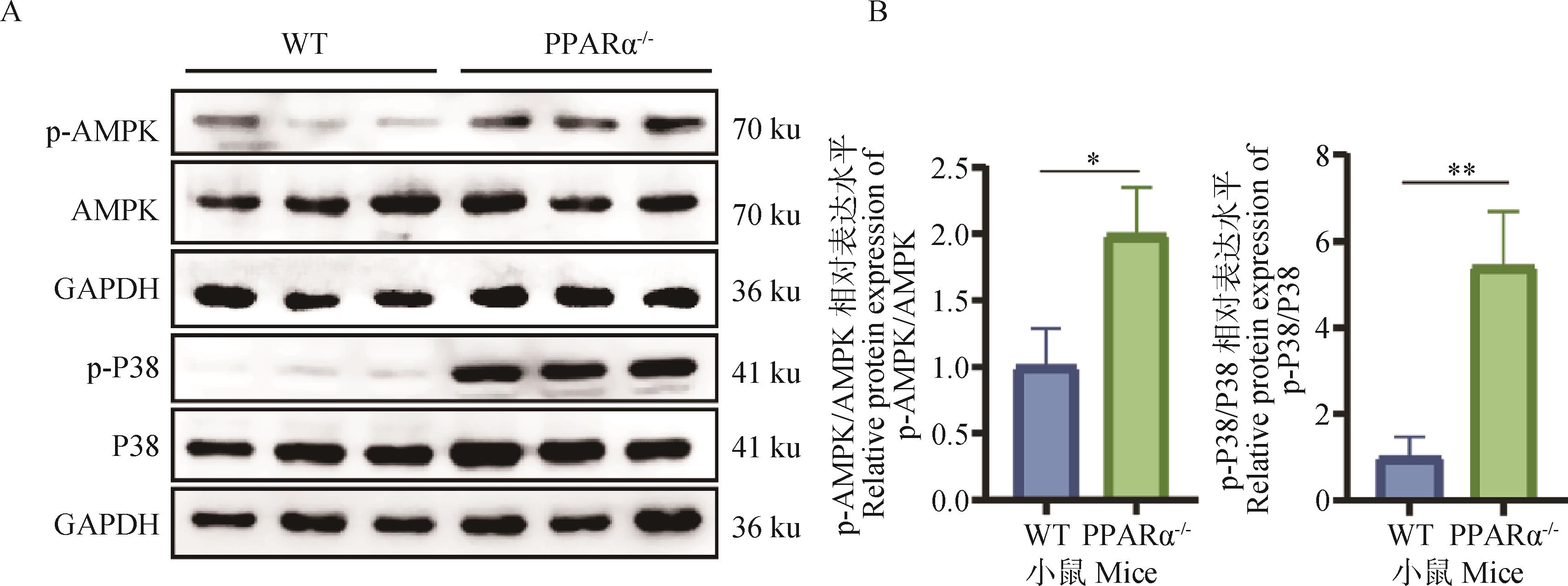
Fig.5
PPARα knockdown activates P38 and AMPK phosphorylation levelsA. Western blot was used to detect the expression and quantification of P38 and AMPK in the gastrocnemius muscle of WT and PPARα-/- mice; B. Quantification of protein bands in panel A. *. P<0.05, **. P<0.01"
| [1] | LIEBER R L,ROBERTS T J,BLEMKER S S,et al.Skeletal muscle mechanics,energetics and plasticity[J].Neuroeng Rehabil,2017,14(1):108. |
| [2] | ESTEVES DE LIMA J,RELAIX F.Master regulators of skeletal muscle lineage development and pluripotent stem cells differentiation[J].Cell Regen,2021,10(1):31. |
| [3] | WU Y J,YANG Y H,DU C X,et al.Berberine attenuates obesity-induced skeletal muscle atrophy via regulation of FUNDC1 in skeletal muscle of mice[J].Sci Rep,2025,15(1):4918. |
| [4] | YAGAI T,NAKAMURA T.Mechanistic insights into the peroxisome proliferator-activated receptor alpha as a transcriptional suppressor[J].Front Med,2022,9:1060244. |
| [5] | TAHRI-JOUTEY M,ANDREOLETTI P,SURAPUREDDI S,et al.Mechanisms mediating the regulation of peroxisomal fatty acid beta-oxidation by PPARα[J].Int J Mol Sci,2021,22(16):8969. |
| [6] | LAMICHANE S,DAHAL L B,KWON S M.Pivotal roles of peroxisome proliferator-activated receptors (PPARs) and their signal cascade for cellular and whole-body energy homeostasis[J].Int J Mol Sci,2018,19(4):949. |
| [7] | FAN S C,GAO Y,QU A J,et al.YAP-TEAD mediates PPAR α-induced hepatomegaly and liver regeneration in mice[J].Hepatol Int,2022,75(1):74-88. |
| [8] | ZHANG L L,LI Y Q,WANG Y,et al.mTORC2 facilitates liver regeneration through sphingolipid-induced PPAR-α-fatty acid oxidation[J].Cell Mol Gatroenter,2022,14(6):1311-1331. |
| [9] | GONCALVES M D,HWANG S K,PAULI C,et al.Fenofibrate prevents skeletal muscle loss in mice with lung cancer[J].Proc Natl Acad Sci,2018,115(4):E743-E752. |
| [10] | AQUILANO K,BALDELLI S,LA BARBERA L,et al.Adipose triglyceride lipase decrement affects skeletal muscle homeostasis during aging through FAs-PPARα-PGC-1α antioxidant response[J].Oncotarget,2016,7(17):23019-23032. |
| [11] | DAI J,XIANG Y,FU D,et al.Ficus carica L.attenuates denervated skeletal muscle atrophy via PPARα/NF-κB pathway[J].Front Physiol,2020,11:580223. |
| [12] | LIU J,CHEN Y,HAN D,et al.Inhibition of the expression of TRIM63 alleviates ventilator-induced diaphragmatic dysfunction by modulating the PPARα/PGC-1α pathway[J].Mitochondrion,2025,83:102025. |
| [13] | BURRI L,THORESEN G H,BERGE R K.The role of PPARα activation in liver and muscle[J].PPAR Res,2010(2010):542359. |
| [14] | STUMP C S,HENRIKSEN E J,WEI Y,et al.The metabolic syndrome:role of skeletal muscle metabolism[J].Ann Med,2006,38(6):389-402. |
| [15] | 任华伟,薛霖莉,曹 靖,等.Myogenin在不同发育阶段小鼠骨骼肌中的表达差异[J].山西农业科学,2019,47(8):1485-1489,1500. |
| REN W H,XUE L L,CAO J,et al.Differences in the expression of myogenin in skeletal muscle of mice at different developmental stages[J].Shanxi Agricultural Sciences,2019,47(8):1485-1489,1500. | |
| [16] | OOST L J,KUSTERMANN M,ARMANI A,et al.Fibroblast growth factor 21 controls mitophagy and muscle mass[J].J Cachexia Sarcopenia,2019,10(3):630-642. |
| [17] | BÖRSCH A,HAM D J,MITTAL N,et al.Molecular and phenotypic analysis of rodent models reveals conserved and species-specific modulators of human sarcopenia[J].Commun Biol,2021,4(1):194. |
| [18] | KEDLIAN VR,WANG Y,LIU T,et al.Human skeletal muscle aging atlas[J].Nat Aging,2024,4(5):727-744. |
| [19] | MANIO M C C,MATSUMURA S,MASUDA D,et al.CD36 is essential for endurance improvement,changes in whole-body metabolism,and efficient PPAR-related transcriptional responses in the muscle with exercise training[J].Physiol Rep,2017,5(10):e13282. |
| [20] | CABRAL-SANTOS C,SILVEIRA,L S,CHIMIN P,et al.Moderate aerobic exercise-induced cytokines changes are disturbed in PPARα knockout mice[J].Cytokine,2020,134:155207. |
| [21] | ATHERTON H J,GULSTON M K,BAILEY N J,et al.Metabolomics of the interaction between PPARα and age in the PPARα‐null mouse[J].Mol Syst Biol,2009,5(1):259. |
| [22] | LI Y,LIU Z Y,YAN H Y,et al.Polygonatum sibiricum polysaccharide ameliorates skeletal muscle aging and mitochondrial dysfunction via PI3K/Akt/mTOR signaling pathway[J].Phytomedicine,2025,136:156316. |
| [23] | MURPHY S A,MIYAMOTO M,KERVADEC A,et al.PGC1/PPAR drive cardiomyocyte maturation at single cell level via YAP1 and SF3B2[J].Nat Commun,2021,12(1):1648. |
| [24] | WONG B W,WANG X W,ZECCHIN A,et al.The role of fatty acid β-oxidation in lymphangiogenesis[J].Nature,2017,542(7639):49-54. |
| [25] | CHANDRASHEKAR P,MANICKAM R,GE X J,et al.Inactivation of PPARβ/δ adversely affects satellite cells and reduces postnatal myogenesis[J].Am J Physiol Endocrinol Metab,2015,309(2):E122-E131. |
| [26] | LEI S,Li C,SHE Y L,et al.Roles of super enhancers and enhancer RNAs in skeletal muscle development and disease[J].Cell Cycle,2023,22(5):495-505. |
| [27] | GE J,LIU K,NIU W,et al.Gold and gold-silver alloy nanoparticles enhance the myogenic differentiation of myoblasts through p38 MAPK signaling pathway and promote in vivo skeletal muscle regeneration[J].Biomaterials,2018,175:19-29. |
| [28] | SALMINEN A,KAARNIRANTA K,KAUPPINEN A.Age-related changes in AMPK activation:Role for AMPK phosphatases and inhibitory phosphorylation by upstream signaling pathways[J].Ageing Res Rev,2016,28:15-26. |
| [29] | THOMSON D M.The role of AMPK in the regulation of skeletal muscle size,hypertrophy,and regeneration[J].Int J Mol Sci,2018,19(10):3125. |
| [30] | BRENNAN C M,EMERSON C P JR,OWENS J,et al.p38 MAPKs - roles in skeletal muscle physiology,disease mechanisms,and as potential therapeutic targets[J].JCI Insight,2021,6(12):e149915. |
| [1] | XIA Chunqiu, MIAO Shu, LI Zhiqing, LIU Lei, WAN Fachun, SHEN Weijun. Valine Regulates Bovine Myoblast Proliferation through the AMPK/mTOR Signaling Pathway [J]. Acta Veterinaria et Zootechnica Sinica, 2025, 56(9): 4491-4506. |
| [2] | SHI Mei, WEI Gege, LI Yihan, WANG Xianzhong, ZHANG Jiaojiao. Metformin Regulates Chicken Growth Metabolism through LKB1/AMPKα2 Signaling Pathway [J]. Acta Veterinaria et Zootechnica Sinica, 2025, 56(9): 4673-4685. |
| [3] | HU Jinling, ZHONG Qiqi, HUANG Cheng, LEI Minggang. AKR1B1 Regulates Proliferation and Differentiation of Porcine Skeletal Muscle Satellite Cells via the AMPK/mTOR/S6 Signaling Pathway [J]. Acta Veterinaria et Zootechnica Sinica, 2025, 56(8): 3722-3733. |
| [4] | WANG Yuqing, XING Ya, ZHOU Xiaoyi, GONG Haizhou, ZHAO Minmeng, LIU Long, GONG Daoqing, GE Jing, GENG Tuoyu. Mitochondrial AMPK (mAMPK) Regulates Mitochondrial Function and Participates in the Formation of Goose Fatty Liver [J]. Acta Veterinaria et Zootechnica Sinica, 2025, 56(7): 3210-3225. |
| [5] | LIU Yumeng, GAO Xing, ZHAO Yali, CAO Di, MANG Lai, ZHANG Xinzhuang. Effects of Selenium Polysaccharides on Oxidative Damage of Equine Skeletal Muscle Satellite Cell [J]. Acta Veterinaria et Zootechnica Sinica, 2025, 56(7): 3357-3367. |
| [6] | LIU Siqi, YANG Zhen, YANG Yanan, CAI Yuan, ZHAO Shengguo. The Effect of Interfering with AdiopR2 on the Thermogenesis of Subcutaneous Inguinal Adipocytes in Tibetan Pigs [J]. Acta Veterinaria et Zootechnica Sinica, 2025, 56(6): 2649-2660. |
| [7] | SHI Shanshan, WAN Qiongfei, XU Yingxin, WANG Qiushuo, ZHANG Linlin, GUO Yiwen, HU Debao, GUO Hong, DING Xiangbin, LI Xin. Sequencing and Bioinformatics Analysis of miRNAs at Different Developmental Stages of Bovine Skeletal Muscle [J]. Acta Veterinaria et Zootechnica Sinica, 2025, 56(6): 2701-2710. |
| [8] | GONG Yuxuan, HEI Wei, BAO Wu, CHEN Jiayi, LI Meng, GUO Xiaohong, LI Bugao. Study on the Regulation of Myogenic Differentiation of Porcine Skeletal Muscle Satellite Cells by Gene TMEM182 [J]. Acta Veterinaria et Zootechnica Sinica, 2025, 56(4): 1676-1688. |
| [9] | HOU Wanchen, XU Tong. Cannabidiol Antagonizes BPA-induced Apoptosis and Autophagy in Porcine Intestinal Epithelial Cells through the BRD4/AMPK/mTOR Signaling Pathway [J]. Acta Veterinaria et Zootechnica Sinica, 2025, 56(4): 1919-1933. |
| [10] | LI Yuanfang, ZHANG Hongyuan, LI Hongtai, LI Zhi, WEI Qianran, WANG Yadong, LI Guoxi, WANG Dandan, LIU Qiaoming. The Effect of Riboflavin Supplementation in Embryonic Eggs on the Development of Skeletal Muscle of Chickens [J]. Acta Veterinaria et Zootechnica Sinica, 2025, 56(3): 1159-1169. |
| [11] | ZHANG Zhengyu, YANG Peihong, GUO Hong, LI Xin, ZHANG Linlin, GUO Yiwen, HU Debao, DING Xiangbin. Effects of Sirt1 Deacetylase on Proliferation and Differentiation of Bovine Skeletal Muscle Satellite Cells [J]. Acta Veterinaria et Zootechnica Sinica, 2025, 56(2): 603-610. |
| [12] | LI Bingzhi, GUO Juntao, WANG Jianfang, YU Shengchen, PAN Yueting, YU Hengwei, LIU Haibing, ZHANG Ke, CHENG Gong, TIAN Wanqiang, ZAN Linsen. Study on the Effect of Interfering with MAPK6 Gene on the Differentiation of Qinchuan Cattle Myoblast Cells [J]. Acta Veterinaria et Zootechnica Sinica, 2025, 56(11): 5475-5488. |
| [13] | ZHANG Shuai, XU Jing, YANG Peihong, GUO Yiwen, HU Debao, LI Xin, DING Xiangbin, GUO Hong, ZHANG Linlin. PFN1-PTEN Inhibits Bovine Skeletal Muscle Satellite Cell Differentiation by Regulating the PI3K/AKT/mTOR Pathway [J]. Acta Veterinaria et Zootechnica Sinica, 2025, 56(11): 5489-5501. |
| [14] | HE Siqi, CHEN Qian, JIANG Lin, MA Yuehui, ZHOU Shenghua, ZHAO Qianjun. The Effect of METTL14 on Myogenic Differentiation of Ovine Skeletal Muscle Satellite Cells Based on Transcriptome Sequencing Analysis [J]. Acta Veterinaria et Zootechnica Sinica, 2025, 56(10): 4925-4937. |
| [15] | XIAO Wei, DONG Jiaqi, ZHANG Xiaosong, ZHOU Ke, WEI Yanming. The Effects of Sheng Mai San on the AMPK-mTOR Pathway and Autophagy in the Lungs of Rats under Heat Stress [J]. Acta Veterinaria et Zootechnica Sinica, 2025, 56(10): 5277-5288. |
| Viewed | ||||||
|
Full text |
|
|||||
|
Abstract |
|
|||||