


Acta Veterinaria et Zootechnica Sinica ›› 2025, Vol. 56 ›› Issue (2): 559-570.doi: 10.11843/j.issn.0366-6964.2025.02.008
• Animal Genetics and Breeding • Previous Articles Next Articles
SU Meng( ), LIU Sha, SONG Danli, GAO Qianmei, ZHENG Maiqing, WEN Jie, ZHAO Guiping, LI Qinghe*()
Received:2024-09-02
Online:2025-02-23
Published:2025-02-26
Contact:
LI Qinghe
E-mail:sm18310082450@163.com;liqonghe@caas.cn
CLC Number:
SU Meng, LIU Sha, SONG Danli, GAO Qianmei, ZHENG Maiqing, WEN Jie, ZHAO Guiping, LI Qinghe. Identification of Candidate Genes Associated with Ascites Syndrome in Broilers Based on Transcriptome Sequencing[J]. Acta Veterinaria et Zootechnica Sinica, 2025, 56(2): 559-570.
Table 1
Gene primer sequences"
| 基因 Gene | 引物序列(5′→3′) Primer sequence | 产物长度/bp Product length |
| NUSAP1 | F: AGATTCGAAGAGTGGCGGTT | 145 |
| R: AACAGTTTATCGGCCCGGAG | ||
| HBAD | F: AACGCTGTGAAGAACGTGGA | 197 |
| R: GACACGGCAGACAGGAACTT | ||
| KIF20A | F: GAAACCCCAACAGCAGCG | 401 |
| R: ACCTATGTGCTGCTTGTCCC | ||
| TFRC | F: TTATCGTGGACGAATCGAGC | 239 |
| R: CCTCACCAGCCTCAAAGGAG | ||
| HBBA | F: GAGCAAGAGCCCAGACCTC | 131 |
| R: ACATTCGGCCACATTGACCT | ||
| β-actin | F: GAGAAATTGTGCGTGACATCA | 152 |
| R: CCTGAACCTCTCATTGCCA |

Fig. 1
Results of comparative analysis of blood biochemistry between ascites chickens and normal chickens a, b, c, d. Comparative analysis results of blood routine between ascites and normal chickens; e, f, g. Comparative analysis results of blood oxygen between ascites and normal chickens"
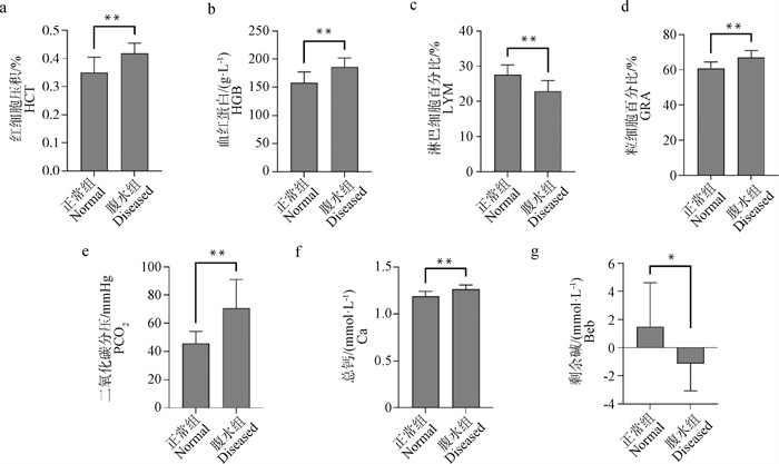
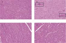
Fig. 2
HE stained pathology section of myocardium (100×) A. Longitudinal section of myocardial tissue from chickens with ascites; B. Transverse section of myocardial tissue from chickens with ascites(The boxed area shows blood cell deposition); C. Longitudinal section of myocardial tissue from normal chickens; D. Transverse section of myocardial tissue from normal chickens"
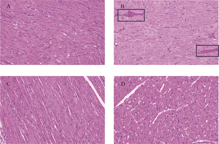
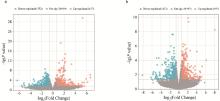
Fig. 3
Volcano map of differentially expressed genes a. Volcano map of differentially expressed genes in lung; b. Volcano map of differentially expressed genes in heart"
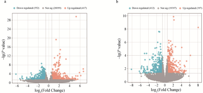

Fig. 4
Venn diagram of differentially expressed genes shared between the heart and lung"
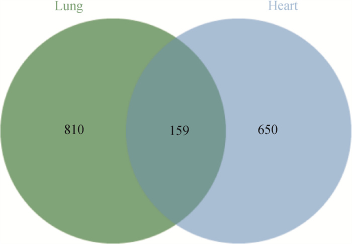

Fig. 5
Results of KEGG enrichment analysis of DEGs a. KEGG pathway enrichment results of DEGs in DEGs in lung; b. KEGG pathway enrichment results of DEGs in heart; c. Heart-lung common DEGs pathway enrichment results"
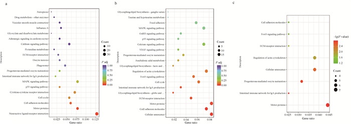

Fig. 6
Results of GO functional annotation analysis of DEGs a. GO functional annotation results of DEGs in lung; b. GO functional annotation results of DEGs in heart; c. Cardiopulmonary co-DEGs functional annotation results"

Table 2
GO terms and KEGG pathways associated with ascites syndrome"
| 富集类型 Category | 编号 ID | 条目/通路 Term/Pathway | P值 P-value | 基因 Gene |
| GO 条目 GO term | GO: 0000278 | 细胞周期 | 8.28×10⁻⁶ | NUSAP1/CCNA2/CDK1 |
| GO 条目 GO term | GO: 0034508 | 着丝粒复合物组装 | 3.23×10⁻⁵ | CENPO/CENPW/CENPN |
| GO 条目 GO term | GO: 0019825 | 氧气结合 | 0.001 3 | HBBA/HBAD/HBA1 |
| KEGG 通路 KEGG pathway | gga04080 | 神经活性配体-受体相互作用 | 8.51×10⁻⁵ | TSPO2/P2RY10/ADRA2BL2/EDN1/GABBR2 |
| KEGG 通路 KEGG pathway | gga04514 | 细胞黏附分子 | 0.001 3 | ITGA4/MHCY11/MHCY4/ |
| KEGG 通路 KEGG pathway | gga04218 | 细胞衰老 | 5.23×10⁻⁵ | MHCY4/MHCY14/CCNB3/ |
| KEGG 通路 KEGG pathway | gga04512 | 细胞外基质-受体相互作用 | 0.001 3 | LAMB3/COL6A6/ITGA2B |
| KEGG 通路 KEGG pathway | gga04814 | 运动蛋白 | 0.000 433 | KIF11/CENPE/KIF20A |

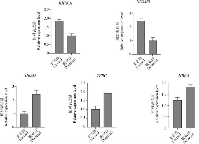
Fig. 7
RT-qPCR validation of candidate genes in White Feather broiler chickens"
| 1 |
GUO F P , LIU P , GUO X Q , et al. Bioinformatics analysis of JAZF1 gene in broilers with ascites syndrome[J]. Pak Vet J, 2021, 41 (1): 19- 24.
doi: 10.29261/pakvetj/2020.072 |
| 2 |
YU J , LIU X Y , WANG K Y , et al. Underlying mechanism of Qiling Jiaogulan Powder in the treatment of broiler ascites syndrome[J]. Poult Sci, 2023, 102 (1): 102144.
doi: 10.1016/j.psj.2022.102144 |
| 3 | 王慧敏, 崔亮, 韩宇峰, 等. 基于RhoA/ROCK通路探究复方中药对腹水综合征肉鸡肝纤维化的影响[J]. 东北农业大学学报, 2023, 54 (5): 47- 54. |
| WANG H M , CUI L , HAN Y F , et al. Effects of compound Chinese medicine on hepatic fibrosis in ascites broilers based on RhoA/ROCK pathway[J]. Journal of Northeast Agricultural University, 2023, 54 (5): 47- 54. | |
| 4 | GUO D Q , ZHANG J , HAN Y F , et al. Transcriptomic study on the lungs of broilers with ascites syndrome[J]. Animals (Basel), 2023, 13 (1): 175. |
| 5 | LI L F , JIA Q F , CHEN L L , et al. Changes in the expression of MIF and other key enzymes of energy metabolism in the myocardia of broiler chickens with ascites syndrome[J]. Animals (Basel), 2022, 12 (19): 2488. |
| 6 | SALEH R Q , ABDUL-ABBAS S J , AZIZ S N , et al. Evaluation of the effect of peppermint extract and probiotics on biochemical factors in the blood of ascites-induced chickens[J]. Arch Razi Inst, 2022, 77 (6): 2243- 2250. |
| 7 |
VAN AS P , ELFERINK M G , CLOSTER A M , et al. The use of blood gas parameters to predict ascites susceptibility in juvenile broilers[J]. Poult Sci, 2010, 89 (8): 1684- 1691.
doi: 10.3382/ps.2009-00430 |
| 8 | 苏志峰, 杨文平, 杜天庆, 等. 施肥深度对生土地玉米根系及根际土壤肥力垂直分布的影响[J]. 中国生态农业学报, 2016, 24 (2): 142- 153. |
| SU Z F , YANG W P , DU T Q , et al. Effect of fertilization depth on maize root and rhizosphere soil fertility vertical distribution in immature loess subsoil[J]. Chinese Journal of Eco-Agriculture, 2016, 24 (2): 142- 153. | |
| 9 |
王亚鑫, 王璟, 田学凯, 等. 多组学技术在畜禽重要经济性状研究中的应用[J]. 畜牧兽医学报, 2024, 55 (5): 1842- 1853.
doi: 10.11843/j.issn.0366-6964.2024.05.003 |
|
WANG Y X , WANG J , TIAN X K , et al. Application of multi-omics technology in the study of important economic traits of livestock and poultry[J]. Acta Veterinaria et Zootechnica Sinica, 2024, 55 (5): 1842- 1853.
doi: 10.11843/j.issn.0366-6964.2024.05.003 |
|
| 10 | LIU P , KE S L , CAO H B , et al. Tanshinone IIA protects against sub-Ascites syndrome in broilers[J]. Infect Genet Evol, 2015, 3 (30): 159- 163. |
| 11 |
WANG Y W , GUO Y M , NING D , et al. Analysis of liver transcriptome in broilers with ascites and regulation by L-carnitine[J]. J Poult Sci, 2013, 50 (2): 126- 137.
doi: 10.2141/jpsa.0120124 |
| 12 |
HASANPUR K , NASSIRI M , HOSSEINI SALEKDEH G . The comparative analysis of phenotypic and whole transcriptome gene expression data of ascites susceptible versus ascites resistant chickens[J]. Mol Biol Rep, 2019, 46 (1): 793- 804.
doi: 10.1007/s11033-018-4534-8 |
| 13 |
ZHANG J B , SCHMIDT C J , LAMONT S J . Distinct genes and pathways associated with transcriptome differences in early cardiac development between fast- and slow-growing broilers[J]. PLoS One, 2018, 13 (12): e0207715.
doi: 10.1371/journal.pone.0207715 |
| 14 |
YANG F , CAO H B , XIAO Q Y , et al. Transcriptome analysis and gene identification in the pulmonary artery of broilers with ascites syndrome[J]. PLoS One, 2016, 11 (6): e0156045.
doi: 10.1371/journal.pone.0156045 |
| 15 | JOHNJS SeqPrep: tool for stripping adaptors and/or merging paired reads with overlap into sing lereads[EB/OL]. 2011. |
| 16 | FU X , ZHANG F L . Role of the HIF-1 signaling pathway in chronic obstructive pulmonary disease[J]. Exp Ther Med, 2018, 16 (6): 4553- 4561. |
| 17 | LAWSON M, JOMOVA K, POPRAC P, et al. Free radicals and antioxidants in human disease[M]//AL-GUBORY K H, LAHER I. Nutritional Antioxidant Therapies: Treatments and Perspectives. Cham: Springer, 2017: 283-305. |
| 18 | 张若旸, 刘杰, 孙英, 等. 代谢重编程在肺动脉高压中的研究进展[J]. 中华结核和呼吸杂志, 2022, 45 (3): 313- 317. |
| ZHANG R Y , LIU J , SUN Y , et al. Metabolic reprogramming in pulmonary hypertension[J]. Chinese Journal of Tuberculosis and Respiratory Diseases, 2022, 45 (3): 313- 317. | |
| 19 | 李玉鹏, 张效生, 王丽, 等. 肉鸡腹水综合征的发病原因及防治研究进展[J]. 天津农业科学, 2021, 27 (7): 33- 37. |
| LI Y P , ZHANG X S , WANG L , et al. Research progress on pathogenesis and prevention of ascites syndrome in broilers[J]. Tianjin Agricultural Sciences, 2021, 27 (7): 33- 37. | |
| 20 | 曾秋凤, 张克英, 陈代文, 等. 饲粮能量水平和来源对肉鸡腹水症的影响及其机理[J]. 动物营养学报, 2010, 22 (6): 1738- 1744. |
| ZENG Q F , ZHANG K Y , CHEN D W , et al. Effects of dietary energy levels and sources on ascites syndrome and its mechanism in broilers[J]. Chinese Journal of Animal Nutrition, 2010, 22 (6): 1738- 1744. | |
| 21 |
HUANG S , INGBER D E . The structural and mechanical complexity of cell-growth control[J]. Nat Cell Biol, 1999, 1 (5): E131- E138.
doi: 10.1038/13043 |
| 22 |
TADZIC R , MIHALJ M , VCEV A , et al. The effects of arterial blood pressure reduction on endocan and soluble endothelial cell adhesion molecules (CAMs) and CAMs ligands expression in hypertensive patients on Ca-channel blocker therapy[J]. Kidney Blood Press Res, 2013, 37 (2-3): 103- 115.
doi: 10.1159/000350064 |
| 23 | XIAO G F , WANG T J , ZHUANG W , et al. RNA sequencing analysis of monocrotaline-induced PAH reveals dysregulated chemokine and neuroactive ligand receptor pathways[J]. Aging (Albany NY), 2020, 12 (6): 4953- 4969. |
| 24 | MUNCIE J M , WEAVER V M . The physical and biochemical properties of the extracellular matrix regulate cell fate[J]. Curr Top Dev Biol, 2018, 130, 1- 37. |
| 25 |
PADHI A , NAIN A S . ECM in differentiation: a review of matrix structure, composition and mechanical properties[J]. Ann Biomed Eng, 2020, 48 (3): 1071- 1089.
doi: 10.1007/s10439-019-02337-7 |
| 26 |
ROMANI P , VALCARCEL-JIMENEZ L , FREZZA C , et al. Crosstalk between mechanotransduction and metabolism[J]. Nat Rev Mol Cell Biol, 2021, 22 (1): 22- 38.
doi: 10.1038/s41580-020-00306-w |
| 27 |
THEOCHARIS A D , SKANDALIS S S , GIALELI C , et al. Extracellular matrix structure[J]. Adv Drug Delivery Rev, 2016, 97, 4- 27.
doi: 10.1016/j.addr.2015.11.001 |
| 28 |
NAJAFI M , FARHOOD B , MORTEZAEE K . Extracellular matrix (ECM) stiffness and degradation as cancer drivers[J]. J Cell Biochem, 2019, 120 (3): 2782- 2790.
doi: 10.1002/jcb.27681 |
| 29 |
SHEN S R , SHEN Y R , ZHANG S , et al. Combinatory evaluation of transcriptome and metabolome profiles of low temperature-induced resistant ascites syndrome in broiler chickens[J]. Sci Rep, 2017, 7 (1): 2389.
doi: 10.1038/s41598-017-02492-8 |
| 30 |
LOUW J J , BASTOS R N , CHEN X W , et al. Compound heterozygous loss-of-function mutations in KIF20A are associated with a novel lethal congenital cardiomyopathy in two siblings[J]. PLoS Genet, 2018, 14 (1): e1007138.
doi: 10.1371/journal.pgen.1007138 |
| 31 |
KRISHNAMOORTHY S , SMITH C D , AL-RUBAYE A A , et al. A quantitative trait locus for ascites on chromosome 9 in broiler chicken lines[J]. Poult Sci, 2014, 93 (2): 307- 317.
doi: 10.3382/ps.2013-03359 |
| 32 |
WIDEMAN R F , RHOADS D D , ERF G F , et al. Pulmonary arterial hypertension (ascites syndrome) in broilers: a review[J]. Poult Sci, 2013, 92 (1): 64- 83.
doi: 10.3382/ps.2012-02745 |
| 33 |
FAZZIO T G , PANNING B . Condensin complexes regulate mitotic progression and interphase chromatin structure in embryonic stem cells[J]. J Cell Biol, 2010, 188 (4): 491- 503.
doi: 10.1083/jcb.200908026 |
| 34 | 葛恒广, 李新丽. 基于生物信息学分析NUSAP1在肺腺癌组织中的表达及临床意义[J]. 现代医药卫生, 2023, 39 (8): 1281- 1287. |
| GE H G , LI X L . The expression and clinical significance of NUSAP1 gene in lung adenocarcinoma and based on bioinformatics analysis[J]. J Mod Med Health, 2023, 39 (8): 1281- 1287. |
| [1] | HU Hanwen, BAO Tugeqin, REN Xiujuan, DING Wenqi, GONG Wendian, JIA Zijie, SHI Lin, MA Muren, Baorigele , DUGARJAVIIN Manglai, BAI Dongyi. Comparative Study on Muscle Fiber Development Phenotype and Gene Expression Profile of Two Mongolian Horse Populations [J]. Acta Veterinaria et Zootechnica Sinica, 2025, 56(2): 643-656. |
| [2] | HANG Zhenyu, WANG Ziyi, ZHANG Lin, XING Tong, ZHAO Liang, GAO Feng. Evaluation and Prediction Equation Construction of Standardized Ileal Amino Acid Digestibility of Corn from Different Sources in 28-day-old White-feathered Broilers [J]. Acta Veterinaria et Zootechnica Sinica, 2025, 56(2): 722-736. |
| [3] | WU Shuang, YIN Na, YU Mohan, PING Yuyu, BAI Hao, CHEN Shihao, CHANG Guobin. The Effect of TRIM39.2 Overexpression on the Transcriptional Expression of Chicken Macrophages [J]. Acta Veterinaria et Zootechnica Sinica, 2025, 56(1): 178-188. |
| [4] | LU Xiu, ZHANG Ming'ai, KONG Min, ZHANG Jing, WANG Binghan, HOU Zhongyi, TENG Xingyi, JIANG Yajing, FAN Wenlei, WANG Baowei. Screening for Candidate Genes Related to Egg Production in Wulong Geese Based on Transcriptome and Proteome Analyses [J]. Acta Veterinaria et Zootechnica Sinica, 2025, 56(1): 232-245. |
| [5] | Tana HE, Xinyun HU, Jielan MI, Li GAO, Yanping ZHANG, Xiaole QI, Hongyu CUI, Guilian YANG, Yulong GAO. Effect of Feeding Lactobacillus salivarius XP132 on the Gut Microbiota of White-feathered Broiler Breeder based on 16S rDNA Analysis [J]. Acta Veterinaria et Zootechnica Sinica, 2024, 55(9): 4091-4099. |
| [6] | Xinman LIU, Hongyuan ZHOU, Rui SANG, Bingjie GE, Kexin YAN, Wei WANG, Minghong YU, Xiaotong LIU, Qian QIU, Xuemei ZHANG. Effect of Taraxasterol on Oxidative Stress in Liver Tissue of Broilers with AFB1 Induced Liver Injury [J]. Acta Veterinaria et Zootechnica Sinica, 2024, 55(9): 4141-4152. |
| [7] | Xiaoxu ZHANG, Hao LI, Pingjie FENG, Hao YANG, Xinyue LI, Ran LÜ, Zhangyuan PAN, Mingxing CHU. Application of Single-Cell Transcriptome Sequencing Technology in Domesticated Animals [J]. Acta Veterinaria et Zootechnica Sinica, 2024, 55(8): 3276-3287. |
| [8] | Jing CHEN, Xuebei WU, Dongzhi MIAO, Chi ZHANG, Zhenyu GUO, Ying WANG. Comparative Analysis of Transcriptome of Pigeon Follicles at Early Stage of Laying Interval Reveals Genes Related to Follicular Development [J]. Acta Veterinaria et Zootechnica Sinica, 2024, 55(8): 3503-3515. |
| [9] | Yinuo WANG, Dan XU, Jianhua YANG, Yang LIU, Yaofu TIAN, Xiaoling ZHAO. Research on a Breeding Method for Predicting Meat Yield Performance of Broilers Based on Ultrasound Measurement of Pectoral Muscle Thickness [J]. Acta Veterinaria et Zootechnica Sinica, 2024, 55(7): 2901-2912. |
| [10] | Wanqing LI, Yaqi ZENG, Xinkui YAO, Jianwen WANG, Xinxin YUAN, Chen MENG, Yuanfang SUN, Xuan PENG, Jun MENG. Comparative Analysis of Blood Transcriptome in Yili Horses Bred for Meat Performance [J]. Acta Veterinaria et Zootechnica Sinica, 2024, 55(7): 2951-2962. |
| [11] | Mingliang HE, Xiaoyang LÜ, Yongqing JIANG, Zhenghai SONG, Yeqing WANG, Huiguo YANG, Shanhe WANG, Wei SUN. Function Analysis of SOX18 in Hu Sheep Hair Follicle Dermal Papilla Cells Based on Transcriptome Sequencing [J]. Acta Veterinaria et Zootechnica Sinica, 2024, 55(6): 2409-2420. |
| [12] | Ming LI, Hongwei CUI, Jie GAO, Lele AN, Songli LI, Zhenghua RAO. Identification and Genomic Analysis of Pathogenic Escherichia coli in Small Intestinal Content of White Feather Broilers [J]. Acta Veterinaria et Zootechnica Sinica, 2024, 55(6): 2692-2700. |
| [13] | Ji WANG, Xinyan ZHOU, Fangrui GUO, Qiurong XU, Dongyi WU, Yan MAO, Zhihang YUAN, Jin'e YI, Lixin WEN, Jing WU. Viola yedoensis Makino Improves the Growth Performance, Meat Quality, and Gut Microbiota of Broilers Exposed to Heat Stress [J]. Acta Veterinaria et Zootechnica Sinica, 2024, 55(6): 2761-2774. |
| [14] | CHEN Zhe, QU Xiaolu, GUO Binbin, SUN Xuefeng, YAN Leyan. Study on Candidate Genes for Green Light Affecting Early Development of Goose Embryo Heart Based on Transcriptome Sequencing [J]. Acta Veterinaria et Zootechnica Sinica, 2024, 55(5): 1978-1988. |
| [15] | LIU Jiahui, WU Kaikai, WANG Lei, ZHANG Kang, HAN Songwei, CHEN Fubin, XU Guowei, GUO Zhiting, GU Xueyan, ZHANG Jingyan, LI Jianxi. Protective Effects of Astragalus Polysaccharides, Saponins and Probiotic Compounds on Intestinal Tract of Broilers Infected with E.coli [J]. Acta Veterinaria et Zootechnica Sinica, 2024, 55(5): 2241-2252. |
| Viewed | ||||||
|
Full text |
|
|||||
|
Abstract |
|
|||||