


Acta Veterinaria et Zootechnica Sinica ›› 2024, Vol. 55 ›› Issue (7): 2983-2994.doi: 10.11843/j.issn.0366-6964.2024.07.018
• Animal Biotechnology and Reproduction • Previous Articles Next Articles
Milan MA1( ), Qi WANG1, Qiu YAN1, Tianan LI1, Xingxu ZHAO1,2,*(), Yong ZHANG1,2
Received:2023-11-06
Online:2024-07-23
Published:2024-07-24
Contact:
Xingxu ZHAO
E-mail:18419142205@163.com;zhaoxx@gsau.edu.cn
CLC Number:
Milan MA, Qi WANG, Qiu YAN, Tianan LI, Xingxu ZHAO, Yong ZHANG. Expression of HIG1 Hypoxia Inducible Domain Family Member 1C in Cryptorchidism of Yak and Its Regulatory Mechanism[J]. Acta Veterinaria et Zootechnica Sinica, 2024, 55(7): 2983-2994.
Table 1
Primer sequences of target and house-keeping genes"
| 引物名称Primer name | 引物序列(5′→3′)Primer sequence | 片段长度/bp Product length | 温度/℃ Tm |
| HIGD1C | F:TCAGCAGATGAAGACGAAGG R:GGACACCACAGTCACAAAGC | 102 | 56 |
| Caspase-3 | F:GGTGGGGTCAGAGCCATAGA R:CATCTTCCACACACACCCGTAG | 84 | 57 |
| Bax | F:CCGAGTGGCGGCTGAAA R:TCCAGATGGTGAGCGAGGC | 287 | 60 |
| Bcl-2 | F:CTGCACCTGACGCCCTTCAC R:GCGTCCCAGCCTCCGTTGT | 236 | 60 |
| GAPDH | F:GCTGGTGCTGAGTATGTGGTG R:GCTGACAATCTTGAGGGTGTTG | 177 | 60 |

Fig. 1
Morphology and sequencing analysis of testis and cryptorchidism tissues A. HE staining of testis and cryptorchidism tissue (200×); B-C. Sequencing scatter plot and histogram of HIGD1C gene; D. Sequencing violin diagrams"


Fig. 2
The expression and distribution of HIGD1C in testis and cryptorchidism A. The relative expression level of HIGD1C mRNA in testis and cryptorchidism; B-C. The relative expression level of HIGD1C protein in testis and cryptorchidism; D. The distribution of HIGD1C protein in testis and cryptorchidism was detected by immunofluorescence (200×)"


Fig. 3
Identification of testicular sertoli cells and verification of HIGD1C overexpression vector A. Immunofluorescence identification of sertoli cells and localization of HIDG1C in sertoli cells (200×); B. Double enzyme digestion identification of pIRES2-EGFP-HIGD1C overexpression vector; C. The relative level of HIGD1C mRNA after overexpression; D-E. The relative level of HIGD1C protein after overexpression"


Fig. 4
The effect of overexpression of HIGD1C on apoptosis A-C. The relative levels of apoptosis-related marker molecules (Bax, Bcl-2 and Caspase-3) mRNA after overexpression of HIGD1C; D-G. The relative levels of apoptosis-related marker molecules (Bax, Bcl-2 and Caspase-3) protein after overexpression of HIGD1C; H-J. Flow cytometry was used to detect the apoptosis rate of primary sertoli cells overexpressing HIGD1C in yak testis"
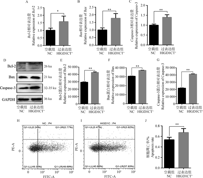

Fig. 5
Detection of HIGD1C knockout efficiency A. The relative level of HIGD1C mRNA after HIGD1C knockout; B-C. The relative level of HIGD1C protein after HIGD1C knockout"


Fig. 6
Effect of HIGD1C knockout on apoptosis A-C. The relative levels of apoptosis-related marker molecules (Bax, Bcl-2 and Caspase-3) mRNA after HIGD1C knockout; D-G. The relative levels of apoptosis-related marker molecules (Bax, Bcl-2 and Caspase-3) protein after HIGD1C knockout; H-J. Flow cytometry was used to detect the apoptosis rate of primary sertoli cells in yak testis after HIGD1C knockout"

| 1 | 张天留, 高雪, 徐凌洋, 等. 高原家养动物环境适应性的研究进展[J]. 畜牧兽医学报, 2020, 51 (7): 1475- 1487. |
| ZHANG T L , GAO X , XU L Y , et al. Research progress on environment adaptation of plateau domestic animals[J]. Acta Veterinaria et Zootechnica Sinica, 2020, 51 (7): 1475- 1487. | |
| 2 |
AMANN R P , VEERAMACHANENI D N R . Cryptorchidism in common eutherian mammals[J]. Reproduction, 2007, 133 (3): 541- 561.
doi: 10.1530/REP-06-0272 |
| 3 |
GURNEY J K , MCGLYNN K A , STANLEY J , et al. Risk factors for cryptorchidism[J]. Nat Rev Urol,, 2017, 14 (9): 534- 548.
doi: 10.1038/nrurol.2017.90 |
| 4 |
PINART E , BONET S , BRIZ M , et al. Morphological and histochemical characteristics of the lamina propria in scrotal and abdominal testes from postpubertal boars: correlation with the appearance of the seminiferous epithelium[J]. J Anat, 2001, 199 (4): 435- 448.
doi: 10.1046/j.1469-7580.2001.19940435.x |
| 5 | SHARMA S , MANCHANDA V , GUPTA R . Testicular microlithiasis in a unilateral undescended testis: a rare phenomenon[J]. Malays J Pathol, 2013, 35 (2): 181- 183. |
| 6 |
PENG Y J , TANG X T , SHU H S , et al. Sertoli cells are the source of stem cell factor for spermatogenesis[J]. Development, 2023, 150 (6): dev200706.
doi: 10.1242/dev.200706 |
| 7 |
MERONI S B , GALARDO M N , RINDONE G , et al. Molecular mechanisms and signaling pathways involved in sertoli cell proliferation[J]. Front Endocrinol (Lausanne), 2019, 10, 224.
doi: 10.3389/fendo.2019.00224 |
| 8 |
JOHNSON L , THOMPSON D L , VARNER D D . Role of sertoli cell number and function on regulation of spermatogenesis[J]. Anim Reprod Sci, 2008, 105 (1-2): 23- 51.
doi: 10.1016/j.anireprosci.2007.11.029 |
| 9 |
FARIAS J G , BUSTOS-OBREGÓN E , ORELLANA R , et al. Effects of chronic hypobaric hypoxia on testis histology and round spermatid oxidative metabolism[J]. Andrologia, 2005, 37 (1): 47- 52.
doi: 10.1111/j.1439-0272.2004.00651.x |
| 10 | ZHANG D C , CHEN R , CAI Y H , et al. Hyperactive reactive oxygen species impair function of porcine sertoli cells via suppression of surface protein ITGB1 and Connexin-43[J]. Zool Res, 2020, 41 (2): 203- 207. |
| 11 |
RIWALDT S , CORYDON T J , PANTALONE D , et al. Role of apoptosis in wound healing and apoptosis alterations in microgravity[J]. Front Bioeng Biotechnol, 2021, 9, 679650.
doi: 10.3389/fbioe.2021.679650 |
| 12 |
NALLURI S , GHOSHAL-GUPTA S , KUTIYANAWALLA A , et al. TIMP-1 inhibits apoptosis in lung adenocarcinoma cells via interaction with Bcl-2[J]. PLoS One, 2015, 10 (9): e0137673.
doi: 10.1371/journal.pone.0137673 |
| 13 | 张勤丽, 牛侨. 细胞凋亡机制概述[J]. 环境与职业医学, 2007, 24 (1): 102- 107. |
| ZHANG Q L , NIU Q . Mechanism of cell apoptosis[J]. Journal of Environmental and Occupational Medicine, 2007, 24 (1): 102- 107. | |
| 14 |
IVANISENKO N V , SEYREK K , HILLERT-RICHTER L K , et al. Regulation of extrinsic apoptotic signaling by c-FLIP: towards targeting cancer networks[J]. Trends Cancer, 2022, 8 (3): 190- 209.
doi: 10.1016/j.trecan.2021.12.002 |
| 15 |
LAUBACH V , KAUFMANN R , BERND A , et al. Extrinsic or intrinsic apoptosis by curcumin and light: still a mystery[J]. Int J Mol Sci, 2019, 20 (4): 905.
doi: 10.3390/ijms20040905 |
| 16 | 张聪慧. 人工隐睾诱发小鼠睾丸组织的自噬与凋亡[D]. 杨凌: 西北农林科技大学, 2016. |
| ZHANG C H. Cryptorchidisminduce autophagy and a poptosis in the mouse testis[D]. Yangling: Northwest A&F University, 2016. (in Chinese) | |
| 17 | GUO Y , TAN J , MIAO Y Y , et al. Effects of microvesicles on cell apoptosis under hypoxia[J]. Oxid Med Cell Longev, 2019, 2019, 5972152. |
| 18 | 陈韦任, 杜辉, 钱赓, 等. Bax抑制因子1通过促进视神经萎缩蛋白1表达抑制小鼠动脉血管钙化[J]. 南方医科大学学报, 2022, 42 (3): 330- 337. |
| CHEN W R , DU H , QIAN G , et al. Bax inhibitor 1 inhibits vascular calcification in mice by activating optic atrophy 1 expression[J]. Journal of Southern Medical University, 2022, 42 (3): 330- 337. | |
| 19 |
CAO X Y , FU M Y , BI R C , et al. Cadmium induced BEAS-2B cells apoptosis and mitochondria damage via MAPK signaling pathway[J]. Chemosphere, 2021, 263, 128346.
doi: 10.1016/j.chemosphere.2020.128346 |
| 20 |
EISENBERG-LERNER A , BIALIK S , SIMON H U , et al. Life and death partners: apoptosis, autophagy and the cross-talk between them[J]. Cell Death Differ, 2009, 16 (7): 966- 975.
doi: 10.1038/cdd.2009.33 |
| 21 | 范小瑞. 猪隐睾睾丸精子发生障碍的分子机制研究[D]. 太谷: 山西农业大学, 2021. |
| FAN X R. Study on the molecular mechanism of spermatogenesis disorder in cryptorchidism boars[D]. Taigu: Shanxi Agricultural University, 2021. (in Chinese) | |
| 22 | KOCAK I , DUNDAR M , HEKIMGIL M , et al. Assessment of germ cell apoptosis in cryptorchid rats[J]. Asian J Androl, 2002, 4 (3): 183- 186. |
| 23 |
TEKAYEV M , BOSTANCIERI N , SAADAT K A S M , et al. Effects of Moringa oleifera lam extract (MOLE) in the heat shock protein 70 expression and germ cell apoptosis on experimentally induced cryptorchid testes of rats[J]. Gene, 2019, 688, 140- 150.
doi: 10.1016/j.gene.2018.11.091 |
| 24 | STAUB C , JOHNSON L . Review: spermatogenesis in the bull[J]. Animal, 2018, 12 (S1): S27- S35. |
| 25 |
WANG J , GAO W J , DENG S L , et al. High temperature suppressed SSC self-renewal through S phase cell cycle arrest but not apoptosis[J]. Stem Cell Res Ther, 2019, 10 (1): 227.
doi: 10.1186/s13287-019-1335-5 |
| 26 |
JOHNSON C , JIA Y , WANG C , et al. Role of caspase 2 in apoptotic signaling in primate and murine germ cells[J]. Biol Reprod, 2008, 79 (5): 806- 814.
doi: 10.1095/biolreprod.108.068833 |
| 27 |
郑航, 郑新民, 李世文, 等. Bcl-2/Bax基因表达对隐睾生殖细胞凋亡的影响[J]. 中国男科学杂志, 2000, 14 (2): 81-82, 85.
doi: 10.3969/j.issn.1008-0848.2000.02.003 |
|
ZHENG H , ZHENG X M , LI S W , et al. Bcl-2/Bax expression and testicular germ cell apoptosis in experimental cryptorchidism[J]. Chinese Journal of Andrology, 2000, 14 (2): 81-82, 85.
doi: 10.3969/j.issn.1008-0848.2000.02.003 |
|
| 28 |
BEDÓ G , VARGAS M , FERREIRO M J , et al. Characterization of Hypoxia induced gene 1: expression during rat central nervous system maturation and evidence of antisense RNA expression[J]. Int J Dev Biol, 2005, 49 (4): 431- 436.
doi: 10.1387/ijdb.041901gb |
| 29 |
CHIRICOSTA L , GUGLIANDOLO A , DIOMEDE F , et al. Moringin pretreatment inhibits the expression of genes involved in mitophagy in the stem cell of the human periodontal ligament[J]. Molecules, 2019, 24 (18): 3217.
doi: 10.3390/molecules24183217 |
| 30 |
ZHU J Y , CHEN M , MU W J , et al. The functional role of higd1a in mitochondrial homeostasis and in multiple disease processes[J]. Genes Dis, 2023, 10 (5): 1833- 1845.
doi: 10.1016/j.gendis.2022.03.018 |
| 31 |
OATLEY M J , RACICOT K E , OATLEY J M . Sertoli cells dictate spermatogonial stem cell niches in the mouse testis[J]. Biol Reprod, 2011, 84 (4): 639- 645.
doi: 10.1095/biolreprod.110.087320 |
| 32 |
ZHENG Y , GAO Q , LI T J , et al. Sertoli cell and spermatogonial development in pigs[J]. J Anim Sci Biotechnol, 2022, 13 (1): 45.
doi: 10.1186/s40104-022-00687-2 |
| 33 |
WEN Y J , MA X X , WANG X L , et al. HnRNPU in sertoli cells cooperates with WT1 and is essential for testicular development by modulating transcriptional factors Sox8/9[J]. Theranostics, 2021, 11 (20): 10030- 10046.
doi: 10.7150/thno.66819 |
| 34 |
LIU B , CUI Y H , CHEN W , et al. Hsa-mir-100-3p controls the proliferation, DNA synthesis, and apoptosis of human sertoli cells by binding to SGK3[J]. Front Cell Dev Biol, 2021, 9, 642916.
doi: 10.3389/fcell.2021.642916 |
| 35 |
TIMÓN-GÓMEZ A , SCHARR A L , WONG N Y , et al. Tissue-specific mitochondrial HIGD1C promotes oxygen sensitivity in carotid body chemoreceptors[J]. Elife, 2022, 11, e78915.
doi: 10.7554/eLife.78915 |
| 36 |
DOBASHI M , FUJISAWA M , YAMAZAKI T , et al. Inhibition of steroidogenesis in leydig cells by exogenous nitric oxide occurs independently of steroidogenic acute regulatory protein (star) mRNA[J]. Arch Androl, 2001, 47 (3): 203- 209.
doi: 10.1080/014850101753145915 |
| 37 |
XU Y R , DONG H S , YANG W X . Regulators in the apoptotic pathway during spermatogenesis: Killers or guards?[J]. Gene, 2016, 582 (2): 97- 111.
doi: 10.1016/j.gene.2016.02.007 |
| 38 | ALLAN L A , CLARKE P R . A mechanism coupling cell division and the control of apoptosis[J]. SEB Exp Biol Ser, 2008, 59, 257- 265. |
| 39 |
YEE Y H , CHONG S J F , PERVAIZ S . The anti-oxidant and pro-oxidant dichotomy of Bcl-2[J]. Biol Chem, 2016, 397 (7): 585- 593.
doi: 10.1515/hsz-2016-0127 |
| 40 |
KVANSAKUL M , CARIA S , HINDS M G . The Bcl-2 family in host-virus interactions[J]. Viruses, 2017, 9 (10): 290.
doi: 10.3390/v9100290 |
| 41 |
叶超群, 黄会芝. 线粒体HIGD 1A在新生儿缺氧缺血性脑损伤中的研究进展[J]. 中国医学创新, 2022, 19 (32): 184- 188.
doi: 10.3969/j.issn.1674-4985.2022.32.040 |
|
YE C Q , HUANG H Z . Research progress of mitochondrial higd 1a in hypoxic-ischemic brain damage in newborn[J]. Medical Innovation of China, 2022, 19 (32): 184- 188.
doi: 10.3969/j.issn.1674-4985.2022.32.040 |
|
| 42 | 周克文, 郑新民. 单侧隐睾对侧睾丸损害与HIF-1α和VEGF的关系[J]. 医学新知杂志, 2006, 16 (5): 283- 285. |
| ZHOU K W , ZHENG X M . Relationship of the damage of contralateral testis of unilateral cryptorchidism to the expression of hypoxia inducible factor-1α and vascular endothelial growth factor[J]. New Medicine, 2006, 16 (5): 283- 285. | |
| 43 |
POWELL J D , ELSHTEIN R , FOREST D J , et al. Stimulation of hypoxia-inducible factor-1 alpha (HIF-1α) protein in the adult rat testis following ischemic injury occurs without an increase in HIF-1α messenger RNA expression[J]. Biol Reprod, 2002, 67 (3): 995- 1002.
doi: 10.1095/biolreprod.101.002576 |
| 44 | ⅡDAT, MINES, FUJIMOTOH, 等. Hypoxia-inducible factor-1α induces cell cycle arrest of endothelial cells[J]. Genes Cells, 2002, 7 (2): 143- 149. |
| 45 | 柴思敏. 哺乳动物睾丸位置的演化和健康"隐睾"的分子进化机制[D]. 南京: 南京师范大学, 2021. |
| CHAI S M. The evolution of mammalian testis position and the molecular evolutionary mechanism of healthy cryptorchidism[D]. Nanjing: Nanjing Normal University, 2021. (in Chinese) |
| [1] | LIU Bohua, FU Hanyu, WANG Yuheng, Suolangsizhu, NIU Jiaqiang, BAO Yuhua, LI Jiakui, XU Yefen. Isolation, Identification and Genome Analysis of Type B Pasteurella multocida Isolated from Yak in Tibetan Nakchu City [J]. Acta Veterinaria et Zootechnica Sinica, 2024, 55(7): 3105-3118. |
| [2] | Jinting LUO, Fafang XU, Lei WANG, Xuan LUO, Yuhong MA, Jianbo ZHANG, Weihua HUANG, Yuejun SHANG, Guofang WU. The Effect of RSP on Cell Proliferation and Apoptosis of Porcine Leydig Cells with Hypoxia [J]. Acta Veterinaria et Zootechnica Sinica, 2024, 55(6): 2441-2450. |
| [3] | DONG Shucan, MAO Shuaixiang, WU Cuiying, LI Yaokun, SUN Baoli, GUO Yongqing, DENG Ming, LIU Dewu, LIU Guangbin. The Effect of the Androgen Receptor Inhibitor Enzalutamide on Proliferation and Apoptosis of Goat Ovarian Granulosa Cells [J]. Acta Veterinaria et Zootechnica Sinica, 2024, 55(5): 2022-2031. |
| [4] | LUO Ting, HAN Zhu, XU Yefen, CAI Lin, SUOLANG Sizhu, XU Jinhua, NIU Jiaqiang. Whole Genome Sequencing and Sequence Analysis on T10 of Mycoplasma bovis Strain from Yaks in Xizang [J]. Acta Veterinaria et Zootechnica Sinica, 2024, 55(5): 2154-2167. |
| [5] | WANG Jiying, YIN Ruiru, XIE Xing, WANG Haiyan, LIU Hudong, HU Hui, XIONG Qiyan, FENG Zhixin, SHAO Guoqing, YU Yanfei. Effects of LDH in Mesomycoplasma (Mycoplasma) hyopneumoniae on Apoptosis of Porcine Bronchial Epithelial Cells [J]. Acta Veterinaria et Zootechnica Sinica, 2024, 55(5): 2195-2205. |
| [6] | LI Qiuyun, TIAN Xinyuan, LIAO Wensheng, ZHANG Huanrong, REN Yupeng, YANG Falong, ZHU Jiangjiang, XIANG Hua. Effects of SOCS2 on Proliferation, Cycle and Apoptosis of Turbinate Bone Cells in Goats [J]. Acta Veterinaria et Zootechnica Sinica, 2024, 55(5): 2226-2240. |
| [7] | HUANG Xianpeng, XING Jiayi, BAI Yuanyuan, JIANG Yuting, MA Zhiwei, FU Wei, LAN Daoliang. Cloning of Six Pluripotent Related Transcription Factors OSKMNL in Yak and Construction of Polycistron Lentiviral Vector [J]. Acta Veterinaria et Zootechnica Sinica, 2024, 55(4): 1579-1591. |
| [8] | LAN Xinrui, ZHAO Baobao, ZHANG Bihan, LIN Xiaoyu, MA Huiming, WANG Yongsheng. Effects of β-sitosterol on Porcine Oocyte Maturation and Embryonic Development in Vitro [J]. Acta Veterinaria et Zootechnica Sinica, 2024, 55(4): 1629-1637. |
| [9] | SHANG Kaiyuan, JIANG Mingfeng, GUAN Jiuqiang, AN Tianwu, ZHAO Hongwen, BAI Qin, WU Weisheng, LI Huade, XIE Rongqing, SHA Quan, LUO Xiaolin, ZHANG Xiangfei. Effects of Maternal Nutritional Regulation in Transition Period on Growth and Development, Serum Biochemistry and Immune Function of Yak Calves [J]. Acta Veterinaria et Zootechnica Sinica, 2024, 55(4): 1638-1648. |
| [10] | HU Qiaoyan, ZHAI Xiangqin, LI Yidan, HAN Jiale, LEI Chuzhao, DANG Ruihua. Effects of bta-miR-101 on Proliferation, Apoptosis and Secretion of Bovine Testicular Sertoli Cells [J]. Acta Veterinaria et Zootechnica Sinica, 2024, 55(3): 1040-1051. |
| [11] | ZUO Zizhen, WANG Haibo, CHAI Zhixin, FU Jianhui, ZHANG Xiangfei, LUO Xiaolin, ZHONG Jincheng. Effects of Rumen-protected Methionine on Meat Quality, Volatile Flavor Compounds and Fatty Acid Composition of Yak Semitendinosus [J]. Acta Veterinaria et Zootechnica Sinica, 2024, 55(3): 1102-1114. |
| [12] | LIU Bin, WANG Meng, PAN Yangyang, WANG Jinglei, XU Gengquan. Effect of LPA on the Expression of HAS2, PTGS2 and PTX3 in Cumulus Cells of Yak (Bos grunniens) [J]. Acta Veterinaria et Zootechnica Sinica, 2024, 55(2): 552-561. |
| [13] | HUO Yuannan, QIU Meijia, ZHANG Jiaojiao, YANG Weirong, WANG Xianzhong. Arginine and Its Metabolites Attenuate Heat Stress-induced Apoptosis of Immature Boar Sertoli Cells [J]. Acta Veterinaria et Zootechnica Sinica, 2024, 55(2): 587-597. |
| [14] | QIU Wenyue, SU Yiman, YE Jiali, ZHANG Xinting, PANG Xiaoyue, WANG Rongmei, XIE Zimao, ZHANG Hui, TANG Zhaoxin, SU Rongsheng. Study on Asiatic Acid Alleviates LPS-induced Acute Kidney Injury by Regulating Apoptosis and Autophagy of Broilers [J]. Acta Veterinaria et Zootechnica Sinica, 2024, 55(2): 809-821. |
| [15] | CHEN Songbiao, LIU Feifei, SHANG Ke, YU Zuhua, HE Lei, WEI Ying, CHEN Jian, ZHANG Chunjie, CHENG Xiangchao, DING Ke. Molecular Mechanism of the “Battle” between Virus Infection and Host Antiviral Immunity-Apoptosis, Necroptosis and Pyroptosis [J]. Acta Veterinaria et Zootechnica Sinica, 2024, 55(1): 59-70. |
| Viewed | ||||||
|
Full text |
|
|||||
|
Abstract |
|
|||||