


Acta Veterinaria et Zootechnica Sinica ›› 2025, Vol. 56 ›› Issue (11): 5852-5863.doi: 10.11843/j.issn.0366-6964.2025.11.041
• Basic Veterinary Medicine • Previous Articles Next Articles
WANG Gongmin( ), WU Gang, CHEN Xueqing, CHEN Xiwen, XU Jiajing, ZHANG Yuanshu*()
Received:2025-02-12
Online:2025-11-23
Published:2025-11-27
Contact:
ZHANG Yuanshu
E-mail:2023807247@stu.njau.edu.cn;zhangyuanshu@njau.edu.cn
CLC Number:
WANG Gongmin, WU Gang, CHEN Xueqing, CHEN Xiwen, XU Jiajing, ZHANG Yuanshu. Analysis of Ferroptosis Induced by Porcine Epidemic Diarrhea Virus[J]. Acta Veterinaria et Zootechnica Sinica, 2025, 56(11): 5852-5863.
Table 1
Cell grouping and treatment"
| 组别Group | 处理Treatment |
| Con | 病毒维持液作用48 h |
| PEDV | 接种PEDV,吸附2 h后换病毒维持液作用48 h |
| PEDV+Fer-1 | 接种PEDV前6 h加入Fer-1处理6 h,接种PEDV,吸附2 h后换病毒维持液并在加入10 μmol·L-1的Fer-1,作用48 h |
Table 2
PEDV N protein primer information"
| 引物名称Primer name | 引物序列(5′→3′) Primer sequence | 产物长度/bp Length |
| PEDV N-F | GGAGTCGTGGTAATGGCAAC | |
| PEDV N-R | CTCTGTTCTGGGAAGCTCCA | 172 |
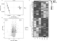
Fig. 1
The expression of differential proteins(n=3) A. Sample PLS-DA model score chart; B. Volcano map of differential protein in infected patients; C. Differential expression protein cluster heat map"

Table 3
Screening of the top 10 differential proteins under PEDV infection (n=3)"
| NCBI accession | 蛋白质Protein | 变化Change | 差异倍数Fold change |
| F1SCY2 | 干扰素诱导的含四肽重复序列的蛋白3 | 上调 | 24.92 |
| I3LS55 | 表皮生长因子受体途径底物8样蛋1 | 上调 | 22.58 |
| A0A287AXN9 | 角蛋白17 | 上调 | 14.33 |
| F1RGY8 | 内质网氧化还原酶1β | 上调 | 13.63 |
| A0A5K1UFA2 | 干扰素诱导的含四肽重复序列的蛋白3 | 上调 | 10.58 |
| A0A5G2REI9 | F-盒蛋白28 | 下调 | -6.80 |
| I3LII4 | 丝氨酸/苏氨酸蛋白激酶 | 下调 | -7.21 |
| F1RT08 | 腺苷甲硫氨酸脱羧酶 | 下调 | -14.28 |
| A0A8W4FQ04 | 铁蛋白 | 下调 | -37.26 |
| P19133 | 铁蛋白轻链 | 下调 | -38.84 |
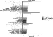
Fig. 2
GO enrichment analysis of differentially expressed proteins"

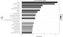
Fig. 3
KEGG enrichment analysis of differentially expressed proteins"


Fig. 4
Results of MDA content detection (n=3)"


Fig. 5
Results of GSH content detection (n=3)"


Fig. 6
Detection of the signature protein of ferroptosis(n=3) A. Protein detection results of TFRC, xCT and GPX4; B, C, D. Relative expression levels of TFRC, xCT and GPX4 proteins; E. Protein detection results of ACSL4, LPCAT3 and Ferritin; F, G, H. Relative expression levels of ACSL4, LPCAT3 and Ferritin proteins; The internal reference for TFRC, xCT and GPX4 is β-Tubulin; The internal reference for ACSL4, LPCAT3 and Ferritin is β-actin; **. P < 0.01, ***. P < 0.001"


Fig. 7
Fe2+ changes of IPEC-J2 cells after PEDV infection A. Fe2+ fluorescence detection; B. Statistics of relative fluorescence intensity; **. P < 0.01"


Fig. 8
MMP changes of IPEC-J2 cells after PEDV infection A.MMP fluorescence detection; B. Statistics of relative fluorescence intensity; The scale is 125 μm; **. P < 0.01"


Fig. 9
Changes of lipid ROS and end product MDA in IPEC-J2 cells infected by PEDV A. Fluorescence detection of lipid ROS, red fluorescence is reduced state, green fluorescence is oxidized state; B. Statistics of relative fluorescence intensity; The scale is 125 μm; **. P < 0.01"


Fig. 10
Replication of PEDV after treatment with the ferroptosis inhibitor Fer-1 c A. Western blot detection results of PEDV N protein after Fer-1 treatment; B. Detection results of PEDV N protein gene level after Fer-1 treatment; C. The change of PEDV titer after Fer-1 treatment. **. P < 0.01"

| 1 |
JUNG K , ANNAMALAI T , LU Z , et al. Comparative pathogenesis of US porcine epidemic diarrhea virus (PEDV) strain PC21A in conventional 9-day-old nursing piglets vs. 26-day-old weaned pigs[J]. Vet Microbiol, 2015, 178 (1-2): 31- 40.
doi: 10.1016/j.vetmic.2015.04.022 |
| 2 |
WANG L , BYRUM B , ZHANG Y . New variant of porcine epidemic diarrhea virus, United States, 2014[J]. Emerg Infect Dis, 2014, 20 (5): 917- 919.
doi: 10.3201/eid2005.140195 |
| 3 |
ZHAO Z , SOKHANSANJ B A , MALHOTRA C , et al. Genetic grouping of SARS-CoV-2 coronavirus sequences using informative subtype markers for pandemic spread visualization[J]. PLoS Comput Biol, 2020, 16 (9): e1008269.
doi: 10.1371/journal.pcbi.1008269 |
| 4 |
LEE C . Porcine epidemic diarrhea virus: An emerging and re-emerging epizootic swine virus[J]. Virol J, 2015, 12, 193.
doi: 10.1186/s12985-015-0421-2 |
| 5 |
GALY B , FERRING-APPEL D , KADEN S , et al. Iron regulatory proteins are essential for intestinal function and control key iron absorption molecules in the duodenum[J]. Cell Metab, 2008, 7 (1): 79- 85.
doi: 10.1016/j.cmet.2007.10.006 |
| 6 |
STOCKWELL B R , FRIEDMANN ANGELI J P , BAYIR H , et al. Ferroptosis: A regulated cell death nexus linking metabolism, redox biology, and disease[J]. Cell, 2017, 171 (2): 273- 285.
doi: 10.1016/j.cell.2017.09.021 |
| 7 |
TANG Z , XU Z , ZHU X , et al. New insights into molecules and pathways of cancer metabolism and therapeutic implications[J]. Cancer Commun (Lond), 2021, 41 (1): 16- 36.
doi: 10.1002/cac2.12112 |
| 8 |
WANG Y , HU M , CAO J , et al. ACSL4 and polyunsaturated lipids support metastatic extravasation and colonization[J]. Cell, 2025, 188 (2): 412- 429.
doi: 10.1016/j.cell.2024.10.047 |
| 9 |
DOLL S , PRONETH B , TYURINA Y Y , et al. ACSL4 dictates ferroptosis sensitivity by shaping cellular lipid composition[J]. Nat Chem Biol, 2017, 13 (1): 91- 98.
doi: 10.1038/nchembio.2239 |
| 10 |
HASHIDATE-YOSHIDA T , HARAYAMA T , HISHIKAWA D , et al. Fatty acid remodeling by LPCAT3 enriches arachidonate in phospholipid membranes and regulates triglyceride transport[J]. Elife, 2015, 4, e06328.
doi: 10.7554/eLife.06328 |
| 11 |
BAI Y , MENG L , HAN L , et al. Lipid storage and lipophagy regulates ferroptosis[J]. Biochem Biophys Res Commun, 2019, 508 (4): 997- 1003.
doi: 10.1016/j.bbrc.2018.12.039 |
| 12 |
KAGAN V E , MAO G , QU F , et al. Oxidized arachidonic and adrenic PEs navigate cells to ferroptosis[J]. Nat Chem Biol, 2017, 13 (1): 81- 90.
doi: 10.1038/nchembio.2238 |
| 13 |
DIXON S J , STOCKWELL B R . The role of iron and reactive oxygen species in cell death[J]. Nat Chem Biol, 2014, 10 (1): 9- 17.
doi: 10.1038/nchembio.1416 |
| 14 |
CHEN X , LI J , KANG R , et al. Ferroptosis: machinery and regulation[J]. Autophagy, 2021, 17 (9): 2054- 2081.
doi: 10.1080/15548627.2020.1810918 |
| 15 |
MUNOZ M , GARCIA-ERCE J A , REMACHA A F . Disorders of iron metabolism. Part 1: molecular basis of iron homoeostasis[J]. J Clin Pathol, 2011, 64 (4): 281- 286.
doi: 10.1136/jcp.2010.079046 |
| 16 |
BRADLEY J M , LE BRUN N E , MOORE G R . Ferritins: furnishing proteins with iron[J]. J Biol Inorg Chem, 2016, 21 (1): 13- 28.
doi: 10.1007/s00775-016-1336-0 |
| 17 |
HOU W , XIE Y , SONG X , et al. Autophagy promotes ferroptosis by degradation of ferritin[J]. Autophagy, 2016, 12 (8): 1425- 1428.
doi: 10.1080/15548627.2016.1187366 |
| 18 |
FRAZER D M , ANDERSON G J . The regulation of iron transport[J]. Biofactors, 2014, 40 (2): 206- 214.
doi: 10.1002/biof.1148 |
| 19 | 程峰, 张庸, 王祥, 等. 谷胱甘肽过氧化物酶GPX4在铁死亡中的作用与机制研究进展[J]. 现代肿瘤医学, 2021, 29 (7): 1254- 1258. |
| CHENG F , ZHANG Y , WANG X , et al. Research progress on the role and mechanism of glutathione peroxidase GPX4 in ferroptosis[J]. Modern Oncology Medicine, 2021, 29 (7): 1254- 1258. | |
| 20 |
LIU Y , WAN Y , JIANG Y , et al. GPX4: The hub of lipid oxidation, ferroptosis, disease and treatment[J]. Biochim Biophys Acta Rev Cancer, 2023, 1878 (3): 188890.
doi: 10.1016/j.bbcan.2023.188890 |
| 21 | 刘磊, 贾少晗, 于鹏. 线粒体在铁死亡中的形态特征和作用[J]. 中国生物化学与分子生物学报, 2023, 39 (6): 769- 777. |
| LIU L , JIA S H , YU P . Morphological characteristics and roles of mitochondria in ferroptosis[J]. Chinese Journal of Biochemistry and Molecular Biology, 2023, 39 (6): 769- 777. | |
| 22 |
LI J , CAO F , YIN H , et al. Ferroptosis: past, present and future[J]. Cell Death Dis, 2020, 11 (2): 88.
doi: 10.1038/s41419-020-2298-2 |
| 23 |
CHEN X , KANG R , KROEMER G , et al. Ferroptosis in infection, inflammation, and immunity[J]. J Exp Med, 2021, 218 (6): e20210518.
doi: 10.1084/jem.20210518 |
| 24 |
GAO J , WANG Q , TANG Y , et al. When ferroptosis meets pathogenic infections[J]. Trends Microbiol, 2023, 31 (5): 468- 479.
doi: 10.1016/j.tim.2022.11.006 |
| 25 |
LIU G , XU X , TAO S , et al. HBx facilitates ferroptosis in acute liver failure via EZH2 mediated SLC7A11 suppression[J]. J Biomed Sci, 2021, 28 (1): 67.
doi: 10.1186/s12929-021-00762-2 |
| 26 |
KAN X , YIN Y , SONG C , et al. Newcastle-disease-virus-induced ferroptosis through nutrient deprivation and ferritinophagy in tumor cells[J]. iScience, 2021, 24 (8): 102837.
doi: 10.1016/j.isci.2021.102837 |
| 27 |
CHENG J , TAO J , LI B , et al. Swine influenza virus triggers ferroptosis in A549 cells to enhance virus replication[J]. Virol J, 2022, 19 (1): 104.
doi: 10.1186/s12985-022-01825-y |
| 28 |
KUNG Y , CHIANG H , LI M , et al. Acyl-coenzyme a synthetase long-chain family member 4 is involved in viral replication organelle formation and facilitates virus replication via ferroptosis[J]. mBio, 2022, 13 (1): e0271721.
doi: 10.1128/mbio.02717-21 |
| 29 |
LI H , LI W , ZHANG S , et al. Enterovirus 71 activates GADD34 via precursor 3CD to promote IRES-mediated viral translation[J]. Microbiol Spectr, 2022, 10 (1): e0138821.
doi: 10.1128/spectrum.01388-21 |
| 30 |
AI Y , MENG Y , YAN B , et al. The biochemical pathways of apoptotic, necroptotic, pyroptotic, and ferroptotic cell death[J]. Mol Cell, 2024, 84 (1): 170- 179.
doi: 10.1016/j.molcel.2023.11.040 |
| 31 |
STOCKWELL B R . Ferroptosis turns 10: Emerging mechanisms, physiological functions, and therapeutic applications[J]. Cell, 2022, 185 (14): 2401- 2421.
doi: 10.1016/j.cell.2022.06.003 |
| 32 | 苏朗驹, 黄宗洋, 黄俊, 等. 猪流行性腹泻病毒核衣壳蛋白的研究进展[J]. 中国动物传染病学报, 2024, 32 (3): 200- 208. |
| SU L J , HUANG Z Y , HUANG J , et al. Research progress of nucleocapsid protein of porcine epidemic diarrhea virus[J]. Chinese Journal of Animal Infectious Diseases, 2024, 32 (3): 200- 208. | |
| 33 |
SU M , SHI D , XING X , et al. Coronavirus porcine epidemic diarrhea virus nucleocapsid protein interacts with p53 to induce cell cycle arrest in S-phase and promotes viral replication[J]. J Virol, 2021, 95 (16): e0018721.
doi: 10.1128/JVI.00187-21 |
| 34 |
ZHAI X , KONG N , ZHANG Y , et al. N protein of PEDV plays chess game with host proteins by selective autophagy[J]. Autophagy, 2023, 19 (8): 2338- 2352.
doi: 10.1080/15548627.2023.2181615 |
| [1] | LI Huimin, LEI Mingkai, RUAN Shengnan, LI Panpan, LI Wentao, HE Qigai. Establishment of Fluorescent Microsphere Immunochromatographic Assay for Porcine Epidemic Diarrhea Virus Antigen Detection [J]. Acta Veterinaria et Zootechnica Sinica, 2025, 56(9): 4572-4580. |
| [2] | YANG Wenzhe, WANG Jinhao, ZHAO Zichen, ZHAO Tong, PAN Feilong, CHEN Fangfang, SHAO Wenqi, LIU Kexiang, ZHAO Shuchen, ZHAO Lijia. Analysis of the Impact of Curcumin on the Ferroptosis Pathway in Alleviating the Inflammatory Response Induced by LPS in Bovine Mammary Epithelial Cells [J]. Acta Veterinaria et Zootechnica Sinica, 2025, 56(9): 4730-4740. |
| [3] | WEN Xue, XU Wanxue, FU Yitong, YANG Jie, FU Hongyu, FAN Ruifeng. Research Progress on the Relationship between Ferroptosis and Inflammation [J]. Acta Veterinaria et Zootechnica Sinica, 2025, 56(8): 3666-3677. |
| [4] | TIAN Ru, FU Xingwei, HU Leyu, ZHU Mingjun, TONG Dewen. Isolation and Pathogenicity Analysis of a GⅡa Porcine Epidemic Diarrhea Virus [J]. Acta Veterinaria et Zootechnica Sinica, 2025, 56(8): 4101-4111. |
| [5] | LI Zhiqiang, CHEN Xueqing, ZHANG Yuanshu. Detection of Angiotensin Converting Enzyme 2 in Intestinal Tissues of Clinically Infected Porcine Epidemic Diarrhea Virus Piglets and Analysis of Its Relationship with Intestinal Pathological Changes [J]. Acta Veterinaria et Zootechnica Sinica, 2025, 56(7): 3463-3473. |
| [6] | WANG Yunke, WANG Na, YUE Ke, HE Kunmiao, ZHANG Xing, LIU Yao, ZHANG Gaiping. Substances with Inhibitory Effects on Porcine Epidemic Diarrhea Virus Replication in vitro [J]. Acta Veterinaria et Zootechnica Sinica, 2025, 56(6): 2577-2589. |
| [7] | WU Chao, MING Wenhan, LU Shuwan, YANG Caimei, LIU Jinsong, MA Xiang, ZHANG Ruiqiang. Innate Immune Evasion Mechanisms of Porcine Epidemic Diarrhea Virus and Advances in Prevention and Control Strategies [J]. Acta Veterinaria et Zootechnica Sinica, 2025, 56(6): 2590-2599. |
| [8] | ZHOU Min, TANG Deyuan, ZENG Zhiyong, WANG Bin, HUANG Tao, HU Wenwen, MAO Yinming, ZHOU Piao, HE Song. Research Progress on the Interaction between Porcine Epidemic Diarrhea Virus Proteins and Host Proteins [J]. Acta Veterinaria et Zootechnica Sinica, 2025, 56(6): 2600-2612. |
| [9] | LI Chengcheng, ZHAO Yongxiang, CAO Qiuxia, SONG Xu, LI Yupeng, FAN Baochao, GUO Rongli, XU Yefen, LI Bin. Tight Junction Protein CLDN4 Promotes Porcine Epidemic Diarrhea Virus Infection [J]. Acta Veterinaria et Zootechnica Sinica, 2025, 56(6): 2826-2835. |
| [10] | WU Peiling, LI Yixuan, WANG Haojie, LI Yafei, LIU Shaomeng, LIU Qingyun, WANG Xiangru. Research Progress of Porcine Epidemic Diarrhea Vaccine for Pigs [J]. Acta Veterinaria et Zootechnica Sinica, 2025, 56(3): 1042-1058. |
| [11] | YU Xinya, HE Haijian, WANG Lei, NI Yuchen, DU Jing, ZHOU Yingshan, DONG Wanyu, WANG Xiaodu. Effect of lncRNA 18850 on Porcine Epidemic Diarrhea Virus Replication [J]. Acta Veterinaria et Zootechnica Sinica, 2025, 56(3): 1366-1375. |
| [12] | ZHANG Dongxuan, WANG Zhihao, QIAO Yan, ZHAO Xiaoxiao, FAN Songjie, ZHANG Chao. Prokaryotic Expression of S1 Protein in Porcine Epidemic Diarrhea Virus and Screening of Its Aptamers [J]. Acta Veterinaria et Zootechnica Sinica, 2025, 56(2): 839-850. |
| [13] | MA Yue, MIAO Yuhang, DING Tao, XIN Jie, MA Wenyan, LI Yanan, ZHOU Xuezhang, DU Jun. Signaling Pathway Analysis of Ferroptosis Induced by Recombinant Candida krusei 14-3-3 Protein in Bovine Mammary Epithelial Cells [J]. Acta Veterinaria et Zootechnica Sinica, 2025, 56(11): 5706-5720. |
| [14] | TANG Jinmeng, YU Shunan, YUAN Yixin, MA Yuchen, DU Wenjuan, YU Linyang, LI Yongtao. Construction of Porcine IPEC-J2 Cell Line Stably Expressing Human IFITM3 and Its Effect on PEDV Proliferation [J]. Acta Veterinaria et Zootechnica Sinica, 2025, 56(10): 5180-5189. |
| [15] | LI Hao, XU Qiaoyu, JIN Xiaohui, CHEN Hongying, LI Qinghao, JIN Xin, MA Junxing, MA Shijie, WEI Zhanyong. Histopathological Changes of Immune Organs and Alterations of Cytokines in Piglets Infected with Porcine Epidemic Diarrhea Virus [J]. Acta Veterinaria et Zootechnica Sinica, 2025, 56(10): 5190-5201. |
| Viewed | ||||||
|
Full text |
|
|||||
|
Abstract |
|
|||||