


Acta Veterinaria et Zootechnica Sinica ›› 2025, Vol. 56 ›› Issue (11): 5575-5587.doi: 10.11843/j.issn.0366-6964.2025.11.018
• Animal Biotechnology and Reproduction • Previous Articles Next Articles
CHEN Qian1,2,3( ), TONG Jiang1,2,3, PAN Yuheng1,2,3, MA Jianfeng1,2,3, SHI Yuqian1,2,3, CHEN Siyu1,2,3, LI Jiaxin1,2,3, ZHOU Ting4, LIU Chendong4, ZHU Li1,2,3, SHEN Linyuan1,2,3,*(), GAN Mailin1,2,3,*()
Received:2025-04-09
Online:2025-11-23
Published:2025-11-27
Contact:
SHEN Linyuan, GAN Mailin
E-mail:chenqian1@stu.sicau.edu.cn;shenlinyuan@sicau.edu.cn;ganmailin@sicau.edu.cn
CLC Number:
CHEN Qian, TONG Jiang, PAN Yuheng, MA Jianfeng, SHI Yuqian, CHEN Siyu, LI Jiaxin, ZHOU Ting, LIU Chendong, ZHU Li, SHEN Linyuan, GAN Mailin. The Impact of Gossypol on Spermatogenic Capacity of the Testis and Apoptosis of Sertoli Cells[J]. Acta Veterinaria et Zootechnica Sinica, 2025, 56(11): 5575-5587.
Table 1
The primer sequences used for RT-qPCR"
| 基因 Gene | 功能 Function | 引物序列(5′→3′) Primers sequence | 产物长度/bp Products length |
| Bcl-2 | 凋亡相关基因 | F:GGAACAGGACGCTCAGACTT | 119 |
| R:GCAGGATAGCAGCACAGGAT | |||
| Bax | F:TGAAGACAGGGGCCTTTTTG | 140 | |
| R:AATTCGCCGGAGACACTCG | |||
| P53 | F:TCACAGCGTCTGTTGACATTT | 210 | |
| R:ACCAAGCTCATTACCCTGACA | |||
| Tnf-α | F:GACGTGGAACTGGCAGAAGAG | 160 | |
| R:TTGGTGGTTTGTGAGTGTGAG | |||
| Ki67 | F:ACCGTGGAGTAGTTTATCTGGG | 126 | |
| R:TGTTTCCAGTCCGCTTACTTCT | |||
| P62 | 自噬相关基因 | F:TCCAGCTTGAACCCTATTGAGA | 166 |
| R:GTCCTGCATCACTGAGCATCT | |||
| Vps15 | F:CTGGCGACAGGAGACTGAC | 188 | |
| R:GGGGAAACCAAGTCCCAGAA | |||
| Atg5 | F:TGTGCTTCGAGATGTGTGGTT | 120 | |
| R:GTCAAATAGCTGACTCTTGGCAA | |||
| Ulk1 | F:AAGTTCGAGTTCTCTCGCAAG | 190 | |
| R:CGATGTTTTCGTGCTTTAGTTCC | |||
| Lc3 | F:GGCTACGGCTACTATCGCAC | 100 | |
| R:AGGAGGGCATGACAAAGGAGA | |||
| Acsl4 | 铁死亡相关基因 | F:CTCACCATTATATTGCTGCCTGT | 163 |
| R:TCTCTTTGCCATAGCGTTTTTCT | |||
| Ho-1 | F:AAGCCGAGAATGCTGAGTTCA | 198 | |
| R:GCCGTGTAGATATGGTACAAGGA | |||
| Ptgs2 | F:TGAGCAACTATTCCAAACCAGC | 74 | |
| R:GCACGTAGTCTTCGATCACTATC | |||
| Gpx4 | F:GCCTGGATAAGTACAGGGGTT | 99 | |
| R:CATGCAGATCGACTAGCTGAG | |||
| Fth1 | F:AGACCGTGATGACTGGGAGA | 138 | |
| R:TCAATGAAGTCACATAAGTGGGGA | |||
| Zo1 | 紧密连接相关基因 | F:CGCGGAGAGAGACAAGATGT | 55 |
| R:CATTGCTGTGCTCTTAGCGG | |||
| Cdh1 | F:AACCCAAGCACGTATCAGGG | 94 | |
| R:GAGTGTTGGGGGCATCATCA | |||
| Occludlin | F:CCCTCTTTCCTTAGGCGACA | 95 | |
| R:CCCAAGATAAGCGAACCTGC | |||
| Claudin5 | F:GTTAAGGCACGGGTAGCACT | 137 | |
| R:TACTTCTGTGACACCGGCAC | |||
| Sycp3 | 精子发生相关基因 | F:ATGATGGAAACTCAGCAGCA | 135 |
| R:GCATGCCTCTTAGCTAATGTTTT | |||
| Sycp1 | F:AGGTTCTGAGGGGAAGCTCA | 98 | |
| R:CCTCCTGGGCCGTTGTC | |||
| Ddx4 | F:GCAGCAAGTGATTCAGGCAA | 146 | |
| R:TCACTGCTTTCACCTCCTTCA | |||
| β-actin | 内参基因 | F:TGGAATCCTGTGGCATCCATGAAAC | 110 |
| R:TGGAATCCTGTGGCATCCATGAAAC |
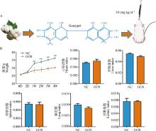
Fig. 1
Effects of gossypol on body weight and some organs of mice A. Schematic diagram of gossypol administration via gastric gavage in mice; B. Comparative chart of body weight growth curves and major visceral index in mice"
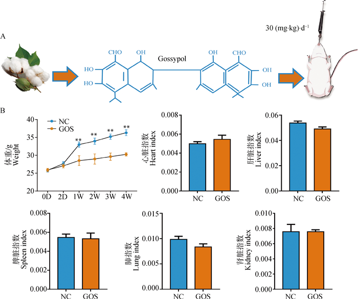
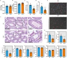
Fig. 2
Effects of gossypol on testis histopathology and sperm quality in mice A. Testis weight in mice; B. Testis organ index in mice; C.Sperm motility in mice; D. Sperm density in mice; E. Sperm motion trajectory diagram in mice; F. Histopathological examination of testis tissue in mice; G. Analysis of sperm kinematic parameters in mice (including 8 parameters: curvilinear velocity (VCL), straight-line velocity (VSL), average path velocity (VAP), beat cross frequency (BCF), amplitude of lateral head displacement (ALH), straightness (STR), linearity (LIN), and wobble (WOB))"

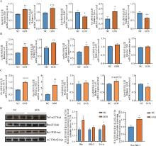
Fig. 3
The changes in the expression levels of genes related to apoptosis, autophagy and ferroptosis in mice testicular tissues, as well as the expression levels of apoptosis-related proteins A. Relative expression levels of apoptosis-related genes Bax, P53, Ki67, Tnf-α and Bcl-2 in mice testicular tissue; B. Relative expression levels of autophagy-related genes Lc3, P62, Atg5, Vps15 and Ulk1 in mice testicular tissue; C. Relative expression levels of ferroptosis-related genes Ho-1, Ptgs2, Fth1, Acsl4, and Gpx4 in mice testicular tissue; D. The relative expression levels of apoptosis-related proteins Bax, Bcl-2, Tnf-α and Bax/Bcl-2 in mice testicular tissues"
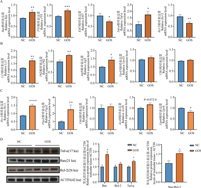
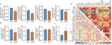
Fig. 4
Changes in expression levels of tight junction and spermatogenesis-related genes in mice testicular tissue and their gene correlation analysis A. Relative expression levels of tight junction-related genes Cdh1, Claudin5, Occludlin, and Zo-1 in mice testicular tissue; B. Relative expression levels of spermatogenesis-related genes Sycp1, Sycp3, and Ddx4 in mice testicular tissue; C. The relative expression levels of apoptosis-related genes Bax/Bcl-2 in the testicular tissue of mice; D. The correlation heatmap of genes related to apoptosis, autophagy, ferroptosis, tight junction and spermatogenesis in the testicular tissue of mice"
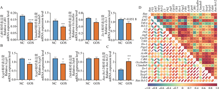
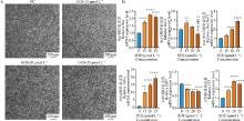
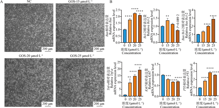
Fig. 5
Effects of varying gossypol concentrations on TM4 cells apoptosis A. TM4 cells were infected with gossypol at different doses; B. Relative expression levels of apoptosis genes Bax, Bcl-2, Tnf-α, Ki67, P53 and Bax/Bcl-2 in TM4 cells"
| 1 |
SHLET S M . Normal testicular function and spermatogenesis[J]. Pediatr Blood Cancer, 2009, 53 (2): 285- 288.
doi: 10.1002/pbc.22000 |
| 2 |
O'SHAUGHNESSY P J , VERHOEVEN G , DE G K , et al. Direct action through the sertoli cells is essential for androgen stimulation of spermatogenesis[J]. Endocrinology, 2010, 151 (5): 2343- 2348.
doi: 10.1210/en.2009-1333 |
| 3 |
SHUPE J , CHENG J , PURI P , et al. Regulation of sertoli-germ cell adhesion and sperm release by FSH and nonclassical testosterone signaling[J]. Mol Endocrinol, 2011, 25 (2): 238- 252.
doi: 10.1210/me.2010-0030 |
| 4 |
RABBANI M , ZHANG X , MANSKE G L , et al. Decoding the spermatogenesis program: New insights from transcriptomic analyses[J]. Annu Rev Genet, 2022, 56, 339- 368.
doi: 10.1146/annurev-genet-080320-040045 |
| 5 | SU J , YANG Y , ZHAO F , et al. Study of spermatogenic and Sertoli cells in the Hu sheep testes at different developmental stages[J]. FASEB J, 2023, Aug (37): 8- e23084. |
| 6 |
JIANG B , YANG D , PENG H . Environmental toxins and reproductive health: unraveling the effects on Sertoli cells and the blood-testis barrier in animalset[J]. Biol Reprod, 2024, 111 (5): 977- 986.
doi: 10.1093/biolre/ioae126 |
| 7 |
RICHBURG J H , BOEKELHEIDE K . Mono-(2-ethylhexyl) phthalate rapidly alters both Sertoli cell vimentin filaments and germ cell apoptosis in young rat testes[J]. Toxicol Appl Pharmacol, 1996, 137 (1): 42- 50.
doi: 10.1006/taap.1996.0055 |
| 8 | 赵龙坡, 孙辉臣. 睾丸支持细胞与生精功能障碍的关系[J]. 华北国防医药, 2005, 17 (5): 318- 321. |
| ZHAO L P , SUN H C . The relationship between Sertoli cells and spermatogenic dysfunction[J]. Medical & Pharmaceutical Journal of Chinese People's Liberation Army, 2005, 17 (5): 318- 321. | |
| 9 |
XIAO Y , ZHANG J , GUAN Y , et al. Research progress on sertoli cell secretion during spermatogenesis[J]. Front Endocrinol (Lausanne), 2025, 15, 1456410.
doi: 10.3389/fendo.2024.1456410 |
| 10 | 邵仕香, 董庆洁, 郭星, 等. 棉酚及其衍生物的医药研究进展[J]. 天津理工学院学报, 2002, 18 (1): 87- 92. |
| SHAO S X , DONG Q J , GUO X , et al. Progress in gossypol and its derivatives as a procreate-resistant drug[J]. Journal of Tianjin University of Technology, 2002, 18 (1): 87- 92. | |
| 11 | 王梅兰. 棉酚——值得开发的避孕药[J]. 厦门科技, 2013 (6): 53- 54. |
| WANG M L . Gossypol-A contraceptive worth developing[J]. Xiamen Science & Technology, 2013 (6): 53- 54. | |
| 12 | 谢淑武, 沈如凌, 林金杏, 等. 雄性不育药物研发相关实验动物模型建立和应用进展[J]. 实验动物与比较医学, 2023, 43 (5): 504- 511. |
| XIE S W , SHEN R L , LIN J X , et al. Progress in Establishment and Application of Laboratory Animal Models Related to Development of Male Infertility Drugs[J]. Laboratory Animal and Comparative Medicine, 2023, 43 (5): 504- 511. | |
| 13 | 李怡佳, 边艳超, 李硕, 等. 男性不育实验动物模型建立的研究进展[J]. 中国医药导报, 2021, 18 (15): 41-44+48. |
| LI Y J , BIAN Y C , LI S , et al. Research progress on the establishment of experimental animal of malesterility[J]. China Medical Herald, 2021, 18 (15): 41-44+48. | |
| 14 | 师慧敏, 刘爽, 赵光卿, 等. 醋酸棉酚对小鼠睾丸抗氧化损伤的作用[J]. 中国兽医杂志, 2021, 57 (11): 62- 65. |
| SHI H M , LIU S , ZHAO G Q , et al. Effect of Gossypol Acetate on Anti-oxidative Damage to Testis in Mice[J]. Chinese Journal of Veterinary Medicine, 2021, 57 (11): 62- 65. | |
| 15 |
LIM W , HAM J , PAPK S , et al. Gossypol induces disruption of spermatogenesis and steroidogenesis in male mice[J]. J Agric Food Chem, 2019, 67 (7): 2075- 2085.
doi: 10.1021/acs.jafc.8b06946 |
| 16 |
AITKEN R J , MUSCLO L , WHITING S , et al. Analysis of the effects of polyphenols on human spermatozoa reveals unexpected impacts on mitochondrial membrane potential, oxidative stress and DNA integrity; implications for assisted reproductive technology[J]. Biochem Pharmacol, 2016, 121, 78- 96.
doi: 10.1016/j.bcp.2016.09.015 |
| 17 | 周德荣. 棉酚对睾丸支持细胞间隙连接蛋白43表达的调节作用的研究[D]. 汕头: 汕头大学, 2009. |
| ZHOU D R. Gossypol repressing the expression ofconnexin43 in Sertoli cells[D]. Shantou: Shantou University, 2009. (in Chinese) | |
| 18 | 王辉, 朴智燕, 马慧, 等. 棉酚短期给药对小鼠的生殖毒性和肾毒性具有可逆性[J]. 南方医科大学学报, 2023, 43 (2): 251- 256. |
| WANG H , PU Z Y , MA H , et al. Short-term exposure to gossypol causes reversible reproductive toxicity and nephrotoxicity in mice[J]. Journal of Southern Medical University, 2023, 43 (2): 251- 256. | |
| 19 | 袁彦波, 赵树臣, 侯振中. 棉酚对雄性小鼠的生殖毒性作用[J]. 中国兽医杂志, 2015, 51 (7): 83- 85. |
| YUAN Y B , ZHAO S C , HOU Z Z . Reproductive toxicity of gossypol on male mice[J]. Chinese Journal of Veterinary Medicine, 2015, 51 (7): 83- 85. | |
| 20 |
LIVAK K J , SCHMITTGEN T D . Analysis of relative gene expression data using real-time quantitative PCR and the 2(-Delta Delta C(T)) Method[J]. Methods, 2001, 25 (4): 402- 408.
doi: 10.1006/meth.2001.1262 |
| 21 | WANG X , HOWELL P C , CHEN F , et al. Chapter 6 gossypol-A polyphenolic compound from cotton plant[J]. Adv Food Nutr Res, 2009, 58, 215- 263. |
| 22 | 郁钧. 仔鹅饲粮中棉籽粕替代豆粕效应及游离棉酚损伤肠肝机制的研究[D]. 扬州: 扬州大学, 2023. |
| YU J. Research on the effect of replacing soybean meal withcottonseed meal in the diet of goose and the mechanismof F'ree gossypol injury to the intestinal and liver[D]. Yangzhou: Yangzhou University, 2023. (in Chinese) | |
| 23 | 李玲, 徐诗强, 杨健. 抗生殖活性化合物棉酚的研究进展[J]. 中华男科学杂志, 2024, 30 (3): 254- 260. |
| LI L , XU S Q , YANG J . The anti-reproductive active compound gossypol: Progress in research[J]. National Journal of Andrology, 2024, 30 (3): 254- 260. | |
| 24 |
WANG J , JIN L , LI X , et al. Gossypol induces apoptosis in ovarian cancer cells through oxidative stress[J]. Mol Biosyst, 2013, 9 (6): 1489- 1497.
doi: 10.1039/c3mb25461e |
| 25 |
ZANG Y , MA J , XU L , et al. Natural product gossypol and its derivatives in precision cancer medicine[J]. Curr Med Chem, 2019, 26 (10): 1849- 1873.
doi: 10.2174/0929867324666170523123655 |
| 26 | YAN M , WANG L , CHENG C Y . Testis toxicants: Lesson from traditional Chinese medicine (TCM)[J]. Adv Exp Med Biol, 2021, 1288, 307- 319. |
| 27 |
OBERSTEIN A , JEFFREY P D , SHI Y . Crystal structure of the Bcl-XL-Beclin 1 peptide complex: Beclin1 is a novel BH3-only protein[J]. J Biol Chem, 2007, 282 (17): 13123- 13132.
doi: 10.1074/jbc.M700492200 |
| 28 | 杨洋, 杨梦婷, 许潇. 醋酸棉酚通过诱导铁死亡抑制脑胶质瘤细胞生长[J]. 江苏大学学报(医学版), 2022, 32 (3): 246- 250. |
| YANG Y , YANG M T , XU X . Inhibitory effect of gossypol acetate on the growth ofglioma cells via induction of ferroptosis[J]. Journal of Jiangsu University(Medicine Edition), 2022, 32 (3): 246- 250. | |
| 29 | 陈可绪, 汪森明, 曹漫明, 等. ApoG2诱导乳腺癌细胞MDA-MB231自噬的最低浓度探讨[J]. 现代肿瘤医学, 2015, 23 (20): 2884- 2890. |
| CHEN K X , WANG S M , CAO M M , et al. Rational use of ApoG2 through autophagy inhibition exploiting and apoptosis inducing in breast cancer MDA-MB231 cells[J]. Journal of Modern Oncology, 2015, 23 (20): 2884- 2890. | |
| 30 | 张学斐. 棉籽蛋白替代鱼粉对大口黑鲈生长性能及肝脏铁死亡的影响[D]. 成都: 四川农业大学, 2023. |
| ZHANG X F. Effects of cottonseed protein replacement of fish meal on growth performance and liver ferroptosis of largemouth bass (Micropterus salmoides)[D]. Chengdu: Sichuan Agricultural University, 2023. (in Chinese) | |
| 31 | 袁彦波. 棉酚致小鼠睾丸生精细胞凋亡以Bax、Bcl-2表达相关性研究[D]. 哈尔滨: 东北农业大学, 2015. |
| YUAN Y B. The correlations study of gossypol on testicular spermatogenic cell apoptosis and expression of Bax, Bcl-2 in mouse[D]. Harbin: Northeast Agricultural University, 2015. (in Chinese) | |
| 32 | 赵树臣, 王海涛, 刘克祥, 等. 棉酚对小鼠睾丸氧化损伤及NF-κB、P53和Caspase-3基因表达的影响[J]. 东北农业大学学报, 2024, 55 (2): 49- 56. |
| ZHAO S C , WANG H T , LIU K X , et al. Effects of gossypol on oxidative damage and expression of NF-kB, P53 and Caspase-3 in mice testis[J]. Journal of Northeast Agricultural University, 2024, 55 (2): 49- 56. | |
| 33 | 孔伟帅, 刘娟, 邓中原, 等. 应用比较毒理基因组学数据库解析棉酚对动物机体的毒性作用机制[J]. 毒理学杂志, 2023, 37 (2): 121-127+136. |
| KONG W S , LIU J , DENG Z Y , et al. Analysis of the mechanism of gossypol toxicity in animals usingcomparative toxicogenomics database[J]. Journal of Toxicology, 2023, 37 (2): 121-127+136. | |
| 34 | 米海潮, 史敏, 崔芳. 铁自噬与铁死亡及其相关疾病[J]. 中国生物化学与分子生物学报, 2022, 38 (9): 1133- 1140. |
| MI H C , SHI M , CUI F . The research on ferritinophagy, ferroptosis and related diseases[J]. Chinese Journal of Biochemistry and Molecular Biology, 2022, 38 (9): 1133- 1140. | |
| 35 |
GAO P , BAUVY C , SOUQUÈRE S , et al. The Bcl-2 homology domain 3 mimetic gossypol induces both Beclin 1-dependent and Beclin 1-independent cytoprotective autophagy in cancer cells[J]. J Biol Chem, 2010, 285 (33): 25570- 25581.
doi: 10.1074/jbc.M110.118125 |
| 36 |
HILL S M , WROBEL L , RUBINSZTEIN D C . Post-translational modifications of Beclin 1 provide multiple strategies for autophagy regulation[J]. Cell Death Differ, 2019, 26 (4): 617- 629.
doi: 10.1038/s41418-018-0254-9 |
| 37 |
LIAN J , KARNAK D , XU L . The Bcl-2-Beclin1 interaction in (-)-gossypol-induced autophagy versus apoptosis in prostate cancer cells[J]. Autophagy, 2010, 6 (8): 1201- 1203.
doi: 10.4161/auto.6.8.13549 |
| 38 |
LIU W , JIN W , ZHU S , et al. Targeting regulated cell death (RCD) with small-molecule compounds in cancer therapy: A revisited review of apoptosis, autophagy-dependent cell death and necroptosis[J]. Drug Discov Today, 2022, 27 (2): 612- 625.
doi: 10.1016/j.drudis.2021.10.011 |
| 39 |
YANG Y , CHEN Y , WU J H , et al. Targeting regulated cell death with plant natural compounds for cancer therapy: A revisited review of apoptosis, autophagy-dependent cell death, and necroptosis[J]. Phytother Res, 2023, 37 (4): 1488- 1525.
doi: 10.1002/ptr.7738 |
| 40 | CHAN K H , ZHENG B X , LEUNG A S , et al. A NRAS mRNA G-quadruplex structure-targeting small-molecule ligand reactivating DNA damage response in human cancer cells for combination therapy with clinical PI3K inhibitors[J]. Int J Biol Macromol, 2024, 279 (Pt 3): 135308. |
| 41 |
RAO M , XIA W , YANG J , et al. Transient scrotal hyperthermia affects human sperm DNA integrity, sperm apoptosis, and sperm protein expression[J]. Andrology, 2016, 4 (6): 1054- 1063.
doi: 10.1111/andr.12228 |
| 42 |
MUSTAFA M , AHMAD R , TANTRY Q I , et al. Apoptosis: A comprehensive overview of signaling pathways, morphological changes, and physiological significance and therapeutic implications[J]. Cells, 2024, 13 (22): 1838.
doi: 10.3390/cells13221838 |
| 43 |
SHEIKH-HAMAD D , CACINI W , BUCKLEY A R , et al. Cellular and molecular studies on cisplatin-induced apoptotic cell death in rat kidney[J]. Arch Toxicol, 2004, 78 (3): 147- 155.
doi: 10.1007/s00204-003-0521-4 |
| [1] | LIU Jiajin, WEN Xiaoqing, LUO Chunhai, JIA Hongdou, WANG Wei, LI Danyang, FU Shixin. The Influence of FoxO1 on Expression of Apoptotic Factors in Dairy Cow Endometrial Epithelial Cells Induced by High NEFA Levels [J]. Acta Veterinaria et Zootechnica Sinica, 2025, 56(9): 4708-4717. |
| [2] | ZHAO Shunran, FU Guixin, PANG Zhaoqi, XIA Wei, LI Junjie, TAO Chenyu. Research Progress on the Mechanism of Porcine Granulosa Cells in Follicular Atresia [J]. Acta Veterinaria et Zootechnica Sinica, 2025, 56(6): 2537-2545. |
| [3] | ZHU Aiwen, WANG Jian, ZHU Gehui, LIU Haixia, PINGCUO Bandan, WANG Jun, DEQING Zhuoga, YAN Wei, HAN Dayong. Zearalenone Induced Proliferation, Apoptosis, Oxidative Stress and NAC Protective Mechanism of Sertoli Cells in Pengbo Semi-fine Wool Sheep [J]. Acta Veterinaria et Zootechnica Sinica, 2025, 56(6): 2752-2764. |
| [4] | CHEN Yun, CHEN Liyuan, SONG Wenjing, ZHANG Xinke, XU Han, WU Jiayi, ZHAO Cuiyan, ZHANG Shouquan. Research Progress on the Mechanism of T-2 Toxin 's Impact on Male Animal Reproduction [J]. Acta Veterinaria et Zootechnica Sinica, 2025, 56(5): 2038-2046. |
| [5] | GAO Zhengjie, LUO Ping, LI Bocheng, WANG Shuilian. Effects of GnIH on Proliferation, Apoptosis and Estrogen Secretion of Mouse Ovarian Granulosa Cells [J]. Acta Veterinaria et Zootechnica Sinica, 2025, 56(5): 2230-2242. |
| [6] | DONG Zhifang, ZHANG Li, ZHU Xiangbo. Repair Effects of Glutathione on Cadmium Induced Oxidative Damage in Pig Kidney PK-15 Cells [J]. Acta Veterinaria et Zootechnica Sinica, 2025, 56(5): 2403-2412. |
| [7] | YE Rungen, LIU Yuanbo, LU Lili, Collins Amponsah Asiamah, SU Ying*. Expression of miR-215-5p in Leizhou Black Duck Tissues and Its Effect on Follicular Granulosa Cells Proliferation and Apoptosis [J]. Acta Veterinaria et Zootechnica Sinica, 2025, 56(4): 1722-1730. |
| [8] | HOU Wanchen, XU Tong. Cannabidiol Antagonizes BPA-induced Apoptosis and Autophagy in Porcine Intestinal Epithelial Cells through the BRD4/AMPK/mTOR Signaling Pathway [J]. Acta Veterinaria et Zootechnica Sinica, 2025, 56(4): 1919-1933. |
| [9] | PANG Siyao, ZHANG Jinlong, SUN Yuhang. Proteomic Analysis of 3D4/21 Cells Infected with H1N1 Swine Influenza Virus under Non-cytotoxic Concentration of AFB1 Exposure [J]. Acta Veterinaria et Zootechnica Sinica, 2025, 56(4): 1947-1957. |
| [10] | LIU Chenlong, JI Huayuan, LU Dan, WAN Mingchun, HU Yao, ZHOU Quanyong. Effect of FST on Proliferation, Apoptosis and Hormone Secretion of Porcine Ovarian Granulosa Cells [J]. Acta Veterinaria et Zootechnica Sinica, 2025, 56(3): 1242-1251. |
| [11] | FENG Yuhuan, ZHANG Yiqian, LIU Xia, ZHOU Xuehui, LIU Yanyan, NIU Luting, NI Xingwei, ZHAO Zhiguo, WANG Yan, YANG Xiaowei, XU Tingting, ZHAO Guangwei. Research Progress on the Interaction Mechanisms between Akabane Virus and Host [J]. Acta Veterinaria et Zootechnica Sinica, 2025, 56(10): 4877-4888. |
| [12] | WANG Lei, BAI Shaocheng, WANG Sen, BAO Zhiyuan, CAI Jiawei, LIU Yan, ZHAO Bohao, WU Xinsheng, CHEN Yang. Effect of SRD5A2 on the Expression of Genes Related to Proliferation, Apoptosis and Steroid Hormone Synthesis in Rabbit Granulosa Cells [J]. Acta Veterinaria et Zootechnica Sinica, 2025, 56(1): 259-268. |
| [13] | 古丽米热·阿布都热依木, Xinru ZHANG, Yangsheng WU, Ying CHEN, Liqin WANG, Xinming XU, Juncheng HUANG, Jiapeng LIN. Effects of FKBP5 on Function of Sheep Follicular Granulosa Cells [J]. Acta Veterinaria et Zootechnica Sinica, 2024, 55(9): 3947-3956. |
| [14] | Yi WANG, Jianfei GONG, Nuo HENG, Yingfan HU, Rui WANG, Huan WANG, Ni ZHU, Wei HE, Zhihui HU, Haisheng HAO, Huabin ZHU, Shanjiang ZHAO. Melatonin Alleviates Palmitic Acid-induced Damage in Bovine Endometrial Epithelial Cells by Improving Mitochondrial Dynamics [J]. Acta Veterinaria et Zootechnica Sinica, 2024, 55(9): 3978-3987. |
| [15] | Zenghua LU, Yan CUI, Sijiu YU, Xuefeng BAI, Hongqin LU, Junfeng HE, Kai LU, Guoliang ZHAI, Zhengman QI. Effect of Erythropoietin on the Expression of Apoptotic Factor in Yak Renal Interstitial Fibroblasts [J]. Acta Veterinaria et Zootechnica Sinica, 2024, 55(8): 3460-3471. |
| Viewed | ||||||
|
Full text |
|
|||||
|
Abstract |
|
|||||