


Acta Veterinaria et Zootechnica Sinica ›› 2025, Vol. 56 ›› Issue (5): 2383-2392.doi: 10.11843/j.issn.0366-6964.2025.05.035
• Basic Veterinary Medicine • Previous Articles Next Articles
CHANG Lingling2( ), ZHANG Yongqiang1(), WANG Yashi2, ZHANG Fengxi2, ZHANG Xinyue2, ZHAO Xiaomin2, GE Shengqiang1, LI Jinming1, WANG Zhiliang1,*()
Received:2024-07-10
Online:2025-05-01
Published:2025-05-27
Contact:
WANG Zhiliang
E-mail:llchang@nwafu.edu.cn;zhangyongqiang@cahec.cn;wangzhiliang@cahec.cn
CLC Number:
CHANG Lingling, ZHANG Yongqiang, WANG Yashi, ZHANG Fengxi, ZHANG Xinyue, ZHAO Xiaomin, GE Shengqiang, LI Jinming, WANG Zhiliang. The Pathological Observation of Major Organs in a Pig Infected by African Swine Fever Virus[J]. Acta Veterinaria et Zootechnica Sinica, 2025, 56(5): 2383-2392.
Table 1
The gross findings in the pig infected by ASFV"
| 器官 Organs | 大体病变描述 Description of gross changes |
| 皮肤及皮下组织 Skin and subcutaneous tissue | 头颈部、躯干及四肢处皮肤散在出血斑;耳朵和四肢发绀;腹部皮下脂肪点状出血 |
| 淋巴结Lymph node | 全身淋巴结肿大出血,呈暗红色,切面有大量血液溢出,淋巴结周围组织水肿 |
| 脾脏Spleen | 脾脏暗红色肿大,切面黑红色脾髓溢出 |
| 胸腺Thymus | 明显萎缩 |
| 肾脏Kidney | 被膜及皮质斑点状出血,切面可见肾盂处分布大片出血区 |
| 输尿管及膀胱Ureteral and bladder | 输尿管黏膜斑点状出血,膀胱浆膜面弥漫性出血 |
| 心脏Heart | 严重心包积液,心包膜点状出血,心内外膜严重片状出血,左心室最严重 |
| 肺脏Lung | 肺脏充血肿胀,两侧心叶区出血,切面湿润,支气管腔有白色泡沫液,混有棕色黏液 |
| 肝脏及胆囊Liver and gallbladder | 肝脏颜色变淡,胆囊浆膜面斑点状出血,且胆囊壁水肿增厚,切面呈暗红色 |
| 肠道Intestine | 空肠、回肠、盲肠、结肠浆膜面点状出血,肠系膜明显出血、水肿,肛门处有血污,呈黑色 |
| 胰腺Pancreas | 胰腺头部片状出血和水肿 |
| 脑Brain | 硬脑膜点状出血,软脑膜明显充血 |
| 唾液腺Salivary gland | 水肿 |
| 甲状腺Thyroid | 被膜面出血斑 |
| 喉头Throat | 会厌软骨点状出血 |
| 胸腔Thoracic cavity | 胸腔积血 |
| 腹腔Abdominal cavity | 腹腔积血 |
| 肌肉Muscle | 躯干及四肢处肌肉局部出血 |
| 其他Other | 扁桃体、肾上腺、脊髓、骨髓等未见明显病变 |
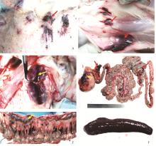
Fig. 1
The gross changes in the pig A. Multifocal ecchymoses on the dermis of the neck (red arrow); B. Incision of skin ecchymosis in Figure A showed hemorrhage in the subcutaneous tissue (red arrow); C. The lymph node is swollen and hemorrhagic, with blood flowing out of its cut surface (yellow arrow); D. Severe hemorrhage in the gastric hilar lymph nodes (yellow arrow), pancreas (red arrow), and the mesenteric area adjacent to the colon (red arrow); E. The mesenteric lymph nodes are enlarged and hemorrhagic(yellow arrow), with multifocal hemorrhagic foci in the mesentery(red arrow); F. The spleen exhibits enlargement, with a dark-red congestion appearance and a soft texture"

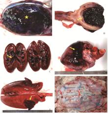
Fig. 2
The gross changes of the ASFV infected pig A. Severe diffuse hemorrhage on the serous surface of the bladder, with dark-red appearance (star); When the bladder was cut longitudinally, there was severe hemorrhage on the mucosa; B. Upon longitudinal incision of the bladder, severe hemorrhage is visible on the mucosal surface (yellow arrow); C. Cross-sections of both kidneys showing congestion and hemorrhage in the renal pelvis (yellow arrow); D. Patchy hemorrhage on the epicardium of the heart (yellow arrow); E. The entire lung is congested and edematous, with patchy hemorrhages in both cardiac lobes (yellow arrow); F. Scattered hemorrhagic foci on the dura mater of the brain (red arrow), with obvious congestion of the pia mater"

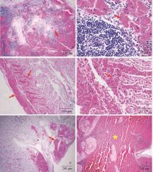
Fig. 3
The histopathological changes in the lymph node, heart and kidney tissues of the ASFV infected pig (HE staining) A. At low magnification, diffuse hemorrhage is visible in the medullary region of the lymph node, filled with numerous red blood cells (red arrow); B. Local magnification of the hemorrhagic area in Figure A (red arrow); C. Diffuse hemorrhage in the endocardium and myocardial layer (red arrow); D. At high magnification, numerous red blood cells are present between the myocardial fibers (red arrow); E. Patchy hemorrhage in the renal pelvis of the kidney (red arrow); F. The submucosal and muscular layers of the bladder are filled with numerous red blood cells (asterisk)"

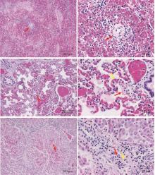
Fig. 4
The histopathological changes in the spleen, lung and liver tissues of the ASFV infected pig (HE staining) A. The splenic parenchyma is almost replaced by red blood cells (red arrow); B. The white pulp area of the spleen is filled with red blood cells, admixed with a small number of lymphocytes (red arrow); C. In the lungs, the small arteries and alveolar capillary walls are congested (red arrow); D. Under high magnification of the lungs, microthrombi are visible in the alveolar capillary walls (yellow arrow), and there is a large amount of pinkish exudate and hemorrhage in the alveolar cavities (red arrow); E. The central area of the hepatic lobules in the liver shows congestion and hemorrhage(red arrow); F. The portal areas of the liver have a large number of neutrophils (red arrow) and lymphocytic infiltration (yellow arrow)"

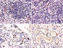
Fig. 5
Distribution of ASFV antigen in the tissues of the ASFV infected pig (Immunohistochemical staining) A. Lymph node, with strongly positive ASFV antigen reaction in the cytoplasm of macrophages within lymph follicles (indicated by the red arrow); B. Spleen, showing strongly positive ASFV antigen in the cytoplasm of macrophages in the red pulp area (indicated by the red arrow); C. Lung, with strongly positive ASFV in the cytoplasm of alveolar macrophages (indicated by the red arrow); D. Kidney, with scattered positive ASFV antigen in the cytoplasm of renal tubular epithelial cells (indicated by the red arrow)"

| 1 |
PENRITH M L , VOSLOO W , JORI F , et al. African swine fever virus eradication in Africa[J]. Virus Res, 2013, 173 (1): 228- 246.
doi: 10.1016/j.virusres.2012.10.011 |
| 2 |
KOLBASOV D , TITOV I , TSYBANOV S , et al. African swine fever virus, Siberia, Russia, 2017[J]. Emerg Infect Dis, 2018, 24 (4): 796- 798.
doi: 10.3201/eid2404.171238 |
| 3 | 王清华, 任炜杰, 包静月, 等. 我国首例非洲猪瘟的确诊[J]. 中国动物检疫, 2018, 35 (9): 1- 4. |
| WANG Q H , REN W J , BAO J Y , et al. The first outbreak of African swine fever was confirmed in China[J]. China Animal Health Inspection, 2018, 35 (9): 1- 4. | |
| 4 |
ZHOU X T , LI N , LUO Y Z , et al. Emergence of African swine fever in China, 2018[J]. Transbound Emerg Dis, 2018, 65 (6): 1482- 1484.
doi: 10.1111/tbed.12989 |
| 5 |
GE S Q , LI J M , FAN X X , et al. Molecular characterization of African swine fever virus, China, 2018[J]. Emerg Infect Dis, 2018, 24 (11): 2131- 2133.
doi: 10.3201/eid2411.181274 |
| 6 |
ZHAO D M , SUN E C , HUANG L Y , et al. Highly lethal genotype Ⅰ and Ⅱ recombinant African swine fever viruses detected in pigs[J]. Nat Commun, 2023, 14 (1): 3096.
doi: 10.1038/s41467-023-38868-w |
| 7 |
SUN E C , ZHANG Z J , WANG Z L , et al. Emergence and prevalence of naturally occurring lower virulent African swine fever viruses in domestic pigs in China in 2020[J]. Sci China Life Sci, 2021, 64 (5): 752- 765.
doi: 10.1007/s11427-021-1904-4 |
| 8 |
ZHAO D M , LIU R Q , ZHANG X F , et al. Replication and virulence in pigs of the first African swine fever virus isolated in China[J]. Emerg Microbes Infect, 2019, 8 (1): 438- 447.
doi: 10.1080/22221751.2019.1590128 |
| 9 | 许春梅, 邵明珠, 王心悦, 等. 非洲猪瘟病毒D250R蛋白生物信息学分析及多克隆抗体制备[J]. 中国畜牧兽医, 2022, 49 (12): 4745- 4755. |
| XU C M , SHAO M Z , WANG X Y , et al. Bioinformatics analysis of African swine fever virus D250R protein and preparation of polyclonal antibodies[J]. China Animal Husbandry & Veterinary Medicine, 2022, 49 (12): 4745- 4755. | |
| 10 |
DIXON L K , STAHL K , JORI F , et al. African swine fever epidemiology and control[J]. Annu Rev Anim Biosci, 2020, 8, 221- 246.
doi: 10.1146/annurev-animal-021419-083741 |
| 11 |
COSTARD S , MUR L , LUBROTH J , et al. Epidemiology of African swine fever virus[J]. Virus Res, 2013, 173 (1): 191- 197.
doi: 10.1016/j.virusres.2012.10.030 |
| 12 | 邓桦, 李慧, 杨鸿, 等. 急性非洲猪瘟的实验病理学研究[J]. 畜牧兽医学报, 2020, 51 (11): 2836- 2848. |
| DENG H , LI H , YANG H , et al. Experimental pathological study of acute African swine fever[J]. Acta Veterinaria et Zootechnica Sinica, 2020, 51 (11): 2836- 2848. | |
| 13 |
SALGUERO F J . Comparative pathology and pathogenesis of African swine fever infection in swine[J]. Front Vet Sci, 2020, 7, 282.
doi: 10.3389/fvets.2020.00282 |
| 14 | NETHERTON C L, SHIMMON G L, HUI J Y K, et al. African swine fever virus host-pathogen interactions[M]//VIJAYAKRISHNAN S, JIU Y M, HARRIS J R. Virus Infected Cells. Cham: Springer, 2023: 283-331. |
| 15 |
CARRASCO L , CHÁCON-M DE LARA F , DE LAS MULAS J M , et al. Ultrastructural changes related to the lymph node haemorrhages in acute African swine fever[J]. Res Vet Sci, 1997, 62 (3): 199- 204.
doi: 10.1016/S0034-5288(97)90190-9 |
| 16 |
AFE A E , SHEN Z J , GUO X R , et al. African swine fever virus interaction with host innate immune factors[J]. Viruses, 2023, 15 (6): 1220.
doi: 10.3390/v15061220 |
| 17 |
VILLEDA C J , WILLIAMS S M , WILKINSON P J , et al. Haemostatic abnormalities in African swine fever/A comparison of two virus strains of different virulence (Dominican Republic '78 and Malta '78)[J]. Arch Virol, 1993, 130 (1-2): 71- 83.
doi: 10.1007/BF01318997 |
| 18 |
FRANZONI G , PEDRERA M , SÁNCHEZ-CORDÓN P J . African swine fever virus infection and cytokine response in vivo: an update[J]. Viruses, 2023, 15 (1): 233.
doi: 10.3390/v15010233 |
| 19 |
BLOME S , GABRIEL C , BEER M . Pathogenesis of African swine fever in domestic pigs and European wild boar[J]. Virus Res, 2013, 173 (1): 122- 130.
doi: 10.1016/j.virusres.2012.10.026 |
| 20 |
GÓMEZ-VILLAMANDOS J C , BAUTISTA M J , SÁNCHEZ-CORDÓN P J , et al. Pathology of African swine fever: the role of monocyte-macrophage[J]. Virus Res, 2013, 173 (1): 140- 149.
doi: 10.1016/j.virusres.2013.01.017 |
| [1] | ZHANG Xiaoling, HE Xinglin, ZHANG Mengdi, LI Pengfei, SUN Yumei, MA Hailong, ZHU Hongmei, ZHANG Mengjia, LI Wentao. Preparation of E2 Protein Nanoparticles from Classical Swine Fever Virus and the Immunogenicity Study in Rabbit [J]. Acta Veterinaria et Zootechnica Sinica, 2025, 56(5): 2301-2311. |
| [2] | CAO Liyan, KONG Xiangyu, YUAN Cong, DUAN Yueyue, MA Guoxiang, SHI Lei, ZHANG Yu, WAN Ying, LI Xiangtong, WANG Yating, DU Yu, ZHENG Haixue, WANG Qi. Identifcation of a Novel Linear B-cell Epitope in the Nucleocapsid Protein of Swine Acute Diarrhea Syndrome Coronavirus [J]. Acta Veterinaria et Zootechnica Sinica, 2025, 56(4): 1854-1864. |
| [3] | PANG Siyao, ZHANG Jinlong, SUN Yuhang. Proteomic Analysis of 3D4/21 Cells Infected with H1N1 Swine Influenza Virus under Non-cytotoxic Concentration of AFB1 Exposure [J]. Acta Veterinaria et Zootechnica Sinica, 2025, 56(4): 1947-1957. |
| [4] | ZHANG Yue, RU Yi, HAO Rongzeng, YANG Rui, ZHAO Longhe, LI Yajun, YANG Yang, ZHANG Rong, JIANG Chenghui, ZHENG Haixue. Preparation and Immunogenicity Evaluation of African Swine Fever Virus H108R Protein [J]. Acta Veterinaria et Zootechnica Sinica, 2025, 56(3): 1344-1354. |
| [5] | MA Xiaoli, LI Duan, ZENG Daoping, LIU Yanling, WANG Xiaomin, PENG Guoliang, SONG Changxu, WANG Lei, XU Zheng. Establishment of a Fully Automated Chemiluminescent Enzyme Immunoassay for Detecting Antibodies against African Swine Fever Virus p72 [J]. Acta Veterinaria et Zootechnica Sinica, 2025, 56(3): 1355-1365. |
| [6] | ZHANG Su, SUN Lifang, LI Lanlan, WU Linjiao, CHEN Leiqing, WU Yunkun. Research Progress on the Interactions of African Swine Fever Virus Structural Proteins with Host Proteins [J]. Acta Veterinaria et Zootechnica Sinica, 2025, 56(1): 95-106. |
| [7] | ZENG Miaomiao, YANG Xiaoman, ZHANG Xin, LIU Dakai, SHI Hongyan, ZHANG Jiyu, ZHANG Liaoyuan, CHEN Jianfei, FENG Tingshuai, LI Xiuwen, SHI Da, FENG Li. Establishment and Preliminary Application of an Indirect ELISA for Swine Acute Diarrhea Syndrome Coronavirus N Protein [J]. Acta Veterinaria et Zootechnica Sinica, 2025, 56(1): 319-326. |
| [8] | Lu FENG, Hong TIAN, Haixue ZHENG, Zhengwang SHI, Juncong LUO, Xiaoyang ZHANG, Juanjuan WEI, Jing ZHOU, Huancheng LIAO, Wanying WANG. A Detection Method of African Swine Fever Virus based on Enzymatic Recombinase Amplification [J]. Acta Veterinaria et Zootechnica Sinica, 2024, 55(9): 4226-4231. |
| [9] | ZHOU Yang, WU Weizi, CAO Weisheng, WANG Fuguang, XU Xiuqiong, ZHONG Wenxia, WU Liyang, YE Jian, LU Shousheng. A Whole Genome Sequencing Method for African Swine Fever Virus based on Nanopore Sequencing Technology was Established [J]. Acta Veterinaria et Zootechnica Sinica, 2024, 55(5): 2080-2089. |
| [10] | HUANG Yuanyuan, WANG Jia, CHEN Jiayu, GAN Yuan, YUAN Ting, FENG Zhixin, SHAO Guoqing, WANG Xianwei, XIONG Qiyan. Isolation and Culture of Swine Peritoneal Mesothelial Cells and Its Preliminary Application [J]. Acta Veterinaria et Zootechnica Sinica, 2024, 55(3): 1249-1256. |
| [11] | LIU Yuanjie, XU Lu, ZHU Yuanyuan, XU Yuan, ZHANG Qianyi, LI Cui, LI Ming, XIA Yingju, WANG Qin, LIU Yebing, ZHAO Qizu, ZOU Xingqi. The Construction and Rescue of Epitope Mutant Strain of Classical Swine Fever Virus C Strain [J]. Acta Veterinaria et Zootechnica Sinica, 2024, 55(2): 698-705. |
| [12] | YAN Wenqian, HOU Jing, YANG Jinke, HAO Yu, YANG Xing, SHI Xijuan, ZHANG Dajun, BIE Xintian, CHEN Guohui, CHEN Lingling, HE Lu, ZHAO Meiyu, ZHAO Siyue, ZHENG Haixue, ZHANG Keshan. Monoclonal Antibody against D1133 L Protein of African Swine Fever Virus Inhibits Its Replication [J]. Acta Veterinaria et Zootechnica Sinica, 2024, 55(2): 854-859. |
| [13] | CHEN Xiaoli, ZHOU Jiahao, ZHOU Jing, QU Qian, WANG Zhihua, XIONG Ying, ZHU Yongqi, JIA Weixin, LÜ Weijie, GUO Shining. Effect of Modified Yuyin Decoction on cGAS-STING Pathway of African Swine Fever Virus Infected PAMs [J]. Acta Veterinaria et Zootechnica Sinica, 2024, 55(12): 5839-5853. |
| [14] | Hongde LU, Haoyang LIU, Shimiao GONG, Zhi YANG, Yuxuan WANG, Luhao WANG, Zhiyuan HE, Hong DONG. Application of Immunochromatographic Technique in the Rapid Detection of Common Swine Viral Diseases [J]. Acta Veterinaria et Zootechnica Sinica, 2024, 55(11): 4900-4911. |
| [15] | Qi ZHANG, Ningning DONG, Xiaomei TAN, Zhengwang SHI, Na LI, Wenqi ZHU, Aoxing TANG, Chuanfeng LI, Jie ZHU, Guangqing LIU, Yan SU, Chunchun MENG. Fusion Expression of African Swine Fever Virus p30 Protein and CRM197 and Evaluation of Its Immunogenicity in Mice [J]. Acta Veterinaria et Zootechnica Sinica, 2024, 55(11): 5230-5237. |
| Viewed | ||||||
|
Full text |
|
|||||
|
Abstract |
|
|||||