


畜牧兽医学报 ›› 2025, Vol. 56 ›› Issue (1): 442-454.doi: 10.11843/j.issn.0366-6964.2025.01.040
王艺( ), 侯露露, 方菲, 高林英, 谢淑敏, 王雨*()
收稿日期:2024-02-19
出版日期:2025-01-23
发布日期:2025-01-18
通讯作者:
王雨
E-mail:wangyi@nefu.edu.cn;wangyu2013@nefu.edu.cn
作者简介:王艺(2001-),女,山东烟台人,本科,主要从事动物中毒病病理学研究,E-mail: wangyi@nefu.edu.cn
基金资助:
WANG Yi(), HOU Lulu, FANG Fei, GAO Linying, XIE Shumin, WANG Yu*()
Received:2024-02-19
Online:2025-01-23
Published:2025-01-18
Contact:
WANG Yu
E-mail:wangyi@nefu.edu.cn;wangyu2013@nefu.edu.cn
摘要:
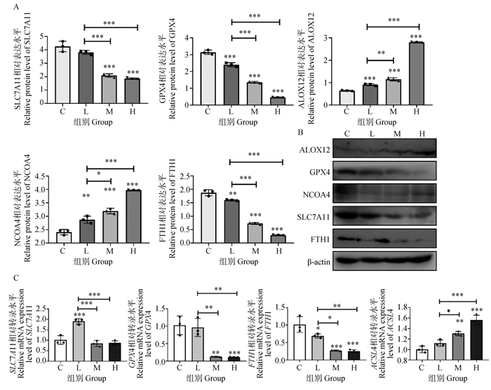
随着人类工业、农业的不断发展,越来越多的氟化物通过饮水、药物、农药和杀菌剂进入生产生活和环境中,会对生活在相应地区的动物产生不利影响。为了探讨氟中毒导致肉鸡小肠损伤的相关作用,本研究通过氟化物饮食暴露建立动物试验模型进行后续验证。试验按照饲料中氟化钠暴露剂量水平分为对照组C(0 mg·kg-1)、低剂量氟组L(500 mg·kg-1)、中剂量氟组M(1 000 mg·kg-1)和高剂量氟组H(2 000 mg·kg-1),分别处理肉鸡6周,分析氟化物暴露对肉鸡小肠的影响。结果显示:随着氟化钠暴露浓度的增加,Nrf2增加(P < 0.001),Keap1减少(P < 0.001),HO-1减少(P < 0.001),NQO1在高浓度组降低(P < 0.01),Nrf2/HO-1通路被激活,但其下游抗氧化酶减少,提示发生了氧化应激;p-AKT和p-mTOR蛋白表达量显著升高(P < 0.001),最后恢复正常甚至减少(P < 0.05),而PI3K显著降低(P < 0.001),提示PI3K/Akt/mTOR通路磷酸化被抑制;Beclin-1显著增加(P < 0.001),ATG12变化不显著,LC3蛋白表达量显著高于对照组(P < 0.05),提示自噬体加速形成和自噬流活化,p62蛋白与自噬底物蛋白结合,含量减少(P < 0.001),表明自噬通路未受阻。同时FTH1降低(P < 0.01),NCOA4升高(P < 0.01),储铁蛋白降解释放出二价铁离子;ASCL4和ALOX12蛋白表达量升高(P < 0.01),促进脂质过氧化过程;SLC7A11和GPX4蛋白表达量显著降低(P < 0.001),细胞清除脂质过氧化物能力降低,易发生铁死亡。综上,氟化钠会干扰Nrf2-Keap1抗氧化途径,导致氧化应激,也会激活自噬,最终导致肉鸡小肠发生铁死亡。
中图分类号:
王艺, 侯露露, 方菲, 高林英, 谢淑敏, 王雨. 氟通过自噬和铁死亡途径诱发肉鸡小肠氧化损伤[J]. 畜牧兽医学报, 2025, 56(1): 442-454.
WANG Yi, HOU Lulu, FANG Fei, GAO Linying, XIE Shumin, WANG Yu. Fluoride Induced Small Intestine Oxidative Damage in Broilers via Autophagy and Ferroptosis[J]. Acta Veterinaria et Zootechnica Sinica, 2025, 56(1): 442-454.
表 1
本研究中使用的引物"
| 基因 Gene | 登录号 Accession No. | 正向序列(5′→3′) Forward sequence | 反向序列(5′→3′) Reverse sequence |
| β-actin | NM_205518.2 | CCGCTCTATGAAGGCTACGC | CTCTCGGCTGTGGTGGTGAA |
| ACSL4 | XM_046917350.1 | CCGTTGTTATCTTCTGCGAGACC | ACGCTCCACATTCATTGAGACCATAG |
| FTH1 | NM_205086.2 | AGAATGTGAACCAGTCGCTGTTAGAG | TGCTTGATGGCTTTCACCTGCTC |
| GPX4 | NM_204220.3 | GCTGTGGAAGTGGCTGAAGGAG | TTGGCCCTCACGGTTGATAAGAAAC |
| SLC7A11 | XM_040670527.2 | AACTGCTGGTTATTCGCCCTG | AAGCCAGCAACTACCAGTCCA |
| AKT | XM_046917866.1 | AGGAGGAAGAGATGATGGAT | GAATGGATGCCGTGAGTT |
| ATG5 | XM_046914043.1 | GGCACCGACCGATTTAGT | GCTGATGGGTTTGCTTTT |
| ATG12 | XM_046936147.1 | GGGACCCTCTATGAGTGTTTTGG | AAGACCACACCTCAGCAACTC |
| Beclin-1 | NM_001006332.1 | CGACTGGAGCAGGAAGAAG | TCTGAGCATAACGCATCTGG |
| mTOR | XM_417614.7 | GCTTATGGCACGGTGTTTCC | GCGATCATGCTCTGTTGCAG |
| p62 | XM_040682727.2 | GTTAATCCTCTGCGTGTGA | AAGAAGAATCGGTGTGCTAA |
| NQO1 | NM_001277620.2 | CGCACCCTGAGAAAACCTCT | TTCTTGAGGGGTCCGGTGAT |
| HO-1 | XM_046921508.1 | GTCGTTGGCAAGAAGCATCC | GGGCCTTTTGGGCGATTTTC |
| Nrf2 | XM_046943490.1 | CAGGGCAATGCTAGTGTGTACTCATC | AGGGTCTTTCTTTGGTGTGTTCATACG |
表 2
本研究中使用的抗体"
| 抗体信息Antibody | 稀释率Dilution | 来源Source |
| β-actin | 1∶2 000 | Abclonal Technology, China |
| ALOX12 | 1∶500 | Boster, China |
| GPX4 | 1∶500 | Boster, China |
| NCOA4 | 1∶500 | Boster, China |
| FTH1 | 1∶500 | Boster, China |
| AKT | 1∶500 | Wanleibio, China |
| ATG12 | 1∶500 | Wanleibio, China |
| Beclin-1 | 1∶500 | Wanleibio, China |
| LC3 | 1∶500 | Wanleibio, China |
| mTOR | 1∶500 | Wanleibio, China |
| PI3K | 1∶500 | Wanleibio, China |
| P62 | 1∶500 | Wanleibio, China |
| Nrf2 | 1∶500 | Wanleibio, China |
| Keap1 | 1∶500 | Wanleibio, China |
| NQO1 | 1∶500 | Wanleibio, China |
| HO-1 | 1∶500 | Wanleibio, China |

图 1
氟化钠对肉鸡小肠肠绒毛形态结构的影响 A. 对照组(C);B. 低氟组(L);C. 中氟组(M);D. 高氟组(H)。扫描文章首页OSID码可查看彩图"

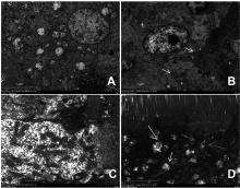
图 2
氟化钠对肉鸡小肠肠绒毛超微结构的电镜图 A. 对照组(C);B. 低氟组(L);C. 中氟组(M);D. 高氟组(H)。绿色代表正常的线粒体结构;红色箭头代表线粒体嵴断裂,黄色箭头代表自噬小体。扫描文章首页OSID码可查看彩图"


图 3
肉鸡小肠中细胞抗氧化途径相关指标 与C组相比,*.P < 0.05,**.P < 0.01,***.P < 0.001,差异不显著P>0.05"


图 4
肉鸡小肠中细胞自噬相关指标 与C组相比,*.P < 0.05,**.P < 0.01,***.P < 0.001,差异不显著P>0.05"


图 5
肉鸡小肠铁死亡途径相关指标 与C组相比,*.P < 0.05,**.P < 0.01,***.P < 0.001,差异不显著P>0.05"
| 1 |
ZUOH,CHENL,KONGM,et al.Toxic effects of fluoride on organisms[J].Life Sci,2018,198,18-24.
doi: 10.1016/j.lfs.2018.02.001 |
| 2 | 邵奎东,张海涛.吉林省地方性氟中毒"十三五"防治进展及今后对策探讨[J].中国地方病防治,2023,38(1):1-7. |
| ZHAOK D,ZHANGH T.Progress in prevention and control of endemic fluorosis in Jilin Province during the 13th Five Year Plan period and future countermeasures[J].Chinese Journal of Control of Endemic Diseases,2023,38(1):1-7. | |
| 3 | 吕纬. 四方针铁矿及其铝氧化物复合体对氟离子的吸附性能与机理[D]. 呼和浩特: 内蒙古大学, 2021. |
| LV W. Adsorption performance and mechanisms of fluoride on the akaganeite and its aluminum oxide complex[D]. Hohhot: Inner Mongolia University, 2021. (in Chinese) | |
| 4 |
OTTAPPILAKKILH,BABUS,BALASUBRAMANIANS,et al.Fluoride induced neurobehavioral impairments in experimental animals: a brief review[J].Biol Trace Elem Res,2023,201(3):1214-1236.
doi: 10.1007/s12011-022-03242-2 |
| 5 |
RAHIMA,ESSAMADIA,EL AMIRIB.A comprehensive review on endemic and experimental fluorosis in sheep: Its diverse effects and prevention[J].Toxicology,2022,465,153025.
doi: 10.1016/j.tox.2021.153025 |
| 6 |
EFEU,DEDES,YVKSEKV,et al.Apoptotic and oxidative mechanisms in liver and kidney tissues of sheep with fluorosis[J].Biol Trace Elem Res,2021,199(1):136-141.
doi: 10.1007/s12011-020-02121-y |
| 7 |
CHOUBISAS L,CHOUBISAD.Status of industrial fluoride pollution and its diverse adverse health effects in man and domestic animals in India[J].Environ Sci Pollut Res,2016,23(8):7244-7254.
doi: 10.1007/s11356-016-6319-8 |
| 8 |
DENGY B,CUIH M,PENGX,et al.Effects of high dietary fluorine on erythrocytes and erythrocyte immune adherence function in broiler chickens[J].Biol Trace Elem Res,2013,155(2):247-252.
doi: 10.1007/s12011-013-9793-6 |
| 9 |
CHENG J,HUP C,XUZ C,et al.The beneficial or detrimental fluoride to gut microbiota depends on its dosages[J].Ecotoxicol Environ Saf,2021,209,111732.
doi: 10.1016/j.ecoenv.2020.111732 |
| 10 |
LUOQ,CUIH M,PENGX,et al.Intestinal IgA+ cell numbers as well as IgA, IgG, and IgM contents correlate with mucosal humoral immunity of broilers during supplementation with high fluorine in the diets[J].Biol Trace Elem Res,2013,154(1):62-72.
doi: 10.1007/s12011-013-9713-9 |
| 11 |
LIUJ,CUIH M,PENGX,et al.Dietary high fluorine induces apoptosis and alters Bcl-2, Bax, and caspase-3 protein expression in the cecal tonsil lymphocytes of broilers[J].Biol Trace Elem Res,2013,152(1):25-30.
doi: 10.1007/s12011-012-9595-2 |
| 12 |
LUOQ,CUIH M,PENGX,et al.Suppressive effects of dietary high fluorine on the intestinal development in broilers[J].Biol Trace Elem Res,2013,156(1-3):153-165.
doi: 10.1007/s12011-013-9845-y |
| 13 |
CHENS Y,XUEY J,SHENY T,et al.Effects of different selenium sources on duodenum and jejunum tight junction network and growth performance of broilers in a model of fluorine-induced chronic oxidative stress[J].Poult Sci,2022,101(3):101664.
doi: 10.1016/j.psj.2021.101664 |
| 14 |
ZHAOW P,WANGH W,LIUJ,et al.Mitochondrial respiratory chain complex abnormal expressions and fusion disorder are involved in fluoride-induced mitochondrial dysfunction in ovarian granulosa cells[J].Chemosphere,2019,215,619-625.
doi: 10.1016/j.chemosphere.2018.10.043 |
| 15 |
JUH,CHENS Y,XUEY J,et al.The role of Nrf2 pathway in alleviating fluorine-induced apoptosis by different selenium sources in the chicken duodenum and jejunum[J].Ecotoxicol Environ Saf,2021,224,112708.
doi: 10.1016/j.ecoenv.2021.112708 |
| 16 | 胡流芳,王迎,任汝静,等.Keap1-Nrf2/ARE信号通路的抗氧化应激作用及其调控机制[J].国际药学研究杂志,2016,43(1):146-152, 166. |
| HUL F,WANGY,RENR J,et al.Anti-oxidative stress actions and regulation mechanisms of Keap1-Nrf2/ARE signal pathway[J].Journal of International Pharmaceutical Research,2016,43(1):146-152, 166. | |
| 17 |
SUNY,ZHENGY F,WANGC X,et al.Glutathione depletion induces ferroptosis, autophagy, and premature cell senescence in retinal pigment epithelial cells[J].Cell Death Dis,2018,9(7):753.
doi: 10.1038/s41419-018-0794-4 |
| 18 |
ZHAOY F,LIY Y,WANGJ M,et al.Fluoride induces apoptosis and autophagy through the IL-17 signaling pathway in mice hepatocytes[J].Arch Toxicol,2018,92(11):3277-3289.
doi: 10.1007/s00204-018-2305-x |
| 19 |
TANGH Y,HOUH Q,SONGL,et al.The role of mTORC1/TFEB axis mediated lysosomal biogenesis and autophagy impairment in fluoride neurotoxicity and the intervention effects of resveratrol[J].J Hazard Mater,2024,467,133634.
doi: 10.1016/j.jhazmat.2024.133634 |
| 20 |
JIANGJ Z,RUANY B,LIUX H,et al.Ferritinophagy is critical for deoxynivalenol-induced liver injury in mice[J].J Agric Food Chem,2024,72(12):6660-6671.
doi: 10.1021/acs.jafc.4c00556 |
| 21 |
DIXONS J,LEMBERGK M,LAMPRECHTM R,et al.Ferroptosis: an iron-dependent form of nonapoptotic cell death[J].Cell,2012,149(5):1060-1072.
doi: 10.1016/j.cell.2012.03.042 |
| 22 | 岑怡,王甜甜,高兴杰,等.铁死亡调控机制及其在肠道疾病中的研究进展[J].生命的化学,2022,42(11):2071-2078. |
| CENY,WANGT T,GAOX J,et al.Regulatory mechanism and research progress of ferroptosis in intestinal diseases[J].Chemistry of Life,2022,42(11):2071-2078. | |
| 23 | 王浩,肖金龙,沈珏,等.细胞死亡的新方式——铁死亡与铜死亡[J].畜牧兽医学报,2024,55(2):461-470. |
| WANGH,XIAOJ L,SHENJ,et al.New ways of cell death—ferroptosis and cuproptosis[J].Acta Veterinaria et Zootechnica Sinica,2024,55(2):461-470. | |
| 24 |
LIX,YANGJ,SHIE B,et al.Riboflavin alleviates fluoride-induced ferroptosis by IL-17A-independent system Xc-/GPX4 pathway and iron metabolism in testicular Leydig cells[J].Environ Pollut,2024,344,123332.
doi: 10.1016/j.envpol.2024.123332 |
| 25 |
ZHANGY,FANGY M,ZHAOS,et al.Fluoride resistance capacity in mammalian cells involves global gene expression changes associate with ferroptosis[J].Chem Biol Interact,2023,381,110555.
doi: 10.1016/j.cbi.2023.110555 |
| 26 |
SONGC,HEPINGH F,SHENY S,et al.AMPK/p38/Nrf2 activation as a protective feedback to restrain oxidative stress and inflammation in microglia stimulated with sodium fluoride[J].Chemosphere,2020,244,125495.
doi: 10.1016/j.chemosphere.2019.125495 |
| 27 | 毕翻,万珑,温建霞,等.氟对仔鼠生长发育、学习记忆及血清氧化应激水平的影响[J].中华地方病学杂志,2020,39(4):243-247. |
| BIF,WANL,WENJ X,et al.Effects of fluoride on growth and development, learning and memory, and oxidative stress in serum of offspring rat[J].Chinese Journal of Endemiology,2020,39(4):243-247. | |
| 28 | 辛涛,许娟,刘红岗.Nrf2/HO-1通路在Erastin诱导的非小细胞肺癌A549细胞铁死亡中的作用[J].山西医科大学学报,2023,54(7):879-884. |
| XINT,XUJ,LIUH G.Role of Nrf2/HO-1 pathway in Erastin-induced ferroptosis in non-small cell lung cancer A549 cells[J].Journal of Shanxi Medical University,2023,54(7):879-884. | |
| 29 | TANGZ,LAIC C,LUOJ,et al.Mangiferin prevents the impairment of mitochondrial dynamics and an increase in oxidative stress caused by excessive fluoride in SH-SY5Y cells[J].J Biochem Mol Toxicol,2021,35(4):e22705. |
| 30 | XUZ R,HANX,OUD M,et al.Targeting PI3K/AKT/mTOR-mediated autophagy for tumor therapy[J].Appl Microbiol Biotechnol,2020,104(2):575-587. |
| 31 | ZHANGJ H,ZHUY C,SHIY,et al.Fluoride-induced autophagy via the regulation of phosphorylation of mammalian targets of rapamycin in mice leydig cells[J].J Agric Food Chem,2017,65(40):8966-8976. |
| 32 | 孔志伟,姚婷婷.自噬相关基因Beclin1和LC3与子宫内膜异位症的研究进展[J].医学综述,2019,25(21):4166-4173. |
| KONGZ W,YAOT T.Research on endometriosis and autophagy associated gene beclin1 and LC3[J].Medical Recapitulate,2019,25(21):4166-4173. | |
| 33 | MIZUSHIMAN.Methods for monitoring autophagy[J].Int J Biochem Cell Biol,2004,36(12):2491-2502. |
| 34 | KABEYAY,MIZUSHIMAN,UENOT,et al.LC3, a mammalian homologue of yeast Apg8p, is localized in autophagosome membranes after processing[J].EMBO J,2000,19(21):5720-5728. |
| 35 | 刘世玉,冯学召,古丽妮尕尔·司马义,等.p62敲除对细胞自噬的影响及其对泛素化修饰蛋白募集的作用[J].新疆医科大学学报,2023,46(9):1119-1123, 1131. |
| LIUS Y,FENGX Z,GULINIGAERS,et al.Effect of p62 knockout on cellular autophagy and its role in recruitment of ubiquitination-modified proteins[J].Journal of Xinjiang Medical University,2023,46(9):1119-1123, 1131. | |
| 36 | 吕晓希,胡卓伟.自噬流的检测方法[J].药学学报,2016,51(1):45-51. |
| LVX X,HUZ W.New methods to detect autophagic flux[J].Acta Pharmaceutica Sinica,2016,51(1):45-51. | |
| 37 | XUW J,HUZ Y,ZHANGJ J,et al.Cross-talk between autophagy and ferroptosis contributes to the liver injury induced by fluoride via the mtROS-dependent pathway[J].Ecotoxicol Environ Saf,2023,250,114490. |
| 38 | 李昕,李平,熊秋宏.自噬和铁死亡的相互联系[J].中国细胞生物学学报,2021,43(1):144-151. |
| LIX,LIP,XIONGQ H.The crosstalk between autophagy and ferroptosis[J].Chinese Journal of Cell Biology,2021,43(1):144-151. | |
| 39 | 李敏,杨前,刘亚轩,等.镉通过诱导自噬促进肾小管上皮细胞铁死亡的研究[J].陆军军医大学学报,2022,44(17):1705-1711. |
| LIM,YANGQ,LIUY X,et al.Cadmium promotes ferroptosis in renal tubular epithelial cells by inducing autophagy[J].Journal of Army Medical University,2022,44(17):1705-1711. | |
| 40 | MANCIASJ D,WANGX X,GYGIS P,et al.Quantitative proteomics identifies NCOA4 as the cargo receptor mediating ferritinophagy[J].Nature,2014,509(7498):105-109. |
| 41 | HOUW,XIEY C,SONGX X,et al.Autophagy promotes ferroptosis by degradation of ferritin[J].Autophagy,2016,12(8):1425-1428. |
| 42 | GAOM F,MONIANP,PANQ H,et al.Ferroptosis is an autophagic cell death process[J].Cell Res,2016,26(9):1021-1032. |
| 43 | SONGX H,LONGD X.Nrf2 and ferroptosis: a new research direction for neurodegenerative diseases[J].Front Neurosci,2020,14,267. |
| 44 | 刘恒,段晓峰.铁自噬及相关基因NCOA4、FTH1在口腔鳞癌中的研究进展[J].中国口腔颌面外科杂志,2023,21(1):87-91. |
| LIUH,DUANX F.Research progress of ferritinophagy and related genes FTH1 and NCOA4 in oral squamous cell carcinoma[J].China Journal of Oral and Maxillofacial Surgery,2023,21(1):87-91. | |
| 45 | DINGK Y,LIUC B,LIL,et al.Acyl-CoA synthase ACSL4: an essential target in ferroptosis and fatty acid metabolism[J].Chin Med J (Engl),2023,136(21):2521-2537. |
| 46 | CHUB,KONN,CHEND L,et al.ALOX12 is required for p53-mediated tumour suppression through a distinct ferroptosis pathway[J].Nat Cell Biol,2019,21(5):579-591. |
| [1] | 李远方, 吴冉, 李帅浩, 魏千然, 王亚东, 王丹丹, 李智, 李国喜, 刘翘铭. G3BP1基因在鸡肌内前脂肪细胞增殖与分化中的作用及其分子标记鉴定[J]. 畜牧兽医学报, 2025, 56(1): 159-167. |
| [2] | 尹琼, 高明超, 姚秀梅, 刘昆煜, 刘玮, 揭泓蔚, 李华, 叶菲. 沐川乌骨鸡胸肌黑色素含量与PMEL17基因的关联性分析[J]. 畜牧兽医学报, 2025, 56(1): 168-177. |
| [3] | 吴双, 尹娜, 余莫涵, 平玉宇, 白皓, 陈世豪, 常国斌. TRIM39.2过表达对鸡巨噬细胞转录表达的影响[J]. 畜牧兽医学报, 2025, 56(1): 178-188. |
| [4] | 王贝贝, 武书庚, 张海华, 张海军, 郝二英, 邱凯. 饲粮添加大豆异黄酮对产蛋后期蛋鸡生产的影响[J]. 畜牧兽医学报, 2025, 56(1): 295-306. |
| [5] | 章琦, 郭江鹏, 倪爱心, 杜洪峰, 陈继兰, 孙研研. 蛋鸡啄羽行为的影响因素与遗传调控基础研究进展[J]. 畜牧兽医学报, 2024, 55(9): 3745-3756. |
| [6] | 杨硕, 霍敏, 苏子轩, 石玉祥. 线粒体质量控制对畜禽氧化应激影响的研究进展[J]. 畜牧兽医学报, 2024, 55(9): 3769-3776. |
| [7] | 付红玉, 李玥, 崔晗, 李玖芝, 许琬雪, 王曦, 樊瑞锋. 长链酯酰辅酶A合成酶4介导铁死亡的发生机制[J]. 畜牧兽医学报, 2024, 55(9): 3792-3801. |
| [8] | 袁紫金, 王婉昕, 邢娅, 李家惠, 薛颖, 葛晶, 赵敏孟, 刘龙, 龚道清, 耿拓宇. HDLBP通过调控氧化应激水平和炎性因子表达参与鹅肥肝的形成[J]. 畜牧兽医学报, 2024, 55(9): 3897-3913. |
| [9] | 张纪桥, 蔡瑛婕, 李雨笑, 曹敞, 李涛, 鲍秀瑜, 张建勤. 不同饲养模式下略阳乌鸡生长性能、免疫、肠道结构及盲肠菌群的对比分析[J]. 畜牧兽医学报, 2024, 55(9): 4001-4011. |
| [10] | 何塔娜, 胡馨匀, 米洁兰, 高立, 张艳萍, 祁小乐, 崔红玉, 杨桂连, 高玉龙. 基于16S rDNA分析饲喂唾液乳杆菌XP132对白羽肉种鸡肠道菌群的影响[J]. 畜牧兽医学报, 2024, 55(9): 4091-4099. |
| [11] | 刘馨蔓, 周鸿缘, 桑锐, 葛冰洁, 闫可心, 王巍, 于明弘, 刘晓童, 邱谦, 张雪梅. 蒲公英甾醇对AFB1性肝损伤肉鸡肝组织氧化应激的影响[J]. 畜牧兽医学报, 2024, 55(9): 4141-4152. |
| [12] | 于秀菊, 胡燕姣, 刘佳悦, 王海东, 朱芷葳, 范阔海, 王蓉蓉, 段承昊, 石佳炜, 杨丽华. 一株鸡源唾液乳杆菌的分离鉴定及其对育雏早期蛋鸡肠道健康的影响[J]. 畜牧兽医学报, 2024, 55(9): 4161-4171. |
| [13] | 娄明, 罗昊玉, 牟芳, 李辉, 王宁. 鸡胰岛素信号通路的研究进展[J]. 畜牧兽医学报, 2024, 55(8): 3288-3296. |
| [14] | 张莉, 于蒙蒙, 王颖, 王素艳, 许壮壮, 刘鹏, 陈运通, 祁小乐, 李留安, 高玉龙. 禽白血病病毒J亚群感染鸡组织中细胞受体chNHE1的表达分析[J]. 畜牧兽医学报, 2024, 55(8): 3631-3639. |
| [15] | 黄晓隆, 盛熙晖, 袁经纬. 基于组学分析的鸡环境适应性研究进展[J]. 畜牧兽医学报, 2024, 55(7): 2809-2824. |
| 阅读次数 | ||||||
|
全文 |
|
|||||
|
摘要 |
|
|||||