


畜牧兽医学报 ›› 2024, Vol. 55 ›› Issue (9): 3897-3913.doi: 10.11843/j.issn.0366-6964.2024.09.015
袁紫金( ), 王婉昕, 邢娅, 李家惠, 薛颖, 葛晶, 赵敏孟, 刘龙, 龚道清, 耿拓宇*()
收稿日期:2024-02-18
出版日期:2024-09-23
发布日期:2024-09-27
通讯作者:
耿拓宇
E-mail:2316066266@qq.com;tygeng@yzu.edu.cn
作者简介:袁紫金(1997-),女,河北承德人,硕士生,主要从事家禽营养调控研究,E-mail:2316066266@qq.com
基金资助:
Zijin YUAN(), Wanxin WANG, Ya XING, Jiahui LI, Ying XUE, Jing GE, Minmeng ZHAO, Long LIU, Daoqing GONG, Tuoyu GENG*()
Received:2024-02-18
Online:2024-09-23
Published:2024-09-27
Contact:
Tuoyu GENG
E-mail:2316066266@qq.com;tygeng@yzu.edu.cn
摘要:
旨在利用活体与细胞模型探究高密度脂蛋白结合蛋白(HDLBP)的亚细胞分布、基因功能及其与鹅肥肝形成的关系。本研究选取70日龄健康朗德鹅公鹅14只,单笼饲养,随机均分为对照组(平均体重为3.71 kg,自由采食)和试验组(平均体重为3.72 kg,填饲20 d)进行活体模型试验。从23日龄朗德鹅胚胎中分离肝细胞并过表达HDLBP基因进行细胞模型试验。首先采用免疫印迹法、免疫荧光技术对鹅原代肝细胞中HDLBP蛋白质进行亚细胞定位分析,其次采用免疫印迹法检测填饲鹅和对照鹅肝脏全细胞HDLBP(wHDLBP)及线粒体中HDLBP(mHDLBP)的蛋白质丰度,然后在鹅原代肝细胞中过表达HDLBP,检测其对细胞中mHDLBP蛋白质丰度、丙二醛(MDA)含量、总超氧化物歧化酶(T-SOD)和谷胱甘肽过氧化物酶(GSH-PX)活性、活性氧类物质(ROS)和线粒体膜电位水平的影响,最后通过转录组测序分析筛选HDLBP过表达影响的差异表达基因与相关信号通路,并在活体模型中对部分差异表达基因进行定量PCR验证。结果表明:HDLBP可结合到线粒体中;填饲组wHDLBP和mHDLBP蛋白质丰度均显著低于对照组(P < 0.01);在鹅原代肝细胞中过表达HDLBP显著增加mHDLBP的蛋白质丰度(P < 0.05),增加MDA(P < 0.01)和ROS(P < 0.05)含量,降低线粒体膜电位(P < 0.05)及T-SOD(P < 0.05)和GSH-PX(P < 0.05)活性;HDLBP过表达所影响的上调差异表达基因主要富集于免疫/炎症相关通路。此外,相对于对照组,填饲组中炎症相关基因IL1R1、TNFSF10、LTC4S、NCF1、SFTPA1及KDR的表达可能受到HDLBP的调控而显著减少(P < 0.05,0.01或0.001)。HDLBP能够与线粒体结合,填饲显著降低鹅肝全细胞和线粒体样中HDLBP的蛋白水平,过表达HDLBP导致线粒体功能损伤、氧化应激和炎性因子的表达增强,因此HDLBP可能通过影响线粒体功能、调控氧化应激和炎症反应为鹅肥肝提供保护。
中图分类号:
袁紫金, 王婉昕, 邢娅, 李家惠, 薛颖, 葛晶, 赵敏孟, 刘龙, 龚道清, 耿拓宇. HDLBP通过调控氧化应激水平和炎性因子表达参与鹅肥肝的形成[J]. 畜牧兽医学报, 2024, 55(9): 3897-3913.
Zijin YUAN, Wanxin WANG, Ya XING, Jiahui LI, Ying XUE, Jing GE, Minmeng ZHAO, Long LIU, Daoqing GONG, Tuoyu GENG. HDLBP Is Involved in Goose Fatty Liver Formation by Regulating the Level of Oxidative Stress and the Expression of Inflammatory Factors[J]. Acta Veterinaria et Zootechnica Sinica, 2024, 55(9): 3897-3913.
表 1
荧光定量引物序列"
| 基因 Gene | 引物序列(5′→3′) Primers sequence | 产物大小/bp Product size |
| HDLBP | F: CCGTGGAGGTGAAGAAGTCC; R: CCTCGCAGTATCACCGTCTC | 139 |
| UBC | F: AGGGTGGATTCTTTCTGG; R: ACTGAGTTTGGAGGGAGC | 243 |
| GAPDH | F: CTGATGCTCCCATGTTCGT; R: CCACGATGCCAAAGTTGTCA | 138 |
| CHP1 | F: TCCAGAAGGAGAGGACCAAGT; R: GGAGCTTGTTACTTCGGCTG | 134 |
| PCAN1 | F: TTGCCTGTGTGGCAGATAGT; R: TGTGCAACTGGACTCTAGCAT | 177 |
| LOC106049357 | F: TGGCAGATGCAGACACAACC; R: CACCCTGGCCTCTCGTAAAT | 140 |
| HRG | F: ACAAGGGATTCTGCAAGGCAC; R: TCTATGTCCATGGTGATGCTGCC | 107 |
| ELOVL6 | F: GGTGGTCGGCACCTAATGAA; R: TCTGGTCACACACTGACTGC | 169 |
| GMPR | F: CTGCCAATCACCCAGAATGC; R: TCAGAGTAGCCGTTTGCCAC | 142 |
| ALDH1A1 | F: GGCGACAAGGCAGATGTAGA; R: CAAGAGCCTTCCTCGCTCTG | 105 |
| MAT1A | F: TGCGCGTTCATACCATTGTG; R: GACCAGTAACACCAGCGTCA | 198 |
| PSAT1 | F: AGAATGTTGGCTGTGCTGGA; R: AAAGAGCCATTCACCGCTTG | 118 |
| LOC106034448 | F: ATCTGCTTGCTCCCTGGATT; R: TGTCCCTTTTCCATCTGCCA | 187 |
| SLC2A9 | F: GCAGGGAAGGGCAATTGGT; R: GACCAGAGGAGAGTCAGGGT | 194 |
| IL1R1 | F: GAACGCCCAGCTCAGAACAT; R: AAAGGATGGCACGAGTTCCG | 140 |
| LTC4S | F: TTCGAGCACAGGTGAACTCC; R: GAGGACGATGCCCTGTATCC | 157 |
| TNFSF10 | F: TGTCCACAGGATAGCAGCAC; R: GGTATCACCAGCTCGCCATT | 180 |
| NCF1 | F: GACACCTTCATCCGGCACA; R: TTTCGGTGAGGTCGTTCCAC | 106 |
| SFTPA1 | F: GGCACACCTCTGAATTACACC; R: TGTGAGGCGATACAGGTTGC | 126 |
| KDR | F: ACCTGACGATGAACCCACAC; R: CCACATTCAGCTCCGTCCTT | 90 |
| PPAP2B | F: TGCTATCATCACGGGAGAGC; R: TGTGAAGGACTGGCTGATGG | 142 |
| SIGLEC1 | F: CTTCAGCACCCAAAAGACCG; R: CAGGACATCCGAAACGCTCA | 229 |
| CCL20 | F: TGAACAGCTCTCCAGTGAAGTC; R: TCCTTAGGGTTTACGCAGGC | 87 |

图 1
HDLBP蛋白质在鹅原代肝细胞中的分布 A. 免疫印迹图;B. 免疫荧光图。β-Actin作为细胞浆的内参基因,而LaminB1作为细胞核的内参基因。DAPI显示细胞核"


图 2
鹅原代肝细胞中HDLBP蛋白质与线粒体的共定位 A. 免疫荧光图,DAPI显示细胞核,Mito-tracker Red显示线粒体,在合并图(Merge)中黄色表明HDLBP蛋白质与线粒体共定位;B. 免疫印迹图(n=3);C. 免疫印迹的量化,字母M表示蛋白Marker,β-Actin作为细胞浆(cyto)的内参基因,而VDAC作为线粒体(mito)的内参基因。*.P < 0.05,下同"


图 3
填饲组和对照组朗德鹅肝脏全细胞样和线粒体样中HDLBP蛋白质的相对含量 A.全细胞样的免疫印迹图(n=6);B. 全细胞样的免疫印迹量化;C. 线粒体样的免疫印迹图(n=4);D. 线粒体样的免疫印迹量化。字母C表示对照组,字母T表示填饲组,GAPDH和VDAC作为内参基因。**.P < 0.01,下同"


图 4
鹅原代肝细胞中HDLBP基因过表达的检测 A. 实时荧光定量检测HDLBP基因的mRNA水平(n=6);B. 蛋白免疫印迹检测HDLBP的蛋白水平(n=4);C. 免疫印迹量化图。字母C表示对照组,OE表示过表达组。***.P < 0.001,下同"


图 5
HDLBP过表达对鹅原代肝细胞中线粒体功能相关指标的影响 A.流式细胞术检测活性氧水平;B. 用FlowJo软件进行定量分析;C. 流式细胞术检测线粒体膜电位;D. 用FlowJo软件进行定量分析,n=3;E. MDA的含量;F. T-SOD活性;G. GSH-PX活性,n=6"
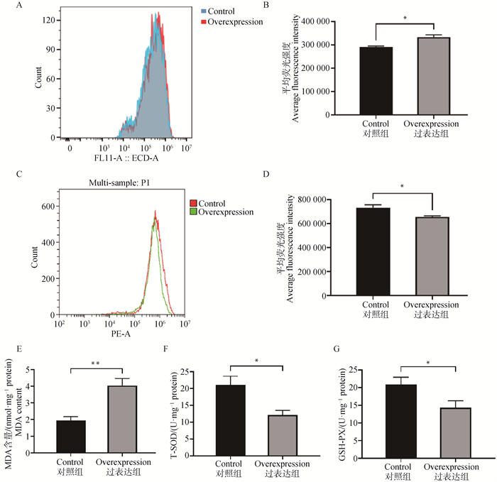
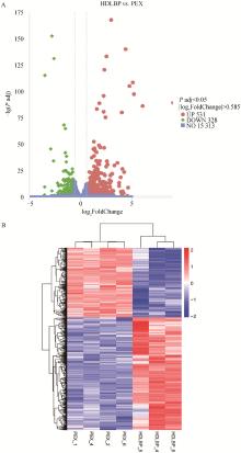
图 6
差异表达基因的火山图(A)及聚类热图(B) A.差异表达基因火山图,上调基因用红点表示,下调基因用绿点表示,灰色虚线表示差异基因筛选标准的阈值线;B. 差异表达基因聚类热图,图中横坐标为样品名,纵坐标为差异基因FPKM归化后的数值,红色表示基因表达水平高,蓝色表示基因表达水平低"

表 2
部分上调和下调的差异表达基因"
| 基因 Gene | 变化倍数的对数 log2(fold change) | Padj | |
| 上调 Up-regulated | CP | 2.97 | 2.38×10-168 |
| TNIP3 | 4.39 | 1.00×10-140 | |
| LOC106046725 | 2.52 | 7.01×10-134 | |
| LOC106046905 | 2.30 | 2.85×10-121 | |
| NOXO1 | 5.09 | 3.23×10-109 | |
| SLCO4C1 | 4.76 | 8.57×10⁻⁶ | |
| STEAP4 | 5.20 | 3.21×10-98 | |
| CHN2 | 2.24 | 1.44×10-96 | |
| SLC13A5 | 9.04 | 3.98×10-90 | |
| 下调 Down-regulated | DPYS | -2.84 | 3.24×10-153 |
| MAP3K7CL | -2.62 | 6.42×10-132 | |
| LOC106030908 | -3.52 | 6.02×10-116 | |
| PCK1 | -1.65 | 4.90×10-69 | |
| HRG | -1.51 | 1.52×10-65 | |
| TPH2 | -2.96 | 2.26×10-46 | |
| LOC106034448 | -1.52 | 6.91×10-43 | |
| GPR1 | -1.39 | 1.94×10-39 | |
| LOC106048076 | -2.67 | 4.63×10⁻³⁵ | |
| CRACR2A | -1.43 | 4.83×10⁻³⁴ |

图 7
差异表达基因的GO功能富集分析 GO富集分析柱状图。图中横坐标为GO条目,纵坐标为GO条目富集的显著性水平,用P adj对数的负数表示。不同颜色分别表示不同的功能分类"
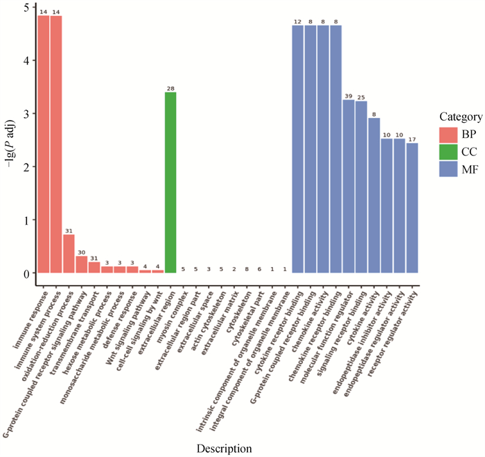

图 8
差异表达基因的KEGG通路富集分析 A. 上调差异表达基因;B. 下调差异表达基因。横坐标为注释到KEGG通路上的差异基因数与差异基因总数的比值,纵坐标为KEGG通路,点的大小表示注释到KEGG通路上的基因数,颜色从红到紫代表富集的显著性水平"


图 9
定量PCR分析验证随机选择的差异表达基因(n=6) A.所选差异表达基因在转录组测序分析中的结果;B. 定量PCR验证的结果。ns表示P>0.05"


图 10
HDLBP过表达对鹅原代肝细胞中免疫/炎症相关基因表达的影响(n=6) A. 免疫/炎症相关基因在转录组测序分析中的结果;B. 定量PCR检测的结果"


图 11
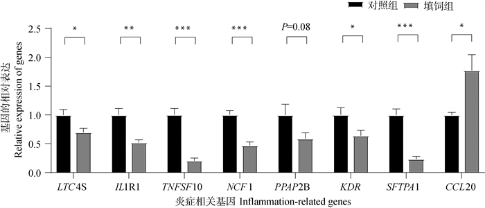
填饲对鹅肝脏中与免疫/炎症相关的差异基因表达的影响(n=6)"
| 1 |
刘同君, 赵盼, 赵敏孟, 等. 内质网应激标记基因Grp78参与鹅肥肝免疫/炎症状态的调控[J]. 畜牧兽医学报, 2019, 50 (4): 727- 737.
doi: 10.11843/j.issn.0366-6964.2019.04.006 |
|
LIU T Q , ZHANG P , ZHAO M M , et al. Endoplasmic reticulum stress marker gene Grp78 Is involved in regulation of immune/inflammatory state of goose fatty liver[J]. Acta Veterinaria et Zootechnica Sinica, 2019, 50 (4): 727- 737.
doi: 10.11843/j.issn.0366-6964.2019.04.006 |
|
| 2 |
WEI R X , NING R , HAN C C , et al. Lipidomics analysis reveals new insights into the goose fatty liver formation[J]. Poult Sci, 2023, 102 (3): 102428.
doi: 10.1016/j.psj.2022.102428 |
| 3 |
LIU L , WANG Q , WANG Q Q , et al. Role of miR29c in goose fatty liver is mediated by its target genes that are involved in energy homeostasis and cell growth[J]. BMC Vet Res, 2018, 14 (1): 325.
doi: 10.1186/s12917-018-1653-3 |
| 4 | 柳序, 刘耀文, 匡佑华, 等. 鹅肥肝的形成及主要影响因素的研究进展[J]. 经济动物学报, 2019, 23 (4): 234- 239. |
| LIU X , LIU Y W , KUANG Y H , et al. Advances on formation and main influencing factors of foie Gras[J]. Journal of Economic Animal, 2019, 23 (4): 234- 239. | |
| 5 |
GENG T Y , YANG B , LI F Y , et al. Identification of protective components that prevent the exacerbation of goose fatty liver: Characterization, expression and regulation of adiponectin receptors[J]. Comp Biochem Physiol B Biochem Mol Biol, 2016, 194-195, 32- 38.
doi: 10.1016/j.cbpb.2016.01.006 |
| 6 |
XU A M , WANG Y , KESHAW H , et al. The fat-derived hormone adiponectin alleviates alcoholic and nonalcoholic fatty liver diseases in mice[J]. J Clin Invest, 2003, 112 (1): 91- 100.
doi: 10.1172/JCI200317797 |
| 7 |
KASER S , MOSCHEN A , CAYON A , et al. Adiponectin and its receptors in non-alcoholic steatohepatitis[J]. Gut, 2005, 54 (1): 117- 121.
doi: 10.1136/gut.2003.037010 |
| 8 |
STOWE D F , CAMARA A K S . Mitochondrial reactive oxygen species production in excitable cells: modulators of mitochondrial and cell function[J]. Antioxid Redox Signal, 2009, 11 (6): 1373- 1414.
doi: 10.1089/ars.2008.2331 |
| 9 |
陈翠英, 邵明亮, 杨莉, 等. 乌丹降脂方对脂肪肝大鼠脂肪肝细胞氧化损伤调节作用研究[J]. 现代中西医结合杂志, 2016, 25 (20): 2180-2182, 2198.
doi: 10.3969/j.issn.1008-8849.2016.20.005 |
|
CHEN C Y , SHAO M L , YANG L , et al. Study on the regulation of Wudan Jiangzhi decoction on peroxidation damage in liver cells of the rats with liver fat[J]. Modern Journal of Integrated Traditional Chinese and Western Medicine, 2016, 25 (20): 2180-2182, 2198.
doi: 10.3969/j.issn.1008-8849.2016.20.005 |
|
| 10 |
PENG K Y , WATT M J , RENSEN S , et al. Mitochondrial dysfunction-related lipid changes occur in nonalcoholic fatty liver disease progression[J]. J Lipid Res, 2018, 59 (10): 1977- 1986.
doi: 10.1194/jlr.M085613 |
| 11 | 沈文婷, 陈明, 许诣, 等. PDTC调节线粒体功能障碍与Klotho蛋白表达对大鼠脓毒症合并急性肾损伤的影响[J]. 广西医科大学学报, 2021, 38 (11): 2097- 2103. |
| SHEN W T , CHEN M , XU Y , et al. The effects of PDTC regulating mitochondrial dysfunction and Klotho protein expression on acute kidney injury in rat with sepsis[J]. Journal of Guangxi Medical University, 2021, 38 (11): 2097- 2103. | |
| 12 | 郭燕, 徐爽, 王亭. PKD1抑制剂CID755673通过诱导线粒体功能障碍加重急性肾损伤[J]. 临床与实验病理学杂志, 2023, 39 (2): 206-211, 215. |
| GUO Y , XU S , WANG T . PKD1 inhibitor CID755673 aggravates acute kidney injury by inducing mitochondrial dysfunction[J]. Chinese Journal of Clinical and Experimental Pathology, 2023, 39 (2): 206-211, 215. | |
| 13 | 朱潇旭, 段小花, 李瑞霞, 等. 线粒体功能障碍与非酒精性脂肪肝发病关系的研究进展[J]. 山东医药, 2018, 58 (29): 108- 111. |
| ZHU X X , DUAN X H , LI R X , et al. Research progress on the relationship between mitochondrial dysfunction and nonalcoholic fatty liver disease[J]. Shandong Medicine, 2018, 58 (29): 108- 111. | |
| 14 |
SUN Q Y , DAI E P , CHEN M , et al. Glucose-induced enhanced anti-oxidant activity inhibits apoptosis in goose fatty liver[J]. J Anim Sci, 2023, 101, skad059.
doi: 10.1093/jas/skad059 |
| 15 | FEICHT J , JANSEN R P . The high-density lipoprotein binding protein HDLBP is an unusual RNA-binding protein with multiple roles in cancer and disease[J]. RNA Biol, 2024, 21 (1): 1- 10. |
| 16 |
CHENG M H K , JANSEN R P . A jack of all trades: the RNA-binding protein vigilin[J]. Wiley Interdiscip Rev RNA, 2017, 8 (6): e1448.
doi: 10.1002/wrna.1448 |
| 17 |
DOMÍNGUEZ-PÉREZ M , SIMONI-NIEVES A , ROSALES P , et al. Cholesterol burden in the liver induces mitochondrial dynamic changes and resistance to apoptosis[J]. J Cell Physiol, 2019, 234 (5): 7213- 7223.
doi: 10.1002/jcp.27474 |
| 18 | ELUSTONDO P , MARTIN L A , KARTEN B . Mitochondrial cholesterol import[J]. Biochim Biophys Acta Mol Cell Biol Lipids, 2017, 1862 (1): 90- 101. |
| 19 |
DUAN Y J , GONG K , XU S W , et al. Regulation of cholesterol homeostasis in health and diseases: from mechanisms to targeted therapeutics[J]. Signal Transduct Target Ther, 2022, 7 (1): 265.
doi: 10.1038/s41392-022-01125-5 |
| 20 | 洪胜辉, 张军, 张蕊, 等. 鹅原代肝细胞的简易、高纯分离及培养[J]. 江苏农业科学, 2012, 40 (4): 56- 58. |
| HONG S H , ZHANG J , ZHANG R , et al. Simple, high-purity isolation and culture of goose primary hepatocytes[J]. Jiangsu Agricultural Sciences, 2012, 40 (4): 56- 58. | |
| 21 |
ZENG S , WU F , CHEN M Y , et al. Inhibition of fatty acid translocase (FAT/CD36) palmitoylation enhances hepatic fatty acid β-oxidation by increasing its localization to mitochondria and interaction with long-chain acyl-CoA synthetase 1[J]. Antioxid Redox Signal, 2022, 36 (16-18): 1081- 1100.
doi: 10.1089/ars.2021.0157 |
| 22 |
MOBIN M B , GERSTBERGER S , TEUPSER D , et al. The RNA-binding protein vigilin regulates VLDL secretion through modulation of ApoB mRNA translation[J]. Nat Commun, 2016, 7 (1): 12848.
doi: 10.1038/ncomms12848 |
| 23 |
GUO X Y , YIN X Z , LIU Z J , et al. Non-alcoholic fatty liver disease (NAFLD) pathogenesis and natural products for prevention and treatment[J]. Int J Mol Sci, 2022, 23 (24): 15489.
doi: 10.3390/ijms232415489 |
| 24 | 周小艺, 邢娅, 龚道清, 等. 线粒体和鹅肥肝形成的关系[J]. 动物营养学报, 2023, 35 (2): 699- 707. |
| ZHOU X Y , XING Y , GONG D Q , et al. Relationship between mitochondria and goose fatty liver formation[J]. Chinese Journal of Animal Nutrition, 2023, 35 (2): 699- 707. | |
| 25 | DRAKE J C , WILSON R J , LAKER R C , et al. Mitochondria-localized AMPK responds to local energetics and contributes to exercise and energetic stress-induced mitophagy[J]. Proc Natl Acad Sci U S A, 2021, 118 (37): e2025932118. |
| 26 | ZHANG Q , DIDONATO J A , KARIN M , et al. BCL3 encodes a nuclear protein which can alter the subcellular location of NF-κB proteins[J]. Mol Cell Biol, 1994, 14 (6): 3915- 3926. |
| 27 | 谢业成, 郭仪琳, 李雪露, 等. BCL3转录共激活因子的亚细胞定位研究[J]. 中国细胞生物学学报, 2021, 43 (8): 1574- 1580. |
| XIE Y C , GUO Y L , LI X L , et al. Investigation on subcellular localization of BCL3 transcription coactivator[J]. Chinese Journal of Cell Biology, 2021, 43 (8): 1574- 1580. | |
| 28 | VON ECKARDSTEIN A , NOFER J R , ASSMANN G . High density lipoproteins and arteriosclerosis.Role of cholesterol efflux and reverse cholesterol transport[J]. Arterioscler Thromb Vasc Biol, 2001, 21 (1): 13- 27. |
| 29 | 郑吉春, 王韫芳, 裴雪涛. 胆汁酸代谢对肝细胞功能的影响[J]. 肝脏, 2007, 12 (4): 308- 310. |
| ZHENG J C , WANG Y F , PEI X T . Effects of bile acids metabolism on hepatocytes function[J]. Chinese Hepatology, 2007, 12 (4): 308- 310. | |
| 30 | MARTIN L A , KENNEDY B E , KARTEN B . Mitochondrial cholesterol: mechanisms of import and effects on mitochondrial function[J]. J Bioenerg Biomembr, 2016, 48 (2): 137- 151. |
| 31 | GOICOECHEA L , DE LA ROSA L C , TORRES S , et al. Mitochondrial cholesterol: Metabolism and impact on redox biology and disease[J]. Redox Biol, 2023, 61, 102643. |
| 32 | AHMAD S , YTTERBERG A J , THULASINGAM M , et al. Phosphorylation of leukotriene C4 synthase at serine 36 impairs catalytic activity[J]. J Biol Chem, 2016, 291 (35): 18410- 18418. |
| 33 | WANG Y S , WANG J , ZHENG W J , et al. Identification of an IL-1 receptor mutation driving autoinflammation directs IL-1-targeted drug design[J]. Immunity, 2023, 56 (7): 1485- 1501.e7. |
| 34 | TANG W H , WANG W M , ZHANG Y X , et al. Tumour necrosis factor-related apoptosis-inducing ligand (TRAIL)-induced chemokine release in both TRAIL-resistant and TRAIL-sensitive cells via nuclear factor kappa B[J]. FEBS J, 2009, 276 (2): 581- 593. |
| 35 | HOLMDAHL R , SAREILA O , OLSSON L M , et al. Ncf1 polymorphism reveals oxidative regulation of autoimmune chronic inflammation[J]. Immunol Rev, 2016, 269 (1): 228- 247. |
| 36 | TOUAT-HAMICI Z , WEIDMANN H , BLUM Y , et al. Role of lipid phosphate phosphatase 3 in human aortic endothelial cell function[J]. Cardiovasc Res, 2016, 112 (3): 702- 713. |
| 37 | CUI Y N , ZHANG P P , LIANG X , et al. Association of KDR mutation with better clinical outcomes in pan-cancer for immune checkpoint inhibitors[J]. Am J Cancer Res, 2022, 12 (4): 1766- 1783. |
| 38 | THORENOOR N , ZHANG X S , UMSTEAD T M , et al. Differential effects of innate immune variants of surfactant protein-A1 (SFTPA1) and SP-A2 (SFTPA2) in airway function after Klebsiella pneumoniae infection and sex differences[J]. Respir Res, 2018, 19 (1): 23. |
| [1] | 杨硕, 霍敏, 苏子轩, 石玉祥. 线粒体质量控制对畜禽氧化应激影响的研究进展[J]. 畜牧兽医学报, 2024, 55(9): 3769-3776. |
| [2] | 黄红艳, 张力允, 黄智荣, 伍仲平, 张续勐, 欧阳宏佳, 陈俊鹏, 林桢平, 田允波, 李秀金, 黄运茂. 狮头鹅群体遗传多样性和体重体尺全基因组关联分析[J]. 畜牧兽医学报, 2024, 55(9): 3914-3924. |
| [3] | 王忆, 巩建飞, 衡诺, 胡樱凡, 王蕊, 王欢, 朱妮, 何维, 胡智辉, 郝海生, 朱化彬, 赵善江. 褪黑素通过改善线粒体动力学缓解棕榈酸诱导的牛子宫内膜上皮细胞损伤[J]. 畜牧兽医学报, 2024, 55(9): 3978-3987. |
| [4] | 刘馨蔓, 周鸿缘, 桑锐, 葛冰洁, 闫可心, 王巍, 于明弘, 刘晓童, 邱谦, 张雪梅. 蒲公英甾醇对AFB1性肝损伤肉鸡肝组织氧化应激的影响[J]. 畜牧兽医学报, 2024, 55(9): 4141-4152. |
| [5] | 王靖萱, 代立志, 王振宇, 刘滢, 禹桐, 严敏, 王瑞龙, 肖建华. 高脂饮食诱导胰岛素抵抗过程中肝脏能量代谢特征的研究[J]. 畜牧兽医学报, 2024, 55(9): 4172-4185. |
| [6] | 王艳, 高亚东, 蒋成辉, 曾巧英. 一株鹅源禽腺病毒4型的分离及致病性[J]. 畜牧兽医学报, 2024, 55(9): 4232-4240. |
| [7] | 李亚楠, 马天文, 马玉辉, 魏成威. 白果内酯调控AMPK-SIRT3正反馈环路介导的线粒体生物发生改善ATDC5软骨细胞炎性损伤[J]. 畜牧兽医学报, 2024, 55(8): 3714-3724. |
| [8] | 孟亚轩, 刘彦, 王晶, 陈国顺, 冯涛. 氧化应激对母畜卵巢功能影响的研究进展[J]. 畜牧兽医学报, 2024, 55(7): 2825-2835. |
| [9] | 李媛媛, 王天玉, 李梦, 张文慧, 王英卉, 赵天瑞, 李浩洁, 赵阳飞, 王金明. 硒代蛋氨酸通过PINK1/Parkin介导的线粒体自噬缓解氟诱导的抑郁样行为[J]. 畜牧兽医学报, 2024, 55(7): 3213-3224. |
| [10] | 陈倩玲, 沙玉柱, 刘秀, 邵鹏阳, 王翻兄, 陈小伟, 杨文鑫, 谢转回, 高敏, 黄薇. 肠道微生物与线粒体互作调控动物脂肪沉积的研究进展[J]. 畜牧兽医学报, 2024, 55(6): 2293-2303. |
| [11] | 陈哲, 曲小露, 郭彬彬, 孙雪峰, 闫乐艳. 基于转录组测序研究绿光影响鹅胚心脏早期发育的候选基因[J]. 畜牧兽医学报, 2024, 55(5): 1978-1988. |
| [12] | 吕世琪, 周荣艳, 田树军, 陈晓勇. 线粒体tRNA-Lys(T7719G)基因变异影响绵羊颗粒细胞凋亡生理机制研究[J]. 畜牧兽医学报, 2024, 55(5): 2011-2021. |
| [13] | 杨小峰, 秦小伟, 吕丽华. MNQ的一种衍生物对LPS体外诱导的牛卵巢卵泡颗粒细胞炎性损伤的保护作用[J]. 畜牧兽医学报, 2024, 55(5): 2032-2041. |
| [14] | 刘思弟, 马贲, 郑言, 邱云桥, 姚泽龙, 曹中赞, 栾新红. 肠道菌群调控动物肠道黏膜免疫和炎症的研究进展[J]. 畜牧兽医学报, 2024, 55(4): 1423-1431. |
| [15] | 李菲菲, 张晨淼, 童津津, 蒋林树. 线粒体自噬调节NLRP3炎症小体活性改善动物健康的作用机制[J]. 畜牧兽医学报, 2024, 55(4): 1446-1455. |
| 阅读次数 | ||||||
|
全文 |
|
|||||
|
摘要 |
|
|||||