


畜牧兽医学报 ›› 2025, Vol. 56 ›› Issue (1): 15-25.doi: 10.11843/j.issn.0366-6964.2025.01.002
孔辰( ), 田云, 高红瑞, 蔡蓓*()
收稿日期:2024-06-24
出版日期:2025-01-23
发布日期:2025-01-18
通讯作者:
蔡蓓
E-mail:15836923037@163.com;caibei1115@163.com
作者简介:孔辰(1999-),男,河南周口人,硕士,主要从事动物遗传与育种研究,E-mail:15836923037@163.com
基金资助:
KONG Chen(), TIAN Yun, GAO Hongrui, CAI Bei*()
Received:2024-06-24
Online:2025-01-23
Published:2025-01-18
Contact:
CAI Bei
E-mail:15836923037@163.com;caibei1115@163.com
摘要:
羊毛是重要的畜牧产品,由毛囊发育而来。毛囊是皮肤组织衍生的微器官,由毛囊干细胞、毛乳头、毛母质、外根鞘和内根鞘等20多种细胞组成,结构及细胞间通讯复杂。因此,提高羊毛产量和品质的前提是充分了解各种细胞相互作用下的毛囊发育特征。目前,细胞培养技术主要分为2D细胞培养和3D细胞培养。传统的2D细胞培养技术培养毛囊相关细胞时,与体内生长状况差距较大,毛发诱导能力逐渐丧失。3D细胞培养是将细胞在模拟组织和器官特异性的3D结构中进行培养,通过模拟天然环境,允许细胞聚集成为一定的形态,并且促进细胞-细胞的交流、细胞-细胞外基质的交流,从而恢复一定的在体功能,弥补了传统2D细胞培养的缺陷。本文简单介绍了3D细胞培养模型及种类、构建方法及在毛囊发育和生命科学相关研究中的应用,并展望了3D细胞培养技术在毛囊发育研究和其它领域的发展前景。
中图分类号:
孔辰, 田云, 高红瑞, 蔡蓓. 3D细胞培养技术及其在毛囊发育研究中的应用[J]. 畜牧兽医学报, 2025, 56(1): 15-25.
KONG Chen, TIAN Yun, GAO Hongrui, CAI Bei. 3D Cell Culture Technology and Its Application in Wool Follicle Development Study[J]. Acta Veterinaria et Zootechnica Sinica, 2025, 56(1): 15-25.
表 1
2D细胞培养与3D细胞培养的比较"
| 培养方法 Culture method | 细胞所处环境 The environment of cells | 细胞生理特点 Physiological characteristics of cells | 基因表达 Gene expression | 观测方法 Methods of observation |
| 2D细胞培养 2D cell culture | 在培养皿或培养瓶中平面单层生长;细胞均匀获取营养素或氧气等 | 仅允许培养一种细胞,培养过程中形态失去在体内的特征、增殖效率高于自然环境,分化能力较低 | 对物理和化学刺激表现更加敏感,基因表达偏离体内细胞的基因表达情况 | 便于观察和检测 |
| 3D细胞培养 3D cell culture | 在人工合成、天然支架材料模拟的ECM中生长,更加类似体内环境;可以产生营养素梯度,嵌入内部的细胞较少获得 | 允许多种细胞共同培养,允许细胞存在异质性。可以是球状体或类器官,有体内细胞特征,可以保留类似于体内的细胞结构;增殖效率和细胞种类以及方法有关,分化能力存在 | 对物理和化学刺激的反应具有一定的抵抗能力;基因表达情况与体内环境类似 | 分析方法有限 |

图 1
有支架细胞培养模型(本图使用Figdraw绘制)"
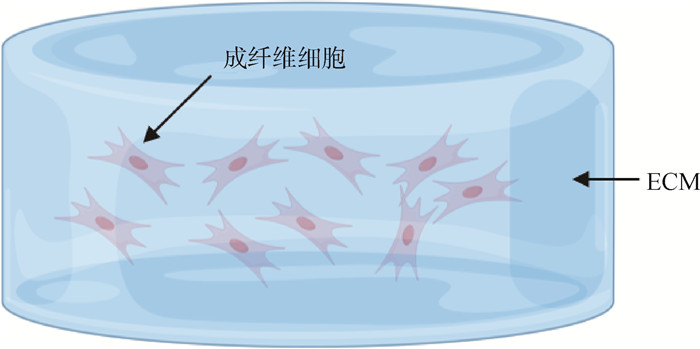

图 2
无支架细胞培养模型 A.超低附着板法;B.颗粒培养法;C.悬挂掉落法;D.磁悬浮法;E.旋转瓶;F.旋转壁容器"

表 2
无支架技术与有支架技术的比较"
| 培养技术Culture technology | 优点Merit | 局限性Limitation |
| 无支架 Scaffold-free | 模型简单、结果具有可重复性、易于制备、可以扩展、对各种细胞系的适用性,可进行高通量筛选 | 形成的球状体与有支架培养出的类器官相比有一定的差距。会在球状体中发现坏死细胞 |
| 有支架 Scaffold | 可通过调整支架的纳米级、微观级、宏观级特征模拟天然组织微环境。能构建结构更加复杂的类器官,与体内组织结构更加相似 | 材料成本高,需要考虑细胞与材料的生物相容性。天然生物材料存在批次性差异,可重复性不好。检查手段较少 |
| 1 |
LIN X Y , ZHU L , HE J . Morphogenesis, growth cycle and molecular regulation of hair follicles[J]. Front Cell Dev Biol, 2022, 10, 899095.
doi: 10.3389/fcell.2022.899095 |
| 2 |
TOPOUZI H , LOGAN N J , WILLIAMS G , et al. Methods for the isolation and 3D culture of dermal papilla cells from human hair follicles[J]. Exp Dermatol, 2017, 26 (6): 491- 496.
doi: 10.1111/exd.13368 |
| 3 |
HIGGINS C A , CHEN J C , CERISE J E , et al. Microenvironmental reprogramming by three-dimensional culture enables dermal papilla cells to induce de novo human hair-follicle growth[J]. Proc Natl Acad Sci U S A, 2013, 110 (49): 19679- 19688.
doi: 10.1073/pnas.1309970110 |
| 4 |
HUH D , HAMILTON G A , INGBER D E . From 3-D cell culture to organs-on-chips[J]. Trends Cell Biol, 2011, 21 (12): 745- 754.
doi: 10.1016/j.tcb.2011.09.005 |
| 5 |
CACCIAMALI A , VILLA R , DOTTI S . 3D cell cultures: evolution of an ancient tool for new applications[J]. Front Physiol, 2022, 13, 836480.
doi: 10.3389/fphys.2022.836480 |
| 6 |
KARAMANOS N K , THEOCHARIS A D , PIPERIGKOU Z , et al. A guide to the composition and functions of the extracellular matrix[J]. FEBS J, 2021, 288 (24): 6850- 6912.
doi: 10.1111/febs.15776 |
| 7 |
YAMADA K M , DOYLE A D , LU J Y . Cell-3D matrix interactions: recent advances and opportunities[J]. Trends Cell Biol, 2022, 32 (10): 883- 895.
doi: 10.1016/j.tcb.2022.03.002 |
| 8 |
ARCURI S , PENNAROSSA G , DE IORIO T , et al. 3D ECM-based scaffolds boost young cell secretome-derived EV rejuvenating effects in senescent cells[J]. Int J Mol Sci, 2023, 24 (9): 8285.
doi: 10.3390/ijms24098285 |
| 9 |
RAIS A , HUSAIN A , HASAN G M , et al. A review on regulation of cell cycle by extracellular matrix[J]. Int J Biol Macromol, 2023, 232, 123426.
doi: 10.1016/j.ijbiomac.2023.123426 |
| 10 |
BERGER FRIDMAN I , KOSTAS J , GREGUS M , et al. High-throughput microfluidic 3D biomimetic model enabling quantitative description of the human breast tumor microenvironment[J]. Acta Biomater, 2021, 132, 473- 488.
doi: 10.1016/j.actbio.2021.06.025 |
| 11 |
HERREROS-POMARES A , ZHOU X , CALABUIG-FARIÑAS S , et al. 3D printing novel in vitro cancer cell culture model systems for lung cancer stem cell study[J]. Mater Sci Eng: C, 2021, 122, 111914.
doi: 10.1016/j.msec.2021.111914 |
| 12 |
COMPERA N , ATWELL S , WIRTH J , et al. Adipose microtissue-on-chip: a 3D cell culture platform for differentiation, stimulation, and proteomic analysis of human adipocytes[J]. Lab Chip, 2022, 22 (17): 3172- 3186.
doi: 10.1039/D2LC00245K |
| 13 | FARUQU F N , LIAM-OR R , ZHOU S , et al. Defined serum-free three-dimensional culture of umbilical cord-derived mesenchymal stem cells yields exosomes that promote fibroblast proliferation and migration in vitro[J]. FASEB J, 2021, 35 (1): e21206. |
| 14 |
GONZÁLEZ-GUALDA E , BAKER A G , FRUK L , et al. A guide to assessing cellular senescence in vitro and in vivo[J]. FEBS J, 2021, 288 (1): 56- 80.
doi: 10.1111/febs.15570 |
| 15 |
KRASNOVA O , KOVALEVA A , SAVELEVA A , et al. Mesenchymal stem cells lose the senescent phenotype under 3D cultivation[J]. Stem Cell Res Ther, 2023, 14 (1): 373.
doi: 10.1186/s13287-023-03599-8 |
| 16 |
HASAN M F , GHIASVAND S , WANG H X , et al. Neural layer self-assembly in geometrically confined rat and human 3D cultures[J]. Biofabrication, 2019, 11 (4): 045011.
doi: 10.1088/1758-5090/ab2d3f |
| 17 |
TVRKER E , YILDIZ V H , ARSLAN YILDIZ A . Biomimetic hybrid scaffold consisting of co-electrospun collagen and PLLCL for 3D cell culture[J]. Int J Biol Macromol, 2019, 139, 1054- 1062.
doi: 10.1016/j.ijbiomac.2019.08.082 |
| 18 |
CHEN K , OZTURK K , LIEFELD T , et al. A phenotypically supervised single-cell analysis protocol to study within-cell-type heterogeneity of cultured mammalian cells[J]. STAR Protoc, 2021, 2 (2): 100561.
doi: 10.1016/j.xpro.2021.100561 |
| 19 |
NAYAK B , BALACHANDER G M , MANJUNATH S , et al. Tissue mimetic 3D scaffold for breast tumor-derived organoid culture toward personalized chemotherapy[J]. Colloids Surf B: Biointerfaces, 2019, 180, 334- 343.
doi: 10.1016/j.colsurfb.2019.04.056 |
| 20 |
KUMAR P , JIMENEZ FRANCO A , ZHAO X B . 3D culture of fibroblasts and neuronal cells on microfabricated free-floating carriers[J]. Colloids Surf B: Biointerfaces, 2023, 227, 113350.
doi: 10.1016/j.colsurfb.2023.113350 |
| 21 | BIDARRA S J, BARRIAS C C. 3D culture of mesenchymal stem cells in alginate hydrogels[M]//TURKSEN K. Stem Cell Niche: Stem Cell Niche. New York, NY: Springer, 2019: 165-180. |
| 22 |
GAO W J , WU D L , WANG Y L , et al. Development of a novel and economical agar-based non-adherent three-dimensional culture method for enrichment of cancer stem-like cells[J]. Stem Cell Res Ther, 2018, 9 (1): 243.
doi: 10.1186/s13287-018-0987-x |
| 23 | NOSENKO M A , MOYSENOVICH A M , ARKHIPOVA A Y , et al. Fibroblasts upregulate expression of adhesion molecules and promote lymphocyte retention in 3D fibroin/gelatin scaffolds[J]. Bioact Mater, 2021, 6 (10): 3449- 3460. |
| 24 |
YAMADA Y , YOSHIDA C , HAMADA K , et al. Development of three-dimensional cell culture scaffolds using laminin peptide-conjugated agarose microgels[J]. Biomacromolecules, 2020, 21 (9): 3765- 3771.
doi: 10.1021/acs.biomac.0c00871 |
| 25 | HAN B. Screening miRNA for functional significance by 3D cell culture system[M]//YING S Y. MicroRNA Protocols. New York, NY: Springer, 2018: 193-201. |
| 26 |
COLLADO-GONZÁLEZ M , PECCI-LLORET M P , GARCÍA-BERNAL D , et al. Biological effects of silk fibroin 3D scaffolds on stem cells from human exfoliated deciduous teeth (SHEDs)[J]. Odontology, 2018, 106 (2): 125- 134.
doi: 10.1007/s10266-017-0310-9 |
| 27 |
BADEKILA A K , PAI V , VIJAYAN V , et al. Engineering alginate/carboxymethylcellulose scaffolds to establish liver cancer spheroids: evaluation of molecular variances between 2D and 3D models[J]. Int J Biol Macromol, 2024, 254, 128058.
doi: 10.1016/j.ijbiomac.2023.128058 |
| 28 |
LONG R M , SHI L R , HE P , et al. 3D cell culture based on artificial cells and hydrogel under microgravity for bottom-up microtissue constructs[J]. Front Bioeng Biotechnol, 2022, 10, 1056652.
doi: 10.3389/fbioe.2022.1056652 |
| 29 |
HU H L , GEHART H , ARTEGIANI B , et al. Long-term expansion of functional mouse and human hepatocytes as 3D organoids[J]. Cell, 2018, 175 (6): 1591- 1606. e19.
doi: 10.1016/j.cell.2018.11.013 |
| 30 |
TAALE M , SCHAMBERGER B , MONCLUS M A , et al. Microarchitected compliant scaffolds of pyrolytic carbon for 3D muscle cell growth[J]. Adv Healthc Mater, 2024, 13 (9): 2303485.
doi: 10.1002/adhm.202303485 |
| 31 |
ELANGO J , ZAMORA-LEDEZMA C , NEGRETE-BOLAGAY D , et al. Retinol-loaded poly(vinyl alcohol)-based hydrogels as suitable biomaterials with antimicrobial properties for the proliferation of mesenchymal stem cells[J]. Int J Mol Sci, 2022, 23 (24): 15623.
doi: 10.3390/ijms232415623 |
| 32 |
MEESUK L , SUWANPRATEEB J , THAMMARAKCHAROEN F , et al. Osteogenic differentiation and proliferation potentials of human bone marrow and umbilical cord-derived mesenchymal stem cells on the 3D-printed hydroxyapatite scaffolds[J]. Sci Rep, 2022, 12 (1): 19509.
doi: 10.1038/s41598-022-24160-2 |
| 33 |
ZHANG M M , XU Y R , CHEN Y , et al. Three-dimensional poly-(ε-caprolactone) nanofibrous scaffolds promote the maturation of human pluripotent stem cells-induced cardiomyocytes[J]. Front Cell Dev Biol, 2022, 10, 875278.
doi: 10.3389/fcell.2022.875278 |
| 34 |
JANSEN L E , KIM H , HALL C L , et al. A poly(ethylene glycol) three-dimensional bone marrow hydrogel[J]. Biomaterials, 2022, 280, 121270.
doi: 10.1016/j.biomaterials.2021.121270 |
| 35 |
DHAMECHA D , LE D N , CHAKRAVARTY T , et al. Fabrication of PNIPAm-based thermoresponsive hydrogel microwell arrays for tumor spheroid formation[J]. Mater Sci Eng: C, 2021, 125, 112100.
doi: 10.1016/j.msec.2021.112100 |
| 36 |
LI L M , LI J D , ZOU Q , et al. Enhanced bone tissue regeneration of a biomimetic cellular scaffold with co-cultured MSCs-derived osteogenic and angiogenic cells[J]. Cell Prolif, 2019, 52 (5): e12658.
doi: 10.1111/cpr.12658 |
| 37 |
DOSH R H , ESSA A , JORDAN-MAHY N , et al. Use of hydrogel scaffolds to develop an in vitro 3D culture model of human intestinal epithelium[J]. Acta Biomater, 2017, 62, 128- 143.
doi: 10.1016/j.actbio.2017.08.035 |
| 38 |
BIAŁKOWSKA K , KOMOROWSKI P , BRYSZEWSKA M , et al. Spheroids as a type of three-dimensional cell cultures—examples of methods of preparation and the most important application[J]. Int J Mol Sci, 2020, 21 (17): 6225.
doi: 10.3390/ijms21176225 |
| 39 |
RAITANEN J , BARTA B , HACKER M , et al. Comparison of radiation response between 2D and 3D cell culture models of different human cancer cell lines[J]. Cells, 2023, 12 (3): 360.
doi: 10.3390/cells12030360 |
| 40 |
MALHÃO F , MACEDO A C , RAMOS A A , et al. Morphometrical, morphological, and immunocytochemical characterization of a tool for cytotoxicity research: 3D cultures of breast cell lines grown in ultra-low attachment plates[J]. Toxics, 2022, 10 (8): 415.
doi: 10.3390/toxics10080415 |
| 41 |
ZHANG S Y , BUTTLER-BUECHER P , DENECKE B , et al. A comprehensive analysis of human dental pulp cell spheroids in a three-dimensional pellet culture system[J]. Arch Oral Biol, 2018, 91, 1- 8.
doi: 10.1016/j.archoralbio.2018.02.008 |
| 42 |
GRIGULL N P , REDEKER J I , SCHMITT B , et al. Chondrogenic potential of pellet culture compared to high-density culture on a bacterial cellulose hydrogel[J]. Int J Mol Sci, 2020, 21 (8): 2785.
doi: 10.3390/ijms21082785 |
| 43 | FOTY R . A simple hanging drop cell culture protocol for generation of 3D spheroids[J]. J Vis Exp, 2011, 6 (51): 2720. |
| 44 |
GUPTA P , KAR S , KUMAR A , et al. Pulsed laser assisted high-throughput intracellular delivery in hanging drop based three dimensional cancer spheroids[J]. Analyst, 2021, 146 (15): 4756- 4766.
doi: 10.1039/D0AN02432E |
| 45 |
ANIL-INEVI M , DELIKOYUN K , MESE G , et al. Magnetic levitation assisted biofabrication, culture, and manipulation of 3D cellular structures using a ring magnet based setup[J]. Biotechnol Bioeng, 2021, 118 (12): 4771- 4785.
doi: 10.1002/bit.27941 |
| 46 |
ADINE C , NG K K , RUNGARUNLERT S , et al. Engineering innervated secretory epithelial organoids by magnetic three-dimensional bioprinting for stimulating epithelial growth in salivary glands[J]. Biomaterials, 2018, 180, 52- 66.
doi: 10.1016/j.biomaterials.2018.06.011 |
| 47 |
SUCOSKY P , OSORIO D F , BROWN J B , et al. Fluid mechanics of a spinner-flask bioreactor[J]. Biotechnol Bioeng, 2004, 85 (1): 34- 46.
doi: 10.1002/bit.10788 |
| 48 | RADTKE A L , HERBST-KRALOVETZ M M . Culturing and applications of rotating wall vessel bioreactor derived 3D epithelial cell models[J]. J Vis Exp, 2012, 3 (62): 3868. |
| 49 |
QIAN X Y , JACOB F , SONG M M , et al. Generation of human brain region-specific organoids using a miniaturized spinning bioreactor[J]. Nat Protoc, 2018, 13 (3): 565- 580.
doi: 10.1038/nprot.2017.152 |
| 50 |
SHIN W , KIM H J . 3D in vitro morphogenesis of human intestinal epithelium in a gut-on-a-chip or a hybrid chip with a cell culture insert[J]. Nat Protoc, 2022, 17 (3): 910- 939.
doi: 10.1038/s41596-021-00674-3 |
| 51 | KALLA J , PFNEISSL J , MAIR T , et al. A systematic review on the culture methods and applications of 3D tumoroids for cancer research and personalized medicine[J]. Cell Oncol (Dordr), 2024, |
| 52 |
RAMÍREZ-CUÉLLAR J , FERRARI R , SANZ R T , et al. LATS1 controls CTCF chromatin occupancy and hormonal response of 3D-grown breast cancer cells[J]. EMBO J, 2024, 43 (9): 1770- 1798.
doi: 10.1038/s44318-024-00080-x |
| 53 |
SASAKI N , ASANO Y , SORAYAMA Y , et al. Promoting biological similarity by collagen microfibers in 3D colorectal cancer-stromal tissue: replicating mechanical properties and cancer stem cell markers[J]. Acta Biomater, 2024, 185, 161- 172.
doi: 10.1016/j.actbio.2024.07.001 |
| 54 |
DU PLESSIS L H , GOUWS C , NIETO D . The influence of viscosity of hydrogels on the spreading and migration of cells in 3D bioprinted skin cancer models[J]. Front Cell Dev Biol, 2024, 12, 1391259.
doi: 10.3389/fcell.2024.1391259 |
| 55 |
DANG B T N , DUWA R , LEE S , et al. Targeting tumor-associated macrophages with mannosylated nanotherapeutics delivering TLR7/8 agonist enhances cancer immunotherapy[J]. J Control Release, 2024, 372, 587- 608.
doi: 10.1016/j.jconrel.2024.06.062 |
| 56 |
WANG C R , NAGAYACH A , PATEL H , et al. Utilizing human cerebral organoids to model breast cancer brain metastasis in culture[J]. Breast Cancer Res, 2024, 26 (1): 108.
doi: 10.1186/s13058-024-01865-y |
| 57 |
YU G X , DING J , YANG N N , et al. Evaluating the pro-survival potential of apoptotic bodies derived from 2D- and 3D- cultured adipose stem cells in ischaemic flaps[J]. J Nanobiotechnology, 2024, 22 (1): 333.
doi: 10.1186/s12951-024-02533-1 |
| 58 |
DING Z G , YAN Z N , YUAN X , et al. Apoptotic extracellular vesicles derived from hypoxia-preconditioned mesenchymal stem cells within a modified gelatine hydrogel promote osteochondral regeneration by enhancing stem cell activity and regulating immunity[J]. J Nanobiotechnology, 2024, 22 (1): 74.
doi: 10.1186/s12951-024-02333-7 |
| 59 |
QUÍLEZ C , JEON E Y , PAPPALARDO A , et al. Efficient generation of skin organoids from pluripotent cells via defined extracellular matrix cues and morphogen gradients in a spindle-shaped microfluidic device[J]. Adv Healthc Mater, 2024, 13 (20): 2400405.
doi: 10.1002/adhm.202400405 |
| 60 |
CHEN W Y , WU P P , JIN C , et al. Advances in the application of extracellular vesicles derived from three-dimensional culture of stem cells[J]. J Nanobiotechnology, 2024, 22 (1): 215.
doi: 10.1186/s12951-024-02455-y |
| 61 |
ELAHI F M , FARWELL D G , NOLTA J A , et al. Preclinical translation of exosomes derived from mesenchymal stem/stromal cells[J]. Stem Cells, 2020, 38 (1): 15- 21.
doi: 10.1002/stem.3061 |
| 62 |
XU Z R , YANG J X , XIN X Y , et al. Merits and challenges of iPSC-derived organoids for clinical applications[J]. Front Cell Dev Biol, 2023, 11, 1188905.
doi: 10.3389/fcell.2023.1188905 |
| 63 |
XIANG J Z , WANG H N , SHI B B , et al. Pig blastocyst-like structure models from embryonic stem cells[J]. Cell Discov, 2024, 10 (1): 72.
doi: 10.1038/s41421-024-00693-w |
| 64 | DIEZ J F F , TEGELER A P , FLESHER C G , et al. Extracellular matrix modulates depot-specific adipogenic capacity in adipose tissue of dairy cattle[J]. J Dairy Sci, 2024, S0022-0302(24)00985-8 |
| 65 |
XU Z B , XU X X , YANG B , et al. 3D sheep rumen epithelial structures driven from single cells in vitro[J]. Vet Res, 2023, 54 (1): 104.
doi: 10.1186/s13567-023-01234-1 |
| 66 |
SCHNEIDER M R , SCHMIDT-ULLRICH R , PAUS R . The hair follicle as a dynamic miniorgan[J]. Curr Biol, 2009, 19 (3): R132- R142.
doi: 10.1016/j.cub.2008.12.005 |
| 67 |
ZHAO B R , LUO H P , HE J M , et al. Comprehensive transcriptome and methylome analysis delineates the biological basis of hair follicle development and wool-related traits in Merino sheep[J]. BMC Biol, 2021, 19 (1): 197.
doi: 10.1186/s12915-021-01127-9 |
| 68 |
ZHANG Y H , TOMANN P , ANDL T , et al. Reciprocal requirements for EDA/EDAR/NF-κB and Wnt/β-catenin signaling pathways in hair follicle induction[J]. Dev Cell, 2009, 17 (1): 49- 61.
doi: 10.1016/j.devcel.2009.05.011 |
| 69 |
GE W , TAN S J , WANG S H , et al. Single-cell transcriptome profiling reveals dermal and epithelial cell fate decisions during embryonic hair follicle development[J]. Theranostics, 2020, 10 (17): 7581- 7598.
doi: 10.7150/thno.44306 |
| 70 |
SULIC A M , DAS ROY R , PAPAGNO V , et al. Transcriptomic landscape of early hair follicle and epidermal development[J]. Cell Rep, 2023, 42 (6): 112643.
doi: 10.1016/j.celrep.2023.112643 |
| 71 |
LI C L , HE X , WU Y , et al. Single-cell transcriptome sequence profiling on the morphogenesis of secondary hair follicles in ordos fine-wool sheep[J]. Int J Mol Sci, 2024, 25 (1): 584.
doi: 10.3390/ijms25010584 |
| 72 |
HOUSCHYAR K S , BORRELLI M R , TAPKING C , et al. molecular mechanisms of hair growth and regeneration: current understanding and novel paradigms[J]. Dermatology, 2020, 236 (4): 271- 280.
doi: 10.1159/000506155 |
| 73 | 刘公言, 白莉雅, 李福昌, 等. 毛囊发育与周期性生长的调控信号通路研究进展[J]. 畜牧与兽医, 2021, 53 (1): 125- 129. |
| LIU G Y , BAI L Y , LI F C , et al. Progress in research on the regulatory signaling pathway of hair follicle development and periodic growth[J]. Acta Veterinaria et Zootechnica Sinica, 2021, 53 (1): 125- 129. | |
| 74 | 马荣, 杜海东, 曹彦龙, 等. 哺乳动物毛囊发生发育调控机制研究进展[J]. 中国农业大学学报, 2024, 29 (5): 40- 55. |
| MA R , DU H D , CAO Y L , et al. Research progress on the regulatory mechanisms of follicle morphogenesis and development in mammals[J]. Journal of China Agricultural University, 2024, 29 (5): 40- 55. | |
| 75 |
ROY S , MEHTA D , PARADKAR A , et al. Dab2 (Disabled-2), an adaptor protein, regulates self-renewal of hair follicle stem cells[J]. Commun Biol, 2024, 7 (1): 525.
doi: 10.1038/s42003-024-06047-2 |
| 76 |
YUSUPOVA M , ANKAWA R , YOSEFZON Y , et al. Apoptotic dysregulation mediates stem cell competition and tissue regeneration[J]. Nat Commun, 2023, 14 (1): 7547.
doi: 10.1038/s41467-023-41684-x |
| 77 |
ZHOU L J , WANG H , JING J , et al. Regulation of hair follicle development by exosomes derived from dermal papilla cells[J]. Biochem Biophys Res Commun, 2018, 500 (2): 325- 332.
doi: 10.1016/j.bbrc.2018.04.067 |
| 78 |
YAN H L , GAO Y , DING Q , et al. Exosomal micro RNAs derived from dermal papilla cells mediate hair follicle stem cell proliferation and differentiation[J]. Int J Biol Sci, 2019, 15 (7): 1368- 1382.
doi: 10.7150/ijbs.33233 |
| 79 |
MARTINO P A , HEITMAN N , RENDL M . The dermal sheath: an emerging component of the hair follicle stem cell niche[J]. Exp Dermatol, 2021, 30 (4): 512- 521.
doi: 10.1111/exd.14204 |
| 80 |
LEE J , RABBANI C C , GAO H , et al. Hair-bearing human skin generated entirely from pluripotent stem cells[J]. Nature, 2020, 582 (7812): 399- 404.
doi: 10.1038/s41586-020-2352-3 |
| 81 |
YOON M , KIM E , SEO S H , et al. KY19382 accelerates cutaneous wound healing via activation of the Wnt/β-catenin signaling pathway[J]. Int J Mol Sci, 2023, 24 (14): 11742.
doi: 10.3390/ijms241411742 |
| 82 |
KANG B M , KWACK M H , KIM M K , et al. Sphere formation increases the ability of cultured human dermal papilla cells to induce hair follicles from mouse epidermal cells in a reconstitution assay[J]. J Invest Dermatol, 2012, 132 (1): 237- 239.
doi: 10.1038/jid.2011.250 |
| 83 |
HIGGINS C A , RICHARDSON G D , FERDINANDO D , et al. Modelling the hair follicle dermal papilla using spheroid cell cultures[J]. Exp Dermatol, 2010, 19 (6): 546- 548.
doi: 10.1111/j.1600-0625.2009.01007.x |
| 84 |
TAN C T , LEO Z Y , LIM C Y . Generation and integration of hair follicle-primed spheroids in bioengineered skin constructs[J]. Biomed Mater, 2022, 17 (6): 061001.
doi: 10.1088/1748-605X/ac99c6 |
| 85 |
LIU Z , HUANG J F , KANG D N , et al. Microenvironmental reprogramming of human dermal papilla cells for hair follicle tissue engineering[J]. Acta Biomater, 2023, 165, 31- 49.
doi: 10.1016/j.actbio.2022.11.004 |
| 86 |
KANG D N , LIU Z , QIAN C M , et al. 3D bioprinting of a gelatin-alginate hydrogel for tissue-engineered hair follicle regeneration[J]. Acta Biomater, 2023, 165, 19- 30.
doi: 10.1016/j.actbio.2022.03.011 |
| [1] | 韩小曼, 孙少宁, 杨洁, 沈宁, 鲍志远, 蔡佳炜, 赵博昊, 陈阳, 吴信生. 家兔MSX2基因的克隆表达及生物信息学分析[J]. 畜牧兽医学报, 2025, 56(6): 2724-2732. |
| [2] | 谢月华, 谢福杰, 索勋, 闫文朝. 3D细胞培养系统在顶复门原虫研究中的应用[J]. 畜牧兽医学报, 2025, 56(4): 1540-1548. |
| [3] | 李云鹏, 韩小曼, 俞咏麒, 蔡佳炜, 赵博昊, 陈阳, 吴信生. 家兔TIMP1基因的克隆表达及其对毛乳头细胞增殖的影响[J]. 畜牧兽医学报, 2025, 56(10): 4988-4997. |
| [4] | 曹馨予, 蔡佳炜, 鲍志远, 姚漱玉, 李云鹏, 陈阳, 吴信生, 赵博昊. ATG14调控家兔毛囊毛乳头细胞自噬进程的功能探究[J]. 畜牧兽医学报, 2024, 55(8): 3472-3481. |
| [5] | 张莉蕊, 张北育, 李玉娟, 刘永需, 赵红, 李福昌, 刘磊. 饲粮蛋氨酸水平对安哥拉兔产毛性能和毛囊发育的影响[J]. 畜牧兽医学报, 2024, 55(7): 3024-3031. |
| [6] | 王小松, 李冬, 李淑, 陈佳力, 刘永需, 赵红, 李福昌, 刘磊. 不同饲粮铜水平对安哥拉兔生产性能及毛囊发育的影响[J]. 畜牧兽医学报, 2024, 55(7): 3032-3039. |
| [7] | 褚婷婷, 张晓宇, 孙磊, 童嘉顺, 张磊, 宋宇轩. 家畜子宫内膜纤维化的细胞分子机制研究进展[J]. 畜牧兽医学报, 2024, 55(6): 2334-2344. |
| [8] | 何明亮, 吕晓阳, 蒋永清, 宋正海, 王叶青, 杨会国, 王善禾, 孙伟. 基于转录组测序分析SOX18在湖羊毛囊毛乳头细胞中的功能[J]. 畜牧兽医学报, 2024, 55(6): 2409-2420. |
| [9] | 康佳, 段香茹, 尹雪姣, 杨若晨, 李太春, 单新雨, 陈美静, 张英杰, 刘月琴. 半胱氨酸、蛋氨酸对体外培养绒山羊次级毛囊生长及毛乳头细胞增殖的影响[J]. 畜牧兽医学报, 2024, 55(2): 515-527. |
| [10] | 陈春, 康昭风, 魏岳, 黎观红, 武艳平, 谢金防. Wnt3a基因多态性与崇仁麻鸡皮肤毛囊性状相关性研究[J]. 畜牧兽医学报, 2023, 54(7): 2810-2823. |
| [11] | 许甜甜, 张彤彤, 王蒙, 王昕. 转录因子Foxq1通过WNT/β-catenin信号通路影响绒山羊毛囊干细胞增殖的研究[J]. 畜牧兽医学报, 2023, 54(6): 2653-2661. |
| [12] | 李玉娟, 张原铭, 张北育, 李福昌, 刘磊. 饲粮赖氨酸水平对安哥拉兔产毛性能及毛囊发育的影响[J]. 畜牧兽医学报, 2023, 54(5): 2013-2019. |
| [13] | 张希宇, 翟频, 王璠, 戴莹莹, 赵博昊, 陈阳, 吴信生. KRT16在长毛兔毛囊发育过程中的表达规律及功能探究[J]. 畜牧兽医学报, 2023, 54(1): 157-167. |
| [14] | 秦枫, 王菁, 翟频, 潘孝青, 杨杰, 邵乐, 李健, 张霞. 不同颜色LED光对苏系长毛兔毛品质及皮肤毛囊发育的影响[J]. 畜牧兽医学报, 2021, 52(9): 2545-2552. |
| [15] | 陈梦月, 张羽晨, 娄江城, 罗传真, 赵红利, 刘晓丽, 胡薛英. 26例犬毛囊肿瘤的组织病理学诊断和免疫组织化学分析[J]. 畜牧兽医学报, 2021, 52(5): 1407-1413. |
| 阅读次数 | ||||||
|
全文 |
|
|||||
|
摘要 |
|
|||||