畜牧兽医学报 ›› 2026, Vol. 57 ›› Issue (1): 443-453.doi: 10.11843/j.issn.0366-6964.2026.01.039

• 基础兽医 • 上一篇    下一篇

过氧化物酶体增殖物激活受体α通过调控AMPK/P38 MAPK通路促进小鼠骨骼肌发育

齐嘉慧(), 付婷婷, 郑敏行, 王璇晶, 吴海洋, 卢嘉茵, 罗小毛, 于秀菊, 王海东(), 闫艺()   

  1. 山西农业大学动物医学学院,太谷 030800
  • 收稿日期:2025-04-18 出版日期:2026-01-23 发布日期:2026-01-26
  • 通讯作者: 王海东,闫艺 E-mail:547475459@qq.com;sxaudywhd@163.com;sxaudywhd@ 163.com;yanyi@sxau.edu.cn
  • 作者简介:齐嘉慧,硕士,主要从事骨骼肌发育相关研究,E-mail:547475459@qq.com
  • 基金资助:
    国家自然科学基金(32102634);山西省研究生教育创新项目(2023KY345)

Peroxisome Proliferator-activated Receptor α Promotes Skeletal Muscle Development in Mice by Regulating the AMPK/P38 MAPK Pathway

QI Jiahui(), FU Tingting, ZHENG Minxing, WANG Xuanjing, WU Haiyang, LU Jiayin, LUO Xiaomao, YU Xiuju, WANG Haidong(), YAN Yi()   

  1. College of Veterinary Medicine,Shanxi Agricultural University,Taigu 030800,China
  • Received:2025-04-18 Online:2026-01-23 Published:2026-01-26
  • Contact: WANG Haidong, YAN Yi E-mail:547475459@qq.com;sxaudywhd@163.com;sxaudywhd@ 163.com;yanyi@sxau.edu.cn

摘要:

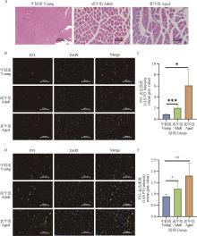
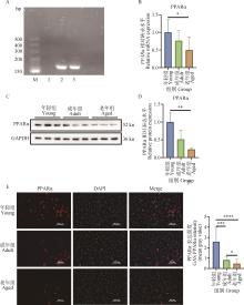
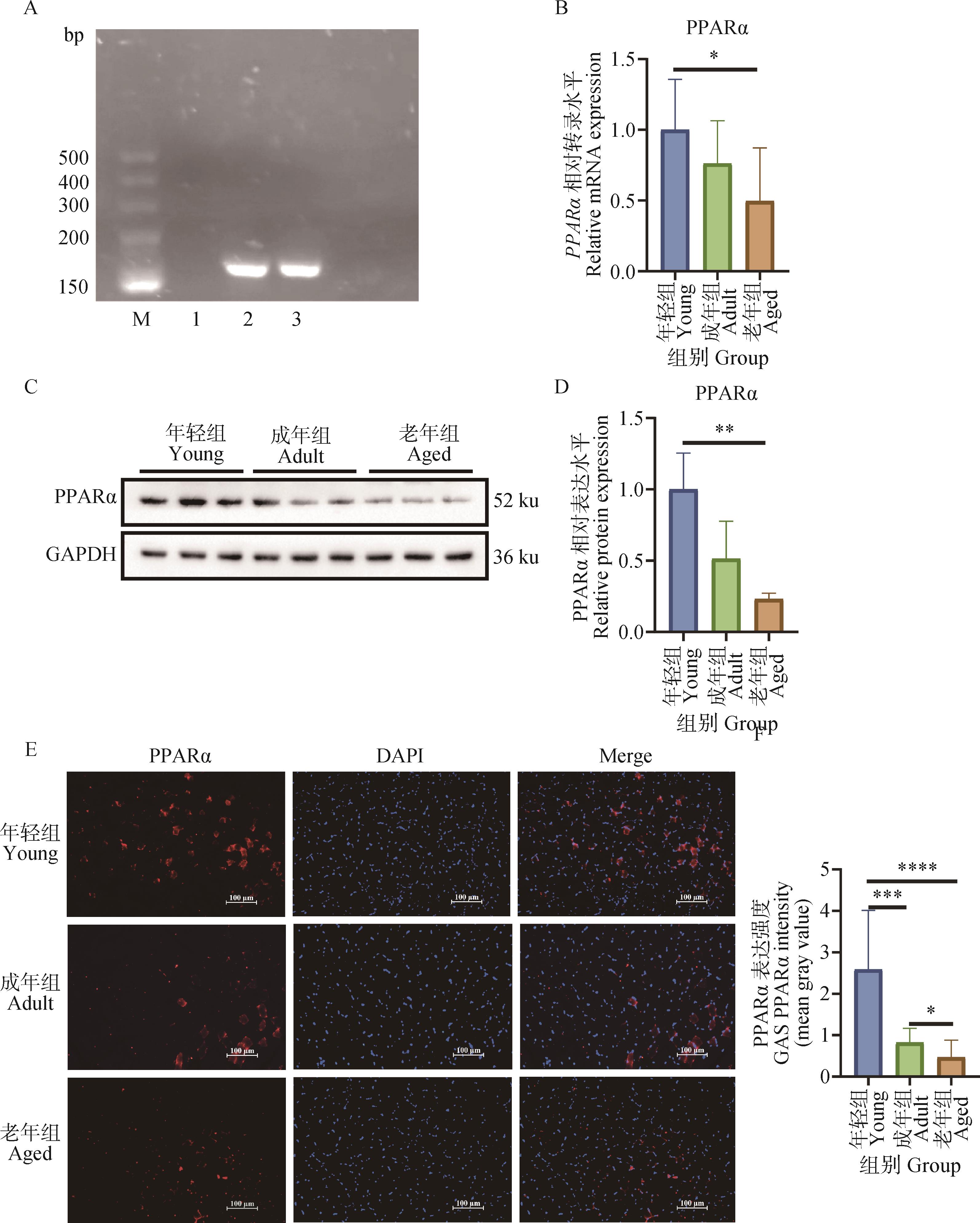
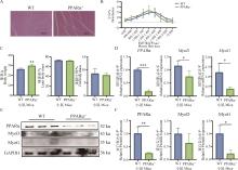
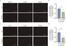
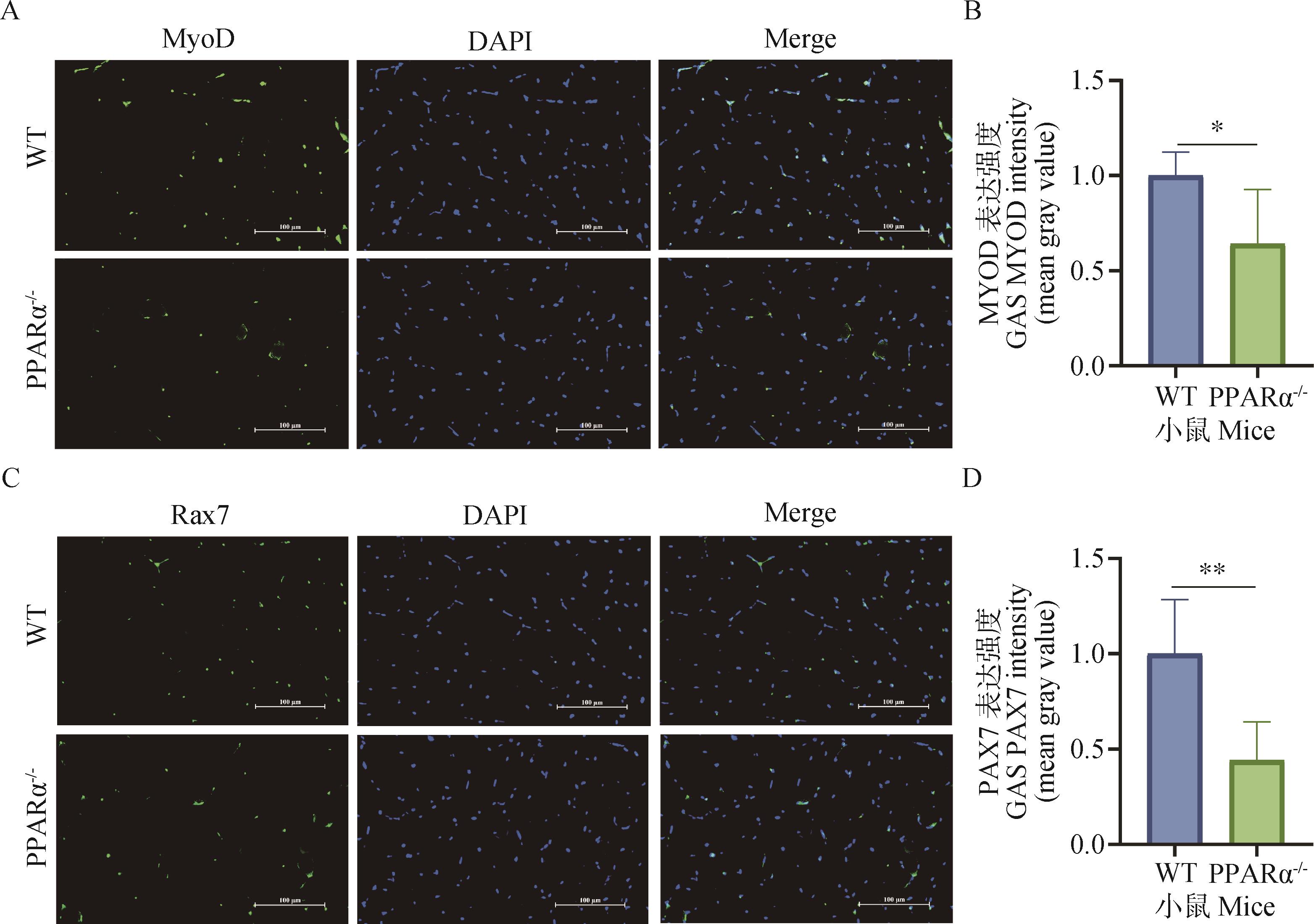
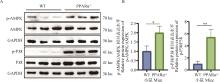
本研究旨在探索过氧化物酶体增殖物激活受体α(PPARα)对骨骼肌发育的影响及分子机制。选取年轻小鼠(6~8周龄)、成年小鼠(3~4月龄)和老年小鼠(18~20月龄)各8只,探究PPARα在不同发育阶段骨骼肌中的表达变化,并构建PPARα全身性敲除小鼠以探究其对骨骼肌发育的影响。通过HE染色观察不同年龄小鼠腓肠肌的形态差异,免疫荧光染色检测不同年龄小鼠腓肠肌中P21和P53的表达变化,确定小鼠的年龄差异;普通PCR反应检测PPARα在骨骼肌中的表达情况,蛋白免疫印迹(Western blot)、实时荧光定量PCR(qRT-PCR)和免疫荧光(IF)检测不同年龄小鼠腓肠肌中PPARα的表达变化;进而采用qRT-PCR、Western blot和IF检测WT和PPARα敲除小鼠(PPARα-/-)腓肠肌中肌肉发育相关指标MyoD、MyoG和Pax7的表达;Western blot检测PPARα下游信号通路AMPK和P38 MAPK的表达。结果表明,PPARα在各个发育阶段的小鼠骨骼肌中均存在表达,且其表达水平随年龄增长呈逐渐下调趋势。通过免疫荧光染色可见,PPARα蛋白广泛分布于小鼠腓肠肌的细胞核与细胞质中。进一步研究发现,PPARα基因敲除导致腓肠肌中生肌调节因子MyoD、MyoG及Pax7的表达显著下降,同时AMPK和P38的磷酸化水平明显上升。上述结果提示PPARα在小鼠骨骼肌发育过程中发挥重要作用,敲除PPARα后通过影响AMPK和P38的磷酸化水平抑制骨骼肌发育。

关键词: 不同发育阶段, 过氧化物酶体增殖物激活受体α(PPARα), 骨骼肌, AMPK, P38 MAPK

Abstract:

The aim of this study was to explore the effect and molecular mechanism of peroxisome proliferator-activated receptor α (PPARα) on skeletal muscle development. We utilized young (6-8 weeks), adult (3-4 months), and old (18-20 months) mice (n=8/group) to examine the expression of PPARα in skeletal muscle during development. The role of PPARα in skeletal muscle development was further investigated using a whole-body knockout mouse model. HE staining was used to observe the morphological differences of gastrocnemius muscle of mice of different ages, and immunofluorescence staining was used to detect the expression changes of P21 and P53 in gastrocnemius muscle of mice of different ages to determine the age difference of mice. The expression of PPARα in skeletal muscle was detected by common PCR, and the expression of PPARα in gastrocnemius muscle of mice of different ages was detected by Western blot, real-time quantitative PCR (qRT-PCR) and immunofluorescence staining (IF). qRT-PCR, Western blot and immunofluorescence staining were used to detect the expression of MyoD, MyoG and Pax7 in gastrocnemius muscles of WT and PPARα knockout mice (PPARα-/-). Western blot was used to detect the expression of PPARα downstream signaling pathways AMPK and P38 MAPK. The results showed that PPARα was expressed in the skeletal muscle of mice at all developmental stages, and its expression level gradually decreased with age. Immunofluorescence staining showed that PPARα protein was widely distributed in the nucleus and cytoplasm of mouse gastrocnemius muscle. Further studies found that PPARα gene knockout led to a significant decrease in the expression of MyoD, MyoG and Pax7 in gastrocnemius muscle, while the phosphorylation levels of AMPK and P38 increased significantly.

Key words: different developmental stages, peroxisome proliferator-activated receptor α (PPARα), skeletal muscle, AMPK, P38 MAPK

中图分类号: