


畜牧兽医学报 ›› 2025, Vol. 56 ›› Issue (8): 3837-3848.doi: 10.11843/j.issn.0366-6964.2025.08.024
董佳宁( ), 胡樱凡, 窦宇飞, 李俊, 石磊*(), 任有蛇*()
收稿日期:2025-01-14
出版日期:2025-08-23
发布日期:2025-08-28
通讯作者:
石磊,任有蛇
E-mail:2250889010@qq.com;shilei@sxau.edu.cn;rys925@126.com
作者简介:董佳宁(2000-),女,河南濮阳人,硕士生,主要从事动物繁殖生理研究,E-mail:2250889010@qq.com
基金资助:
DONG Jianing(), HU Yingfan, DOU Yufei, LI Jun, SHI Lei*(), REN Youshe*()
Received:2025-01-14
Online:2025-08-23
Published:2025-08-28
Contact:
SHI Lei, REN Youshe
E-mail:2250889010@qq.com;shilei@sxau.edu.cn;rys925@126.com
摘要:
旨在探讨微量元素硒通过PI3K/AKT/FoxO1通路对绵羊睾丸间质细胞自噬的影响及其分子机制。本研究采用3只8月龄健康雄性杜湖杂交绵羊的睾丸进行睾丸间质细胞的分离与培养,根据团队前期研究结果,建立不同浓度亚硒酸钠处理组(0、2、4和8 μmol·L-1)处理细胞18 h,每个处理组6个重复,试验重复3次。使用CCK-8检测细胞增殖活性;采用MDC检测细胞自噬水平、qRT-PCR和Western blot检测细胞内自噬相关基因(ATG5、P62和LC3)表达以及PI3K/AKT/FoxO1信号通路关键因子(PI3K、AKT、p-AKT、FoxO1、p-FoxO1和核内FoxO1)的表达水平;在此基础上,通过添加AKT特异性激活剂SC79或转染siFoxO1,探究PI3K/AKT/FoxO1信号通路在高硒(8 μmol·L-1)调控绵羊睾丸间质细胞自噬的作用机制。结果表明:2 μmol·L-1组细胞活力最高(P < 0.05),而8 μmol·L-1组细胞活力最低(P < 0.05),2 μmol·L-1组ROS含量显著低于其他各组(P < 0.05),高硒处理导致ROS的积累。使用MDC检测自噬,随着硒浓度升高,在8 μmol·L-1时自噬水平最高(P < 0.05),显著增加细胞自噬相关因子(ATG5和LC3-Ⅱ/Ⅰ)的丰度(P < 0.05),显著降低P62表达(P < 0.05)。与对照组相比,8 μmol·L-1组显著抑制(P < 0.05)PI3K和p-AKT活性,显著增加(P < 0.05)FoxO1以及核内FoxO1的表达,显著降低p-FoxO1的丰度(P < 0.05)。为了进一步探讨PI3K/AKT/FoxO1信号通路在高硒诱导细胞自噬中的作用,使用SC79(15 mg·L-1)预处理1 h,或者转染siFoxO1发现在间质细胞中LC3-Ⅱ/Ⅰ比值和ATG5丰度显著降低(P < 0.05),P62显著上调减轻高硒诱导的自噬(P < 0.05)。同时,SC79显著抑制高硒诱导的FoxO1去磷酸化和大量核转位(P < 0.05)。综上所述,高硒通过抑制PI3K/AKT通路调控FoxO1磷酸化及其核易位,诱导绵羊间质细胞氧化应激和自噬。研究结果有助于阐明高硒对绵羊睾丸间质细胞产生负面影响的分子机制。
中图分类号:
董佳宁, 胡樱凡, 窦宇飞, 李俊, 石磊, 任有蛇. 高硒通过PI3K/AKT/FoxO1信号通路对绵羊睾丸间质细胞自噬的影响[J]. 畜牧兽医学报, 2025, 56(8): 3837-3848.
DONG Jianing, HU Yingfan, DOU Yufei, LI Jun, SHI Lei, REN Youshe. Effects of Excessive Selenium on Autophagy of Leydig Cells in Sheep through PI3K/AKT/FoxO1 Pathway[J]. Acta Veterinaria et Zootechnica Sinica, 2025, 56(8): 3837-3848.
表 1
小干扰RNA序列"
| 小干扰RNA siRNA | 正义序列(5′→3′)Sense | 反义序列(5′→3′)Antisense |
| si-NC | UUCUCCGAACGUGUCACGUTT | ACGUGACACGUUCGGAGAATT |
| si-FoxO1-1 | GCUGUCAAUGCCGACUUCATT | UGAAGUCGGCAUUGACAGCTT |
| si-FoxO1-2 | GCAAUGACGACUUUGACAATT | UUGUCAAAGUCGUCAUUGCTT |
| si-FoxO1-3 | CGGUGAAGACAGCUUUACATT | UGUAAAGCUGUCUUCACCGTT |
表 2
qRT-PCR引物信息"
| 基因Gene | 序列号GenBank No. | 引物序列(5′→3′)Primer sequence | 产物长度/bp Product length |
| ATG5 | XM_042253359.1 | F: GCCTTTCATCCAGAAGCTGT R: TCATCACCTGGCTCCTCTTC | 174 |
| LC3-Ⅱ | XM_004014953.4 | F: ATGCCGTCCGAGAAAACCTT R: GCTGCTCTCGGATAAGTCGG | 79 |
| P62 | XM_015104932.3 | F: CGCAGAACAGAGTTACGAAGG R: TCCCATTCCAGTCATCTTGTC | 183 |
| PI3K | XM_042233720.2 | F: GGCGGCTGAGTATCGTGAGATTG R: CGGACACCTTTCTGAGTCAACCAC | 114 |
| AKT | XM_027960258.2 | F: CAGGAGGAGGAGACGATGGACTTC R: CCCAGCAGCTTCAGGTACTCAAAC | 134 |
| FoxO1 | XM_027973596.2 | F: TGTCCTACGCCGACCTCATCAC R: GCACGCTCTTGACCATCCACTC | 96 |
| β-actin | NM_001009784.3 | F: AAGACCTCTACGCCAACACG R: GCCAGGGCAGTGATCTCTTT | 91 |
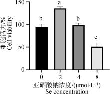
图 1
CCK-8检测绵羊睾丸间质细胞活力 柱上标注不同字母代表差异显著(P < 0.05),相同字母代表差异不显著(P>0.05),下同"
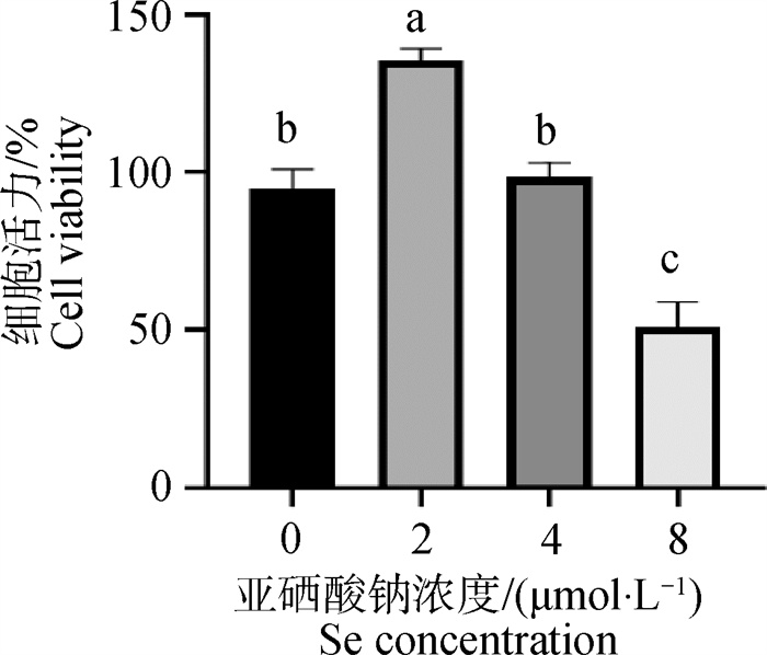
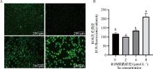
图 2
不同浓度亚硒酸钠处理后睾丸间质细胞的ROS水平 A. DCFH-DA染色后荧光显微镜下细胞图像;B. DCFH-DA荧光强度分析"
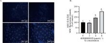
图 3
不同浓度亚硒酸钠处理后睾丸间质细胞的自噬水平 A. MDC染色后荧光显微镜下睾丸间质细胞图像; B. MDC荧光强度分析"
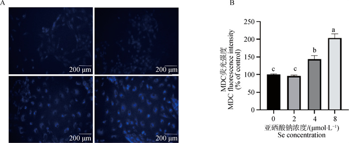
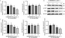
图 4
高硒抑制睾丸间质细胞中PI3K/AKT信号通路 A. qRT-PCR检测基因的mRNA水平;B. 蛋白Western blot结果;C. 灰度值分析"
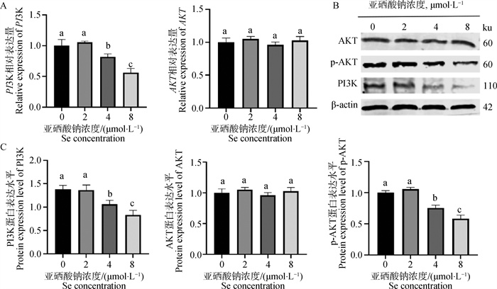

图 5
AKT激活剂SC79对高硒诱导睾丸间质细胞的自噬基因表达的影响 A. 用不同浓度SC79预处理的CCK-8评估细胞活力;B. Western blot检测SC79(15 mg·L-1)处理对细胞中p-AKT表达的影响;C. qRT-PCR检测自噬基因的mRNA水平;D. 灰度值分析;E. 蛋白Western blot结果"
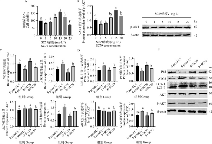

图 6
AKT激活剂SC79在睾丸间质细胞中对FoxO1水平、活性和易位的调节 A、B. 细胞用亚硒酸钠处理18 h。C、D. 使用SC79预处理1 h。通过qRT-PCR检测FoxO1的mRNA水平,通过Western blot分析FoxO1和p-FoxO1的蛋白水平,细胞核中FoxO1的Western blot结果和分析。E、F. 蛋白Western blot结果"
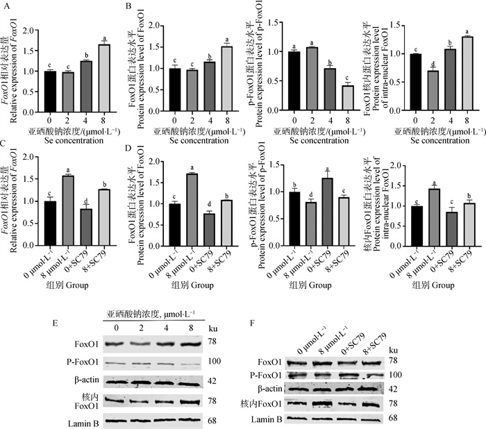
图 7
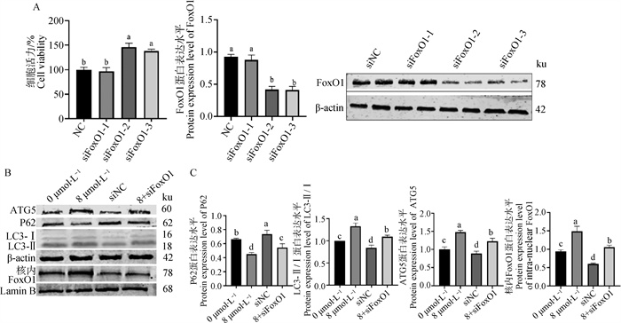
siFoxO1对自噬基因蛋白表达的影响 A. siFoxO1的筛选和验证,细胞用NC或siFoxO1转染6 h,然后用高硒处理18 h;B.蛋白Western blot结果;C. 灰度值分析"
| 1 |
CALIKA,EMAMIN K,WHITEM B,et al.Influence of dietary vitamin E and selenium supplementation on broilers subjected to heat stress, part Ⅰ: growth performance, body composition and intestinal nutrient transporters[J].Poult Sci,2022,101(6):101857.
doi: 10.1016/j.psj.2022.101857 |
| 2 |
DALIAA M,LOHT C,SAZILIA Q,et al.The effect of dietary bacterial organic selenium on growth performance, antioxidant capacity, and Selenoproteins gene expression in broiler chickens[J].Bmc Vet Res,2017,13(1):254.
doi: 10.1186/s12917-017-1159-4 |
| 3 |
黄靓,胡聪,孙久鹏,等.生物活性硒对不同品种育肥猪生长性能、组织硒含量、抗氧化能力和肉品质的影响[J].动物营养学报,2023,35(10):6301-6317.
doi: 10.12418/CJAN2023.579 |
|
HUANGL,HUC,SUNJ P,et al.Effects of bioactive Selenium on growth performance, tissue Selenium content, antioxidant ability and meat quality of different breeds of finishing pigs[J].Chinese Journal of Animal Nutrition,2023,35(10):6301-6317.
doi: 10.12418/CJAN2023.579 |
|
| 4 | LIC,LIUX,LIJ,et al.Selenomethionine inhibited HADV-induced apoptosis mediated by ROS through the JAK-STAT3 signaling pathway[J].Nutrients,2024,16(12):2969. |
| 5 |
LIUS,YAOS,YANGH,et al.Autophagy: regulator of cell death[J].Cell Death Dis,2023,14(10):648.
doi: 10.1038/s41419-023-06154-8 |
| 6 |
YAMAMOTOH,MATSUIT.Molecular mechanisms of macroautophagy, microautophagy, and chaperone-mediated autophagy[J].Nippon Med Sch,2024,91(1):2-9.
doi: 10.1272/jnms.JNMS.2024_91-102 |
| 7 | ZHANGC,ZHANGH L,LIUS L,et al.P62/SQSTM1 mediates the autophagy-lysosome degradation of CDK2 protein undergoing PI3Kα/AKT T308 inhibition[J].Biochem Bioph Res Co,2022,62(7):11-15. |
| 8 |
HOCHFELDW E,LEES,RUBINSZTEIND C.Therapeutic induction of autophagy to modulate neurodegenerative disease progression[J].Acta Pharmacol Sin,2013,34(5):600-604.
doi: 10.1038/aps.2012.189 |
| 9 |
GLAVIANOA,FOOA S C,LAMH Y,et al.PI3K/AKT/mTOR signaling transduction pathway and targeted therapies in cancer[J].Mol Cancer,2023,22(1):138.
doi: 10.1186/s12943-023-01827-6 |
| 10 | FANGY,OUS,WUT,et al.Lycopene alleviates oxidative stress via the PI3K/Akt/Nrf2pathway in a cell model of Alzheimer's disease[J].Peer J,2020,8(6):9308. |
| 11 |
DE FELICIM,KLINGERF G.PI3K/PTEN/AKT signaling pathways in germ cell development and their involvement in germ cell tumors and ovarian dysfunctions[J].Int J Mol Sci,2021,22(18):9838.
doi: 10.3390/ijms22189838 |
| 12 |
SANZ-CASTILLOB,HURTADOB,VARA-CIRUELOSD,et al.The MASTL/PP2A cell cycle kinase-phosphatase module restrains PI3K-Akt activity in an mTORC1-dependent manner[J].Embo J,2023,42(2):110833.
doi: 10.15252/embj.2022110833 |
| 13 |
ZHUY,WUF,HUJ,et al.LDHA deficiency inhibits trophoblast proliferation via the PI3K/AKT/FoxO1/CyclinD1 signaling pathway in unexplained recurrent spontaneous abortion[J].FASEB J,2023,37(2):22744.
doi: 10.1096/fj.202201219RR |
| 14 |
LUOM,SUZ,GAOH,et al.Cirsiliol induces autophagy and mitochondrial apoptosis through the AKT/FoxO1 axis and influences methotrexate resistance in osteosarcoma[J].Transl Med,2023,21(1):907.
doi: 10.1186/s12967-023-04682-7 |
| 15 |
ZIRKINB R,PAPADOPOULOSV.Leydig cells: formation, function, and regulation[J].Biol Reprod,2018,99(1):101-111.
doi: 10.1093/biolre/ioy059 |
| 16 | YUANF,BAIK,HOUY,et al.Small Molecule cocktails promote fibroblast-to-Leydig-like cell conversion for hypogonadism therapy[J].Pharmaceutics,2023,15(10):56-72. |
| 17 | 王晓蕾. MiR-200a/Nrf2信号通路在硒调控绵羊睾丸间质细胞增殖和凋亡的作用机制[D]. 晋中: 山西农业大学, 2022. |
| WANG X L. Mechanism of MiR-200a/Nrf2 pathway in selenium regulating the proliferation and apoptosis of Leydig cells in sheep [D]. Jinzhong: Shanxi Agricultural University, 2022. (in Chinese) | |
| 18 | 双燕, 李航, 杨振鸿. 城巴断裂带高硒背景区土壤元素地球化学特征[C]. 杭州: 中国矿物岩石地球化学学会第17届学术年会论文摘要集, 2019: 918-919. |
| SHUANG Y, LI H, YANG Z H. Geochemical characteristics of soil elements in the high selenium background area of Chengba fault zone [C]. Hangzhou: Abstract Collection of the 17th Annual Conference of Mineralogical, Petrogeochemical Society of China, 2019: 918-919. (in Chinese) | |
| 19 |
ZHUK,YANGS,LIT,et al.Advances in the study of the mechanism by which Selenium and Selenoproteins boost immunity to prevent food allergies[J].Nutrients,2022,14(15):3133.
doi: 10.3390/nu14153133 |
| 20 | 董小雨,苑景达,付绍印,等.微量元素硒对羊生长发育及繁殖性能的调控作用[J].中国畜牧杂志,2025,61(4):55-60. |
| DONGX Y,YUANJ D,FUS Y,et al.Effects of Selenium on growth and reproductive performance of sheep[J].Chinese Journal of Animal Husbandry and Veterinary Medicine,2025,61(4):55-60. | |
| 21 |
WANGS,LIUX,LEIL,et al.Selenium deficiency induces apoptosis, mitochondrial dynamic imbalance, and inflammatory responses in calf liver[J].Biol Trace Elem Res,2022,200(11):4678-4689.
doi: 10.1007/s12011-021-03059-5 |
| 22 |
ZHANGY,LIUJ,LIX,et al.Dietary selenium excess affected spermatogenesis via DNA damage and telomere-related cell senescence and apoptosis in mice[J].Food Chem Toxicol,2023,171,113556.
doi: 10.1016/j.fct.2022.113556 |
| 23 | 李万栋,李光梅,景建武,等.微量元素硒在反刍动物生产中的应用研究进展[J].中国畜牧杂志,2022,58(11):44-51. |
| LIW D,LIG M,JINGJ W,et al.Effects of Selenium on growth and reproductive performance of sheep[J].Chinese Journal of Animal Husbandry and Veterinary Medicine,2022,58(11):44-51. | |
| 24 | SHIL,SONGR,YAOX,et al.Effects of selenium on the proliferation, apoptosis and testosterone production of sheep Leydig cells in vitro[J].Theriogenology,2017,9(3):24-32. |
| 25 | XUZ J,LIUM,NIUQ J,et al.Both selenium deficiency and excess impair male reproductive system via inducing oxidative stress-activated PI3K/AKT-mediated apoptosis and cell proliferation signaling in testis of mice[J].Free Radical Bio Med,2023,19(7):15-22. |
| 26 |
UDDINM H,RITUJ R,PUTNALAS K,et al.Selenium toxicity in fishes: A current perspective[J].Chemosphere,2024,364,143214.
doi: 10.1016/j.chemosphere.2024.143214 |
| 27 | WANGM,WANGY,WANGS,et al.Selenium alleviates cadmium-induced oxidative stress, endoplasmic reticulum stress and programmed necrosis in chicken testes[J].Sci Total Environ,2023,86(3):160601. |
| 28 |
RENY,WANGR,WENGS,et al.Multifaceted role of redox pattern in the tumor immune microenvironment regarding autophagy and apoptosis[J].Mol Cancer,2023,22(1):130-136.
doi: 10.1186/s12943-023-01831-w |
| 29 |
WANGY,LVJ,LIUG,et al.ZnO NPs impair the viability and function of porcine granulosa cells through autophagy regulated by ROS production[J].Antioxidants-Basel,2024,13(11):1295.
doi: 10.3390/antiox13111295 |
| 30 | LUOD,HEF,LIUJ,et al.Pseudolaric acid B suppresses NSCLC progression through the ROS/AMPK/mTOR/autophagy signalling pathway[J].Biomed Pharmacother,2024,17(5):116614. |
| 31 |
MEIL,CHENX,WEIF,et al.Tethering ATG16L1 or LC3 induces targeted autophagic degradation of protein aggregates and mitochondria[J].Autophagy,2023,19(11):2997-3013.
doi: 10.1080/15548627.2023.2234797 |
| 32 |
ZHONGZ,UMEMURAA,SANCHEZ-LOPEZE,et al.NF-κB Restricts Inflammasome activation via elimination of damaged mitochondria[J].Cell,2016,164(5):896-910.
doi: 10.1016/j.cell.2015.12.057 |
| 33 |
ZHUH,ZHONGY,CHENR,et al.ATG5 knockdown attenuates ischemia-reperfusion injury by reducing excessive autophagy-induced ferroptosis[J].Transl Stroke Res,2024,15(1):153-164.
doi: 10.1007/s12975-022-01118-0 |
| 34 |
LIY,CHENH,LIAOJ,et al.Long-term copper exposure promotes apoptosis and autophagy by inducing oxidative stress in pig testis[J].Environ Sci Pollut Res Int,2021,28(39):55140-55153.
doi: 10.1007/s11356-021-14853-y |
| 35 |
YUDUSHKINI.Getting the Akt together: guiding intracellular Akt ativity by PI3K[J].Biomolecules,2019,9(2):67.
doi: 10.3390/biom9020067 |
| 36 |
KMAL,BARUAHT J.The interplay of ROS and the PI3K/Akt pathway in autophagy regulation[J].Biotechnol Appl Biochem,2022,69(1):248-264.
doi: 10.1002/bab.2104 |
| 37 | SOLINASG,BECATTINNIB.PI3K and AKT at the Interface of signaling and metabolism[J].Curr Top Microbiol,2022,43(6):311-336. |
| 38 | WANGQ,ZHANGX,WANGJ,et al.Effect of high selenium on insulin signaling pathway PI3K-AKT-mTOR in L02 cells[J].Wei Sheng Yan Jiu,2024,53(1):77-87. |
| 39 |
LIS T,CHENN N,QIAOY B,et al.SC79 rescues osteoblasts from dexamethasone though activating Akt-Nrf2 signaling[J].Biochem Bioph Res Co,2016,479(1):54-60.
doi: 10.1016/j.bbrc.2016.09.027 |
| 40 |
LUOH,HUANGQ,HUANGD,et al.HABP2 encapsulated by peripheral Blood-Derived exosomes suppresses astrocyte autophagy to exacerbate neuroinflammatory injury in Mice with ischemic stroke[J].ACS Chem Neurosci,2023,14(12):2347-2361.
doi: 10.1021/acschemneuro.3c00089 |
| 41 | DUY,HUANGF,GUANL,et al.Role of PI3K/Akt/mTOR pathway-mediated macrophage auto-phagy in affecting the phenotype transformation of lung fibroblasts induced by silica dust exposure[J].Zhong Nan Da Xue Xue Bao Yi Xue Ban,2023,48(8):1152-1162. |
| 42 |
LIX,BAIC,WANGH,et al.LncRNA MEG3 regulates autophagy and pyroptosis via FoxO1 in pancreatic β-cells[J].Cell Signal,2022,92,110247.
doi: 10.1016/j.cellsig.2022.110247 |
| 43 |
ZHANGY,WANGM,TANGL,et al.FoxO1 silencing in Atp7b (-/-) neural stem cells attenuates high copper-induced apoptosis via regulation of autophagy[J].J Neurochem,2024,168(9):2762-2774.
doi: 10.1111/jnc.16136 |
| 44 |
IOANNILLIL,CICCARONEF,CIRIOLOM R.Adipose tissue and FoxO1: bridging physiology and mechanisms[J].Cells,2020,9(4):849.
doi: 10.3390/cells9040849 |
| 45 | LEEH,LEEJ.Anti-diabetic effect of hydroxybenzoic acid derivatives in free fatty acid-induced HepG2 cells via miR-1271/IRS1/PI3K/AKT/FOXO1 pathway[J].J Food Biochem,2021,45(12):13993. |
| 46 | MOEINIFARDM,HASSANZ M,FALLAHIANF,et al.Britannin induces apoptosis through AKT-FoxO1 pathway in human pancreatic cancer cells[J].Biomed Pharmacother,2017,9(4):1101-1110. |
| 47 | 陈宇. lncRNA FPMAL竞争抑制FoxO1磷酸化提高奶牛妊娠早期子宫容受性的分子机制研究[D]. 武汉: 华中农业大学, 2023. |
| CHEN Y. Molecular mechanism of lncRNA FPMAL promoting uterinereceptivity in early pregnancy of dairy cows by competitiveinhibition of FoxO1 phosphorylation [D]. Wuhan: Huazhong Agricultural University, 2023. (in Chinese) |
| [1] | 刘雨蒙, 高星, 赵雅丽, 曹迪, 芒来, 张心壮. 硒多糖缓解马骨骼肌卫星细胞氧化损伤作用的研究[J]. 畜牧兽医学报, 2025, 56(7): 3357-3367. |
| [2] | 赵顺然, 付桂鑫, 庞钊琪, 夏威, 李俊杰, 陶晨雨. 猪颗粒细胞在卵泡闭锁中的作用机制研究进展[J]. 畜牧兽医学报, 2025, 56(6): 2537-2545. |
| [3] | 王莹, 张姣姣, 王鲜忠, 权富生. 卵巢颗粒细胞自噬研究进展[J]. 畜牧兽医学报, 2025, 56(4): 1508-1517. |
| [4] | 侯宛辰, 徐童. 大麻二酚通过BRD4/AMPK/mTOR信号通路拮抗双酚A诱导的猪肠上皮细胞凋亡和自噬[J]. 畜牧兽医学报, 2025, 56(4): 1919-1933. |
| [5] | 王艺, 侯露露, 方菲, 高林英, 谢淑敏, 王雨. 氟通过自噬和铁死亡途径诱发肉鸡小肠氧化损伤[J]. 畜牧兽医学报, 2025, 56(1): 442-454. |
| [6] | 李相辰, 王林楠, 于正青, 张莉, 杨晨晨, 宋亮丽. 槲皮素抑制自噬恢复LTA诱导的奶牛乳腺上皮细胞紧密连接功能[J]. 畜牧兽医学报, 2024, 55(9): 3887-3896. |
| [7] | 王怡, 高娟, 胡悦旻, 杨跃飞, 范博钧, 鞠辉明. 短期血清饥饿胁迫对猪骨骼肌卫星细胞代谢及自噬发生的影响[J]. 畜牧兽医学报, 2024, 55(8): 3408-3417. |
| [8] | 曹馨予, 蔡佳炜, 鲍志远, 姚漱玉, 李云鹏, 陈阳, 吴信生, 赵博昊. ATG14调控家兔毛囊毛乳头细胞自噬进程的功能探究[J]. 畜牧兽医学报, 2024, 55(8): 3472-3481. |
| [9] | 赵彤, 杨文哲, 潘飞龙, 赵树臣, 刘克祥, 吕占军, 赵立佳. 双酚A通过上调Apoa1基因的表达抑制TM3细胞睾酮合成[J]. 畜牧兽医学报, 2024, 55(8): 3516-3525. |
| [10] | 李媛媛, 王天玉, 李梦, 张文慧, 王英卉, 赵天瑞, 李浩洁, 赵阳飞, 王金明. 硒代蛋氨酸通过PINK1/Parkin介导的线粒体自噬缓解氟诱导的抑郁样行为[J]. 畜牧兽医学报, 2024, 55(7): 3213-3224. |
| [11] | 杜红旭, 苏利娟, 何政科, 谭晓燕, 张旭, 马琪, 曹立亭, 陈红伟, 甘玲. 五味子多糖纳米硒的体外抗氧化和肠道菌群调节作用研究[J]. 畜牧兽医学报, 2024, 55(7): 3234-3245. |
| [12] | 罗金婷, 许发芳, 王磊, 罗璇, 马玉红, 张剑搏, 黄伟华, 尚月军, 吴国芳. 红景天多糖对低氧环境下猪睾丸间质细胞增殖及凋亡的影响[J]. 畜牧兽医学报, 2024, 55(6): 2441-2450. |
| [13] | 魏雅婷, 徐泽君, 陈虹宇, 王献伟, 陈其新, 刘深贺. 外源维生素E和硒调控动物精液品质的研究进展[J]. 畜牧兽医学报, 2024, 55(4): 1389-1400. |
| [14] | 李菲菲, 张晨淼, 童津津, 蒋林树. 线粒体自噬调节NLRP3炎症小体活性改善动物健康的作用机制[J]. 畜牧兽医学报, 2024, 55(4): 1446-1455. |
| [15] | 罗通旺, 吴亚, 王书杰, 宋厚辉, 邵春艳. 镉致肝损伤机制及硒拮抗镉肝毒性的研究进展[J]. 畜牧兽医学报, 2024, 55(4): 1456-1466. |
| 阅读次数 | ||||||
|
全文 |
|
|||||
|
摘要 |
|
|||||