


Acta Veterinaria et Zootechnica Sinica ›› 2025, Vol. 56 ›› Issue (9): 4559-4571.doi: 10.11843/j.issn.0366-6964.2025.09.034
• Preventive Veterinary Medicine • Previous Articles Next Articles
LIU Junjun1( ), GUO Donghui1,2,3(), LIU Huanhuan1, SONG Runze1, ZHAO Saiya1, YANG Junyao1, WEI Zhanyong2, XIANG Yuqiang1,2,3,*(), CHEN Liying1,2,3,*()
Received:2024-12-17
Online:2025-09-23
Published:2025-09-30
Contact:
XIANG Yuqiang, CHEN Liying
E-mail:ljj_box@163.com;guodonghui0808@126.com;yuqiangxiang@henau.edu.cn;chliying@henau.edu.cn
CLC Number:
LIU Junjun, GUO Donghui, LIU Huanhuan, SONG Runze, ZHAO Saiya, YANG Junyao, WEI Zhanyong, XIANG Yuqiang, CHEN Liying. Rapid Visual Detection for PDCoV/TGEV IgG Antibodies Using Smartphone-Assisted Colorimetric Sensing Platform based on Immunomagnetic Beads[J]. Acta Veterinaria et Zootechnica Sinica, 2025, 56(9): 4559-4571.
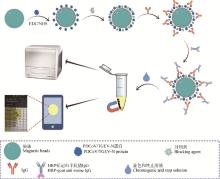
Fig. 1
Schematic illustration of colorimetric smartphone sensing platforms based on immunomagnetic beads for the detection of porcine coronavirus IgG antibodies EDC. N-(3-dimethylaminopropyl)-N′-ethylcarbodiimide hydrochloride; NHS. N-Hydroxysuccinimide; PDCoV. Porcine deltacoronavirus; TGEV. Transmissible gastroenteritis virus"
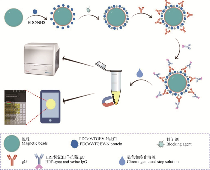

Fig. 2
Optimization of the detection conditions A. S/N values of different dosages of magnetic beads; B. S/N values of different recombinant PDCoV N protein concentrations; C. S/N values of different antigen coating time; D. S/N values of different blocking agents; E. S/N values of incubation time between diluted PDCoV positive serum and immunomagnetic beads; F. S/N values of different HRP-labeled goat anti-swine IgG dilutions; G. S/N values of different enzyme-labeled antibody incubation times; H. S/N values of different chromogenic development times"
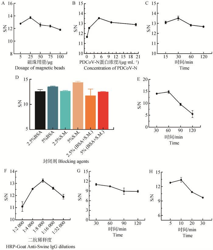
Fig. 3
Critical value analysis of the indirect ELSIA method for detection of PDCoV and TGEV IgG antibodies based on immunomagnetic beads A. ROC curve of PDCoV-N-IMB method results based on multimode microplate reader; B. ROC curve of TGEV-N-IMB method results based on multimode microplate reader; C. ROC curve of PDCoV-N-IMB method results based on smartphone; D. ROC curve of TGEV-N-IMB method results based on smartphone"
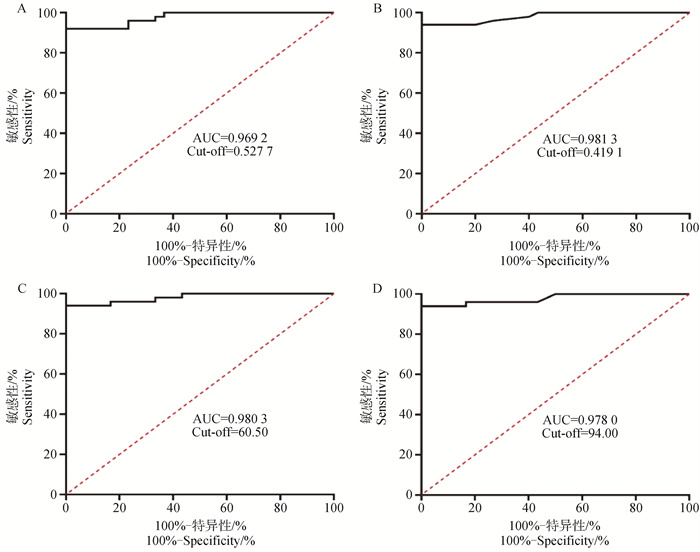
Fig. 4
Sensitivity of the IMB-based indirect ELISA A. Sensitivity of PDCoV-N-IMB indirect ELISA based on multimode microplate reader; B. Sensitivity of TGEV-N-IMB indirect ELISA based on multimode microplate reader; C. Sensitivity of PDCoV-N-IMB indirect ELISA based on smartphone; D. Sensitivity of PDCoV-N-IMB indirect ELISA based on smartphone"
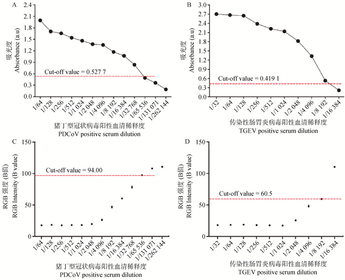
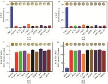
Fig. 5
Specificity of the IMB-based indirect ELISA for PDCoV and TGEV A. Specificity of PDCoV-N-IMB indirect ELISA based on multimode microplate reader; B. Specificity of TGEV-N-IMB indirect ELISA based on multimode microplate reader; C. Specificity of PDCoV-N-IMB indirect ELISA based on smartphone; D. Specificity of PDCoV-N-IMB indirect ELISA based on smartphone"
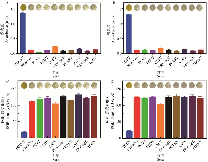
Table 1
Comparisons of the detection result of PDCoV-N-IMB based indirect ELISA"
| 血清中和试验 Serum neutralization test | 酶标仪检测结果(OD450 nm) Microplate reader test result (OD450 nm) | 智能手机检测结果(RGB) Smartphone test result (RGB) | ||||||
| 阳性 Positive | 阴性 Negative | 符合率/% Coincidence rate | 阳性 Positive | 阴性 Negative | 符合率/% Coincidence rate | |||
| 阳性血清 Positive sera | 50 | 46 | 4 | 92 | 46 | 4 | 92 | |
| 阴性血清 Negative sera | 30 | 0 | 30 | 100 | 0 | 30 | 100 | |
| 合计 Total | 80 | 46 | 34 | 95 | 46 | 34 | 95 | |
Table 2
Comparisons of the detection result of TGEV-N-IMB based indirect ELISA"
| 血清中和试验 Serum neutralization test | 酶标仪检测结果(OD450 nm) Microplate reader test result (OD450 nm) | 智能手机检测结果(RGB) Smartphone test result (RGB) | ||||||
| 阳性 Positive | 阴性 Negative | 符合率/% Coincidence rate | 阳性 Positive | 阴性 Negative | 符合率/% Coincidence rate | |||
| 阳性血清 Positive sera | 50 | 47 | 3 | 94 | 47 | 3 | 94 | |
| 阴性血清 Negative sera | 30 | 0 | 30 | 100 | 0 | 30 | 100 | |
| 合计 Total | 80 | 47 | 33 | 96 | 46 | 34 | 96.25 | |
| 1 |
HUH,JUNGK,VLASOVAA N,et al.Isolation and characterization of porcine deltacoronavirus from pigs with diarrhea in the United States[J].J Clin Microbiol,2015,53(5):1537-1548.
doi: 10.1128/JCM.00031-15 |
| 2 |
HANF,SHANF,HOUJ,et al.Establishment and application of PDCoV antigen-specific DAS-ELISA detection method[J].BMC Vet Res,2024,20(1):342.
doi: 10.1186/s12917-024-04201-w |
| 3 |
KONGF Z,WANGQ H,KENNEYS P,et al.Porcine deltacoronaviruses: Origin, evolution, cross-species transmission and zoonotic potential[J].Pathogens,2022,11(1):79.
doi: 10.3390/pathogens11010079 |
| 4 |
WANGN X,WANGZ,MAM Y,et al.Expression of codon-optimized PDCoV-RBD protein in baculovirus expression system and immunogenicity evaluation in mice[J].Int J Biol Macromol,2023,252,126113.
doi: 10.1016/j.ijbiomac.2023.126113 |
| 5 |
ZHAOF J,LIUL T,XUM L,et al.Assessments of different inactivating reagents in formulating transmissible gastroenteritis virus vaccine[J].Virol J,2020,17(1):163.
doi: 10.1186/s12985-020-01433-8 |
| 6 |
NIEDERWERDERM C,HESSER A.Swine enteric coronavirus disease: A review of 4 years with porcine epidemic diarrhoea virus and porcine deltacoronavirus in the United States and Canada[J].Transbound Emerg Dis,2018,65(3):660-675.
doi: 10.1111/tbed.12823 |
| 7 |
常新见,周金柱,殷杰,等.2017-2019年华东地区猪场主要病毒性腹泻病原调查[J].畜牧兽医学报,2020,51(12):3141-3150.
doi: 10.11843/j.issn.0366-6964.2020.12.023 |
|
CHANGX J,ZHOUJ Z,YINJ,et al.Investigation on pathogens of major viral diarrhea in pig farms in east china from 2017 to 2019[J].Acta Veterinaria et Zootechnica Sinica,2020,51(12):3141-3150.
doi: 10.11843/j.issn.0366-6964.2020.12.023 |
|
| 8 |
WANGZ,QUK,LIJ H,et al., Prevalence and potential risk factors of PDCoV in pigs based on publications during 2015-2021 in China: Comprehensive literature review and meta-analysis[J].Microb Pathog,2023,179,106118.
doi: 10.1016/j.micpath.2023.106118 |
| 9 |
WANGS,LIW J,WANGZ S,et al.Emerging and reemerging infectious diseases: global trends and new strategies for their prevention and control[J].Sig Transduct Target Ther,2024,9(1):223.
doi: 10.1038/s41392-024-01917-x |
| 10 |
OKDAF S,LAWSONX,LIUA,et al.Development of monoclonal antibodies and serological assays including indirect ELISA and fluorescent microsphere immunoassays for diagnosis of porcine deltacoronavirus[J].BMC Vet Res,2016,12,95.
doi: 10.1186/s12917-016-0716-6 |
| 11 |
WANGW L,ZHANGY N,YANGH C.Development of a nucleocapsid protein-based blocking ELISA for the detection of porcine deltacoronavirus antibodies[J].Viruses,2022,14(8):1815.
doi: 10.3390/v14081815 |
| 12 | 路浩,丁雪燕,袁晋,等.猪肠道冠状病毒检测方法的研究进展[J].中国兽医科学,2024,54(10):1391-1398. |
| LUH,DINGX Y,YUANJ,et al.Research progress on detection methods of swine enteric coronavirus[J].Chinese Veterinary Science,2024,54(10):1391-1398. | |
| 13 |
LABORIEE,LE-MINHV,MAIT D,et al.Analytical methods of antibody surface coverage and orientation on bio-functionalized magnetic beads: application to immunocapture of TNF-α[J].Anal Bioanal Chem,2021,413(25):6425-6434.
doi: 10.1007/s00216-021-03608-w |
| 14 |
VINDUSKAV,GALLOPSC E,O'CONNORR,et al.Exosomal surface protein detection with quantum dots and immunomagnetic capture for cancer detection[J].Nanomaterials (Basel),2021,11(7):1853.
doi: 10.3390/nano11071853 |
| 15 |
黄婧洁,李苗,陈莹娴,等.基于免疫磁珠净化间接竞争酶联免疫吸附试验检测氟喹诺酮类药物[J].畜牧兽医学报,2023,54(2):766-778.
doi: 10.11843/j.issn.0366-6964.2023.02.033 |
|
HUANGJ J,LIM,CHENY X,et al.Indirect competitive enzyme-linked immunosorbent assay for the detection of fluorquinolones based on immunomagnetic bead purification[J].Acta Veterinaria et Zootechnica Sinica,2023,54(2):766-778.
doi: 10.11843/j.issn.0366-6964.2023.02.033 |
|
| 16 |
XUT,ZHOUY C,LIUZ Y,et al.Prevalence and genetic diversity of porcine epidemic diarrhea virus in Southwest China during 2020-2022[J].Sci Rep,2024,14(1):29124.
doi: 10.1038/s41598-024-80844-x |
| 17 |
ZHANGF F,LUOY Y,LINC,et al.Epidemiological monitoring and genetic variation analysis of pathogens associated with porcine viral diarrhea in southern China from 2021 to 2023[J].Front Microbiol,2024,15,1303915.
doi: 10.3389/fmicb.2024.1303915 |
| 18 | 王挺,李风云,葛生虎,等.2020年~2021年河北省4种猪腹泻病毒流行情况调查及PEDV分离株S1和ORF3基因序列的遗传变异分析[J].中国预防兽医学报,2023,45(5):473-479. |
| WANGT,LIF Y,GES H,et al.Epidemic survey of four diarrhea viruses in pig farms and genetic variation analysis of S1 and ORF3 gene sequences of PEDV strains in Hebei Province from 2020 to 2021[J].Chinese Journal of Preventive Veterinary Medicine,2023,45(5):473-479. | |
| 19 | 韦学雷,梁青青,曹贝贝,等.猪Delta冠状病毒、猪传染性胃肠炎病毒和猪流行性腹泻病毒多重RT-PCR检测方法的建立及应用[J].中国兽医学报,2018,38(1):11-16. |
| WEIX L,LIANGQ Q,CAOB B,et al.Establishment and application of a multiplex RT-PCR assay for simultaneous detection of porcine delta coronavirus, porcine transmissible gastroenteritis virus and porcine epidemic diarrhea virus[J].Chinese Journal of Veterinary Medicine,2018,38(1):11-16. | |
| 20 | 辛忠昊,焦安琪,朱彤,等.四种常见猪肠道病毒多重RT-PCR检测方法的建立及临床应用[J].微生物学通报,2022,49(12):5126-5137. |
| XINZ H,JIAOA Q,ZHUT,et al.Establishment and clinical application of multiplex RT-PCR assay for four common porcine enteroviruses[J].Microbiological Bulletin,2022,49(12):5126-5137. | |
| 21 | 闫晓光,袁晋,胡文阳,等.猪冠状病毒通用荧光定量PCR检测方法的建立[J].中国兽医学报,2023,43(1):16-22. |
| YANX G,YUANJ,HUW Y,et al.Establishment of universal fluorescence quantitative PCR detection method for four common porcine enteroviruses[J].Chinese Journal of Veterinary Medicine,2023,43(1):16-22. | |
| 22 |
LAZOVC M,PAPETTIA,BELSHAMG J,et al.Multiplex real-time RT-PCR assays for detection and differentiation of porcine enteric coronaviruses[J].Pathogens,2023,12(8):1040.
doi: 10.3390/pathogens12081040 |
| 23 | 张利卫,曹贝贝,李炳晓,等.新发猪Delta冠状病毒和猪流行性腹泻病毒SYBR Green Ⅰ双重荧光RT-PCR检测方法的建立及应用[J].中国兽医学报,2018,38(4):618-624. |
| ZHANGL W,CAOB B,LIB X,et al.Establishment and application of a duplex SYBR Green Ⅰ real-time RT-PCR assay for simultaneous detection of emerging PDCoV and PEDV[J].Chinese Journal of Veterinary Medicine,2018,38(4):618-624. | |
| 24 | 舒佳新,陈传君,谢礼,等.重组酶聚合酶扩增技术快速检测猪肉及其制品中ASFV、PDCoV和SVA[J].中国食品卫生杂志,2023,35(7):1013-1020. |
| SHUJ X,CHENC J,XIEL,et al.Rapid detection of ASFV, PDCoV, and SVA in pork and its products by recombinant enzyme polymerase amplification[J].Chinese Journal of Food Hygiene,2023,35(7):1013-1020. | |
| 25 |
CONGX,ZHUY J,LIUX C,et al.Establishment of a recombinase polymerase amplification (RPA) fluorescence assay for the detection of swine acute diarrhea syndrome coronavirus (SADS-CoV)[J].BMC Vet Res,2022,18(1):369.
doi: 10.1186/s12917-022-03465-4 |
| 26 |
EL-THOLOTHM,BAIH,MAUKM G,et al.A portable, 3D printed, microfluidic device for multiplexed, real time, molecular detection of the porcine epidemic diarrhea virus, transmissible gastroenteritis virus, and porcine deltacoronavirus at the point of need[J].Lab Chip,2021,21(6):1118-1130.
doi: 10.1039/D0LC01229G |
| 27 | 韩郁茹,石达,张记宇,等.猪急性腹泻综合征冠状病毒RT-LAMP快速检测方法的建立与应用[J].中国预防兽医学报,2021,43(1):35-39. |
| HANY R,SHID,ZHANGJ Y,et al.Development and application of RT-LAMP method for rapid detection of SADS-CoV[J].Chinese Journal of Preventive Veterinary Medicine,2021,43(1):35-39. | |
| 28 | 臧冉,徐飞飞,郑丹阳,等.PEDV与TGEV双重荧光RT-LAMP检测方法的建立与评价[J].中国兽医学报,2024,44(8):1600-1610. |
| ZANGR,XUF F,ZHENGD Y,et al.Establishment and evaluation of a dual fluorescent RT-LAMP assay for PEDV and TGEV detection[J].Chinese Journal of Veterinary Medicine,2024,44(08):1600-1610. | |
| 29 |
ZHANGY X,SONGY,RENH J,et al.Preparation of a single-chain antibody against nucleocapsid protein of porcine deltacoronavirus by phage display technology[J].Viruses,2022,14(4):772.
doi: 10.3390/v14040772 |
| 30 |
CHENY W,ZHANGY Z,WANGX,et al.Transmissible gastroenteritis virus: An update review and perspective[J].Viruses,2023,15(2):359.
doi: 10.3390/v15020359 |
| [1] | TAO Lihan, LIN Cui, WU Chengcheng, KANG Zhaofeng, HUANG Jianzhen. Research Progress on the Structure and Function of Proteins Encoded by Porcine Deltacoronavirus [J]. Acta Veterinaria et Zootechnica Sinica, 2025, 56(8): 3678-3689. |
| [2] | ZHANG Xuan, YANG Xue, LI Xinke, ZHENG Nan, MENG Lu. The Regulatory Effects of Sodium Butyrate on Ileal Development, Inflammatory Factors and Physical Barrier Function in Young Mice [J]. Acta Veterinaria et Zootechnica Sinica, 2025, 56(3): 1278-1289. |
| [3] | ZHAO Long, LIN Jingyi, DOU Wei, XU Tingxuan, GU Qingyun, GAO Haihui, LI Shengqing, GUO Kangkang. In vitro Screening of Tibetan Medicine with Inhibitory Effects on Bovine Coronavirus Replication [J]. Acta Veterinaria et Zootechnica Sinica, 2025, 56(2): 826-838. |
| [4] | ZENG Miaomiao, YANG Xiaoman, ZHANG Xin, LIU Dakai, SHI Hongyan, ZHANG Jiyu, ZHANG Liaoyuan, CHEN Jianfei, FENG Tingshuai, LI Xiuwen, SHI Da, FENG Li. Establishment and Preliminary Application of an Indirect ELISA for Swine Acute Diarrhea Syndrome Coronavirus N Protein [J]. Acta Veterinaria et Zootechnica Sinica, 2025, 56(1): 319-326. |
| [5] | Lindan LÜ, Hao MU, Xia HU, Mingni LIU, Shaomei LI, Xing LI, Zhenhui SONG, Liu YANG. Establishment and Preliminary Application of RAA Assay for the Detection of Porcine Transmissible Gastroenteritis Virus based on S Gene [J]. Acta Veterinaria et Zootechnica Sinica, 2024, 55(8): 3590-3599. |
| [6] | HU Zeqi, LI Runcheng, TAN Zuming, XIE Xiuyan, WANG Jiangping, QIN Lejuan, LI Rong, GE Meng. Establishment and Preliminary Application of PEDV, PoRVA and PDCoV TaqMan Triple RT-qPCR Assay [J]. Acta Veterinaria et Zootechnica Sinica, 2024, 55(5): 2267-2272. |
| [7] | GUO Xuelian, LI Yongqin, LI Ruiqian, LI Hao, JIN Shuangyuan, WANG Xueyan, DU Jiawei, XU Lihua. Biological Functions of Bovine Respiratory Syncytial Virus G and F Proteins [J]. Acta Veterinaria et Zootechnica Sinica, 2024, 55(4): 1478-1487. |
| [8] | LUO Xiaofen, XIE Xiaodong, ZHAO Chao, HU Qian, WANG Yongxuan, RAN Fangfei, HU Pengfei, WEN Ming, ZHU Erpeng, CHENG Zhentao. Initial Identification of Adhesion-related Proteins of Mycoplasma bovis of Guizhou Strains [J]. Acta Veterinaria et Zootechnica Sinica, 2024, 55(4): 1672-1683. |
| [9] | Qiuyuan FENG, Hanwei YU, Yunfeng YANG, Hui ZHANG, Weiyi HANWU, Xintong SUN, Xiaolong SHAO, Junlin LIU, Guohua CHEN, Zhizhong JING, Yixia CHEN. Regulation of the NF-κB Signaling Pathway by Goat Poxvirus Ankyin Protein ORF140 [J]. Acta Veterinaria et Zootechnica Sinica, 2024, 55(11): 5191-5199. |
| [10] | Fukang LIU, Ligang YUAN, Da ZHANG, Aoxing TANG, Guangqing LIU, Jie ZHU. Preparation and Application of N Protein Polyclonal Antibody of Feline Infectious Peritonitis Virus SH2021 Strain [J]. Acta Veterinaria et Zootechnica Sinica, 2024, 55(10): 4773-4778. |
| [11] | Lü Qiao, ZHAO Zhongyi, YIN Dewei, LIU Yumeng, WANG Wei, ZHENG Min, HU Shiyue, ZHAO Chenchen, ZHANG Xinyu, LEI Xiaoxiao, LU Jingyi, SUN Wenchao, LAN Tian. Establishment and Preliminary Application of a Real-time RT-RAA for Detection of Transmissible Gastroenteritis Virus [J]. Acta Veterinaria et Zootechnica Sinica, 2023, 54(5): 2208-2214. |
| [12] | WANG Zi, WANG Nianxiang, TIAN Changming, ZHAO Fujie, LIU Lintao, MA Mengyao, JIA Xinhao, LIU Guoxing, ZHENG Lanlan. Using Mouse to Evaluate the Immune Effect of Bridged Diphenylalanine Dipeptide with Inactivated Porcine Deltacoronavirus [J]. Acta Veterinaria et Zootechnica Sinica, 2023, 54(4): 1590-1597. |
| [13] | HUANG Jingjie, LI Miao, CHEN Yingxian, ZHONG Yalan, ZHANG Tingting, JIANG Tingchaonan, LI Jiancheng. Indirect Competitive Enzyme-Linked Immunosorbent Assay for the Detection of Fluorquinolones based on Immunomagnetic Bead Purification [J]. Acta Veterinaria et Zootechnica Sinica, 2023, 54(2): 766-778. |
| [14] | ZHONG Lemiao, LIU Binghui, WU Chunlin, WU Yijian. Preparation and Evaluation of Subunit Vaccine based on Adhesin Protein of Mycoplasma gallisepticum [J]. Acta Veterinaria et Zootechnica Sinica, 2023, 54(12): 5171-5183. |
| [15] | HE Yeqing, ZUO Baijiao, LI Qinghao, SUN Juan, JIN Xin, XIA Lu, WEI Zhanyong. Histopathological Observation of Immune Organs of Piglets Infected with Transmissible Gastroenteritis Virus [J]. Acta Veterinaria et Zootechnica Sinica, 2023, 54(11): 4872-4879. |
| Viewed | ||||||
|
Full text |
|
|||||
|
Abstract |
|
|||||