


Acta Veterinaria et Zootechnica Sinica ›› 2024, Vol. 55 ›› Issue (11): 5101-5113.doi: 10.11843/j.issn.0366-6964.2024.11.026
• Animal Nutrition and Feeds • Previous Articles Next Articles
Yue HU1( ), Ting YI1, Jiayun QIAO2,*(), Yupeng LI3,4,5, Haihua LI1,*()
Received:2024-01-25
Online:2024-11-23
Published:2024-11-30
Contact:
Jiayun QIAO, Haihua LI
E-mail:734853801@qq.com;qiaojy1979@126.com;lihaihuaok@126.com
CLC Number:
Yue HU, Ting YI, Jiayun QIAO, Yupeng LI, Haihua LI. Establishment and Molecular Mechanism Study of Diquat Induced Oxidative Damage Model in Broilers Chickens[J]. Acta Veterinaria et Zootechnica Sinica, 2024, 55(11): 5101-5113.
Table 1
Histological scoring standard of liver and small intestine"
| 评分 Score | 肝组织学评分标准 Liver histological scoring standard | 小肠组织学评分标准 Histological scoring standard of small intestine | ||
| 肿胀 Swelling | 空泡样变 Vacuolar degeneration | 坏死 Necrosis | ||
| 0 | 无 | 无 | 无 | 肠绒毛正常 |
| 1 | 轻度 | 轻度 | 轻度 | 绒毛上皮细胞轻微脱落,炎性细胞增多 |
| 2 | 中度 | 中度 | 中度 | 绒毛上皮细胞中度脱落,炎性细胞弥漫性增多,肠黏膜下层出现水肿 |
| 3 | 严重 | 严重 | 严重 | 绒毛上皮细胞严重脱落,炎性细胞大量聚集,肠黏膜下层病理性水肿 |
Table 2
Primer sequences"
| 引物名称 Primer name | 上(F)/下(R)游引物序列(5′→3′) Forward(F)/reverse(R) primer sequence(5′→3′) | 片段大小/bp Product length | 退火温度/℃ Annealing temperature | 序列号 GenBank No. |
| 闭锁小蛋白1 ZO-1 | F:TAAAGCCATTCCTGTAAGCC R:GTTTCACCTTTCTCTTTGTCC | 243 | 62 | XM_040706827 |
| 密封蛋白 OCLDN | F:TCATCGCCTCCATCGTCTAC R:TCTTACTGCGCGTCTTCTGG | 240 | 62 | NM_205128 |
| 核因子E2相关因子2 Nrf2 | F:CCCAGCTTGCAACGTATGAAC R:GGCTTCACTGAACTGCTCCT | 228 | 61 | NM_001396902 |
| 血红素加氧酶 HO-1 | F:CCACGAGTTCAAGCTGGTCA R:AGCCTCAGGACATGGGATCT | 199 | 60 | NM_205344 |
| 甘油醛-3-磷酸脱氢酶 GAPDH | F:CCCCCATGTTTGTGATGGGT R:TGATGGCATGGACAGTGGTC | 162 | 60 | NM_204305 |
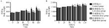
Fig. 1
Analysis of body weight changes in broilers after injection A. Body weight changes of broilers in the first stage; B. Body weight changes of broilers in the second stage. Value columns with different small letters mean significant difference (P≤0.05), while with different capital letters mean extremely significant difference (P < 0.01), and with the same or no letters mean no significant difference (P>0.05). The same as below"

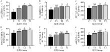
Fig. 2
The content of biochemical indicators in broilers serum A-C. The activities of DAO, ALT and AST in the serum of broilers in the first stage; D-F. The activities of DAO, ALT and AST in the serum of broilers in the second stage"

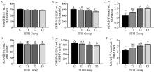
Fig. 3
The content of oxidative damage indicators in broilers serum A-C. The activities of SOD、GSH-Px and the level of MDA in the serum of broilers in the first stage; D-F. The activities of SOD、GSH-Px and the level of MDA in the serum of broilers in the second stage"


Fig. 4
The content of inflammatory indicators in broilers serum A-B. The contents of TNF-α and IL-6 in the serum of broilers in the first stage; C-D. The contents of TNF-α and IL-6 in the serum of broilers in the second stage"


Fig. 5
The first stage histological sections in broilers(100×, scale bar=200 μm)"


Fig. 6
The second stage histological sections in broilers(100×, scale bar=200 μm)"


Fig. 7
Histological scoring results A. Histological scoring of broilers in the first stage; B. Histological scoring of broilers in the second stage"


Fig. 8
Relative expression of tight junction protein mRNA in the liver and jejunum A-B. Relative expression of tight junction protein mRNA in broilers liver and jejunum in the first stage; C-D. Relative expression of tight junction protein mRNA in broilers liver and jejunum in the second stage"


Fig. 9
Relative expression of key proteins mRNA in the Nrf2/HO-1 signaling pathway in liver and jejunum A-B. Relative expression of key proteins mRNA in Nrf2/HO-1 signaling pathway in broilers liver and jejunum in the first stage; C-D. Relative expression of key proteins mRNA in Nrf2/HO-1 signaling pathway in broilers liver and jejunum in the second stage"

| 1 |
SURAI P F , KOCHISH I I , FISININ V I , et al. Antioxidant defence systems and oxidative stress in poultry biology: an update[J]. Antioxidants (Basel), 2019, 8 (7): 235.
doi: 10.3390/antiox8070235 |
| 2 |
ABO GHANIMA M M , ABD EL-HACK M E , OTHMAN S I , et al. Impact of different rearing systems on growth, carcass traits, oxidative stress biomarkers, and humoral immunity of broilers exposed to heat stress[J]. Poult Sci, 2020, 99 (6): 3070- 3078.
doi: 10.1016/j.psj.2020.03.011 |
| 3 |
WANG A Q , ZHANG K X , FU C Y , et al. Alleviation effect of conjugated linoleic acid on estradiol benzoate induced fatty liver hemorrhage syndrome in Hy-line male chickens[J]. J Anim Sci, 2023, 101, skad045.
doi: 10.1093/jas/skad045 |
| 4 |
ZHA P P , WEI L Y , LIU W H , et al. Effects of dietary supplementation with chlorogenic acid on growth performance, antioxidant capacity, and hepatic inflammation in broiler chickens subjected to diquat-induced oxidative stress[J]. Poult Sci, 2023, 102 (3): 102479.
doi: 10.1016/j.psj.2023.102479 |
| 5 |
CAO S T , WU H , WANG C C , et al. Diquat-induced oxidative stress increases intestinal permeability, impairs mitochondrial function, and triggers mitophagy in piglets[J]. J Anim Sci, 2018, 96 (5): 1795- 1805.
doi: 10.1093/jas/sky104 |
| 6 | 王曼, 李敏, 刘宏伟, 等. 博落回提取物对氧化应激蛋雏鸡生长性能和氧化功能的影响[J]. 中国畜牧杂志, 2022, 58 (9): 290- 293. |
| WANG M , LI M , LIU H W , et al. Effect of Macleaya cordata extract on growth performance and oxidative function of oxidative stress egg chicks[J]. Chinese Journal of Animal Science, 2022, 58 (9): 290- 293. | |
| 7 | 吴悦, 王怡梦, 王萌萌, 等. 致病性大肠杆菌感染小鼠肠炎模型的建立及其分子机制研究[J]. 动物营养学报, 2021, 33 (10): 5817- 5826. |
| WU Y , WANG Y M , WANG M M , et al. Establishment of enteritis model in mice infected with enterotoxigenic Escherichia coli and its molecular mechanism[J]. Chinese Journal of Animal Nutrition, 2021, 33 (10): 5817- 5826. | |
| 8 |
PANG X Y , MIAO Z Q , DONG Y Y , et al. Dietary methionine restriction alleviates oxidative stress and inflammatory responses in lipopolysaccharide-challenged broilers at early age[J]. Front Pharmacol, 2023, 14, 1120718.
doi: 10.3389/fphar.2023.1120718 |
| 9 |
FERROCINO I , BIASATO I , DABBOU S , et al. Lactiplantibacillus plantarum, Lactiplantibacillus pentosus and inulin meal inclusion boost the metagenomic function of broiler chickens[J]. Anim Microbiome, 2023, 5 (1): 36.
doi: 10.1186/s42523-023-00257-5 |
| 10 | 刘雪姣, 李海花, 王怡梦, 等. 沙门菌感染小鼠氧化应激模型的建立及其分子机制研究[J]. 中国畜牧兽医, 2021, 48 (10): 3834- 3844. |
| LIU X J , LI H H , WANG Y M , et al. Establishment of oxidative stress model in mice infected with Salmonella and its molecular mechanism[J]. China Animal Husbandry & Veterinary Medicine, 2021, 48 (10): 3834- 3844. | |
| 11 | 魏庆, 魏朝阳, 王克玮, 等. 植物精油和柠檬酸复合物对肉鸡肠道屏障功能及炎症反应的影响[J]. 中国畜牧兽医, 2021, 48 (11): 4014- 4024. |
| WEI Q , WEI Z Y , WANG K W , et al. Effects of plant essential oil-citric acid complex on intestinal barrier function and inflammatory response of broilers[J]. China Animal Husbandry & Veterinary Medicine, 2021, 48 (11): 4014- 4024. | |
| 12 |
DIERYCK I , DE BACKERE J , PAESHUYSE J . Effect of hatching system and prophylactic antibiotic use on serum levels of intestinal health biomarker diamine oxidase in broilers at an early age[J]. Animal, 2022, 16 (4): 100493.
doi: 10.1016/j.animal.2022.100493 |
| 13 |
PARK J E , OH S H , CHA Y S . Lactobacillus brevis OPK-3 from kimchi prevents obesity and modulates the expression of adipogenic and pro-inflammatory genes in adipose tissue of diet-induced obese mice[J]. Nutrients, 2020, 12 (3): 604.
doi: 10.3390/nu12030604 |
| 14 |
WANG C H , GONG B , PENG D Q , et al. Agarwood extract mitigates alcoholic fatty liver in C57 mice via anti-oxidation and anti-inflammation[J]. Mol Med Rep, 2023, 28 (5): 210.
doi: 10.3892/mmr.2023.13097 |
| 15 |
ZHAO Z L , XU X G , CHANG Y C , et al. Protective effect of mussel polysaccharide on cyclophosphamide-induced intestinal oxidative stress injury via Nrf2-Keap1 signaling pathway[J]. Food Sci Nutr, 2023, 11 (7): 4233- 4245.
doi: 10.1002/fsn3.3453 |
| 16 |
SANTOS E V , FONTES D O , BENFATO M D S , et al. Mycotoxin deactivator improves performance, antioxidant status, and reduces oxidative stress in nursery pigs fed diets containing mycotoxins[J]. J Anim Sci, 2021, 99 (10): skab277.
doi: 10.1093/jas/skab277 |
| 17 |
MONTOYA T , APARICIO-SOTO M , CASTEJÓN M L , et al. Peracetylated hydroxytyrosol, a new hydroxytyrosol derivate, attenuates LPS-induced inflammatory response in murine peritoneal macrophages via regulation of non-canonical inflammasome, Nrf2/HO1 and JAK/STAT signaling pathways[J]. J Nutr Biochem, 2018, 57, 110- 120.
doi: 10.1016/j.jnutbio.2018.03.014 |
| 18 | 黄思琪, 曲红焱, 黄大鹏, 等. L-精氨酸对冷应激仔猪生长性能、免疫功能及肝脏、肾脏中肿瘤坏死因子-α、干扰素-γ基因表达量的影响[J]. 动物营养学报, 2019, 31 (1): 131- 139. |
| HUANG S Q , QU H Y , HUANG D P , et al. Effects of L-arginine on growth performance, immune function and genes expression levels of tumor necrosis factor-α and interferon-γ in liver and kidney of cold-stressed piglets[J]. Chinese Journal of Animal Nutrition, 2019, 31 (1): 131- 139. | |
| 19 | 刘颖, 田旭, 冯晓梦, 等. 黄芪多糖对肠炎雏鸡小肠黏膜损伤的保护作用[J]. 中国畜牧兽医, 2023, 50 (1): 76- 85. |
| LIU Y , TIAN X , FENG X M , et al. Protective effect of Astragalus polysaccharide on intestinal mucosal injury in chicks with enteritis[J]. China Animal Husbandry & Veterinary Medicine, 2023, 50 (1): 76- 85. | |
| 20 |
TACKE F , WEISKIRCHEN R . Non-alcoholic fatty liver disease (NAFLD)/non-alcoholic steatohepatitis (NASH)-related liver fibrosis: mechanisms, treatment and prevention[J]. Ann Transl Med, 2021, 9 (8): 729.
doi: 10.21037/atm-20-4354 |
| 21 |
CHANG S Y , LEE J H , OH H J , et al. Effect of different ratios of phytogenic feed additives on growth performance, nutrient digestibility, intestinal barrier integrity, and immune response in weaned pigs challenged with a pathogenic Escherichia Coli[J]. J Anim Sci, 2023, 101, skad148.
doi: 10.1093/jas/skad148 |
| 22 |
XUN W J , FU Q Y , SHI L G , et al. Resveratrol protects intestinal integrity, alleviates intestinal inflammation and oxidative stress by modulating AhR/Nrf2 pathways in weaned piglets challenged with diquat[J]. Int Immunopharmacol, 2021, 99, 107989.
doi: 10.1016/j.intimp.2021.107989 |
| 23 | 杨国峰, 贺佳仪, 狄斌, 等. 复合酸化剂对伊拉兔空肠消化酶活性、免疫功能和紧密连接蛋白表达的影响[J]. 动物营养学报, 2023, 35 (3): 1948- 1956. |
| YANG G F , HE J Y , DI B , et al. Effects of compound acidifier on digestive enzyme activity, immune function and tight junction protein expression in jejunum of Ira rabbits[J]. Chinese Journal of Animal Nutrition, 2023, 35 (3): 1948- 1956. | |
| 24 |
CHU T J , YU R Y , GU Y P , et al. Kaempferol protects gut-vascular barrier from high glucose-induced disorder via NF-κB pathway[J]. J Nutr Biochem, 2024, 123, 109496.
doi: 10.1016/j.jnutbio.2023.109496 |
| 25 |
BECKER S L , LI Q Y , BURROUGH E R , et al. Effects of an F18 enterotoxigenic Escherichia coli challenge on growth performance, immunological status, and gastrointestinal structure of weaned pigs and the potential protective effect of direct-fed microbial blends[J]. J Anim Sci, 2020, 98 (5): skaa113.
doi: 10.1093/jas/skaa113 |
| 26 |
WANG L , HE C Q . Nrf2-mediated anti-inflammatory polarization of macrophages as therapeutic targets for osteoarthritis[J]. Front Immunol, 2022, 13, 967193.
doi: 10.3389/fimmu.2022.967193 |
| 27 |
BELLAVER B , SOUZA D G , SOUZA D O , et al. Hippocampal astrocyte cultures from adult and aged rats reproduce changes in glial functionality observed in the aging brain[J]. Mol Neurobiol, 2017, 54 (4): 2969- 2985.
doi: 10.1007/s12035-016-9880-8 |
| 28 |
CHEN C J , HAN X , WANG G , et al. Nrf2 deficiency aggravates the kidney injury induced by subacute cadmium exposure in mice[J]. Arch Toxicol, 2021, 95 (3): 883- 893.
doi: 10.1007/s00204-020-02964-3 |
| 29 |
GUL S , ATTAULLAH S , ALSUGOOR M H , et al. Folicitin abrogates scopolamine induced hyperlipidemia and oxidative stress mediated neuronal synapse and memory dysfunction in mice[J]. Heliyon, 2023, 9 (6): e16930.
doi: 10.1016/j.heliyon.2023.e16930 |
| 30 | LI J J , DAI W Q , MO W H , et al. Fucoidan ameliorates ferroptosis in ischemia-reperfusion-induced liver injury through Nrf2/HO-1/GPX4 activation[J]. J Clin Transl Hepatol, 2023, 11 (6): 1341- 1354. |
| 31 |
PRAKASH A N , PRASAD N , PUPPALA E R , et al. Loganic acid protects against ulcerative colitis by inhibiting TLR4/NF-κB mediated inflammation and activating the SIRT1/Nrf2 anti-oxidant responses in-vitro and in-vivo[J]. Int Immunopharmacol, 2023, 122, 110585.
doi: 10.1016/j.intimp.2023.110585 |
| 32 |
TANG X P , XIONG K N , LI M J . Effects of dietary epidermal growth factor supplementation on liver antioxidant capacity of piglets with intrauterine growth retardation[J]. J Anim Sci, 2023, 101, skad323.
doi: 10.1093/jas/skad323 |
| 33 |
EL-BAZ A M , KHODIR A E , ADEL EL-SOKKARY M M , et al. The protective effect of Lactobacillus versus 5-aminosalicylic acid in ulcerative colitis model by modulation of gut microbiota and Nrf2/HO-1 pathway[J]. Life Sci, 2020, 256, 117927.
doi: 10.1016/j.lfs.2020.117927 |
| [1] | YANG Xiaofeng, QIN Xiaowei, LÜ Lihua. Protective Effect of a Derivative of MNQ Against LPS-Induced Inflammatory Injury in Bovine Ovarian Follicular Granulosa Cells in Vitro [J]. Acta Veterinaria et Zootechnica Sinica, 2024, 55(5): 2032-2041. |
| [2] | DAI Fan, LIU Zhanyou, ZHANG Xuyang, LI Wu. Aconitate Decarboxylase 1 Could Regulate the Inflammatory Response Caused by BCG [J]. Acta Veterinaria et Zootechnica Sinica, 2024, 55(4): 1696-1706. |
| [3] | Hui ZHANG, Doukun LU, Yiqiu ZHANG, Gang ZHAO, Yingyu CHEN, Xi CHEN, Changmin HU, Aizhen GUO. Establishment of Cattle Precision-Cut Lung Tissue Slices (PCLS) Models Infected with Mycoplasma bovis in vitro [J]. Acta Veterinaria et Zootechnica Sinica, 2024, 55(10): 4638-4645. |
| [4] | ZHAO Yangfei, YU Yanghuan, WANG Jinming, ZHANG Jianhai, SUN Zilong, NIU Ruiyan, WANG Jundong. Effects of IL-17A Knockout on Fluoride-Induced Hepatic Inflammation and Hepatocyte Apoptosis [J]. Acta Veterinaria et Zootechnica Sinica, 2023, 54(7): 3108-3117. |
| [5] | XUE Linli, SUN Rui, HAO Xiaojing, CAO Xiaorui, WANG Haidong, LU Jiayin. The Promoting Effect Analysis of Danshensu on Skeletal Muscle Repair and Regeneration after Skeletal Muscle Injury in Mice based on a Mouse Skeletal Muscle Injury Model [J]. Acta Veterinaria et Zootechnica Sinica, 2023, 54(12): 5252-5263. |
| [6] | TANG Ziwen, CHENG Huaqin, LIU Dongju, PHAGMO Droma, YANG Xue, LI Jian, YIN Shi. The Protective Effect of Proanthocyanidins on Oxidative Damage of Yak Granulosa Cells Induced by Zearalenone [J]. Acta Veterinaria et Zootechnica Sinica, 2022, 53(9): 3006-3017. |
| [7] | WEI Jiayuan, ZHU Qian, YANG Yaxing, SHEN Ming. Inhibition of Porcine Follicular Granulosa Cell Proliferation by Cobalt Chloride Induced DNA Oxidative Damage [J]. Acta Veterinaria et Zootechnica Sinica, 2022, 53(9): 2982-2992. |
| [8] | ZHANG Pengguang, YAN Enfa, WANG Liqi, MA Chenghong, ZHANG Xin, YIN Jingdong. Effects of Dietary Supplementation of L-malic Acid on Inflammatory Response and Intestinal Health Status in Weaned Piglets [J]. Acta Veterinaria et Zootechnica Sinica, 2022, 53(12): 4306-4314. |
| [9] | HUANG Xiaoyu, YANG Qiaoli, YAN Zunqiang, WANG Pengfei, SHI Hairen, GUN Shuangbao. Characterization of circRNA Expression Profiles Involved in Intestines of Clostridium Perfringens Type C-infected Diarrheal Piglet [J]. Acta Veterinaria et Zootechnica Sinica, 2022, 53(11): 4058-4070. |
| [10] | XIE Zhiming, WU Chunmei, CHEN Shaoyuan, WANG Zhao, WANG Yan. Study on Bupleurum Polysaccharide Reduces Lead-induced Liver and Kidney Injury in Mice by Inhibiting Oxidative Stress and Inflammation [J]. Acta Veterinaria et Zootechnica Sinica, 2021, 52(9): 2660-2672. |
| [11] | HUANG Chengyu, XU Dequan, FENG Zhe, ZHOU Ling, LIU Min. The Addition of Melatonin Reduces the Oxidative Damage of Pig Spermatogonial Stem Cells [J]. Acta Veterinaria et Zootechnica Sinica, 2021, 52(7): 1880-1890. |
| [12] | JIA Peilu, ZHANG Hao, CHEN Ya'nan, JI Shuli, WANG Tian. Effect of Piceatannol on the Antioxidant Capacity, Mucosal Morphology and Barrier Function of the Jejunum of Weaned Piglets under Oxidative Stress [J]. Acta Veterinaria et Zootechnica Sinica, 2021, 52(6): 1616-1624. |
| [13] | LIU Yankun, LIN Yan, ZHU Weiyun. Advances in the Bacteriophage Translocation and the Effect of Bacteriophage on the Immunity [J]. Acta Veterinaria et Zootechnica Sinica, 2021, 52(3): 588-595. |
| [14] | XIANG Yi, ZHANG Hua, WANG Li, WEI Yong, EMU Quzhe. Effects of Aspergillus terreus on Oxidative Damage and Ferroptosis Related Indicators in Mice Liver [J]. Acta Veterinaria et Zootechnica Sinica, 2021, 52(12): 3619-3626. |
| [15] | PEI Ruonan, YANG Fan, LIAO Jianzhao, MA Feiyang, MA Xinyan, LIN Yuyin, YAO Qifa, TANG Zhaoxin, LIANG Zhaoping. The Effect of High Dietary Copper on Oxidative Damage and Expression of Nrf2 Signaling Pathway Related Genes in the Kidneys of Broilers [J]. ACTA VETERINARIA ET ZOOTECHNICA SINICA, 2019, 50(9): 1912-1919. |
| Viewed | ||||||
|
Full text |
|
|||||
|
Abstract |
|
|||||