


Acta Veterinaria et Zootechnica Sinica ›› 2025, Vol. 56 ›› Issue (8): 3837-3848.doi: 10.11843/j.issn.0366-6964.2025.08.024
• Animal Biotechnology and Reproduction • Previous Articles Next Articles
DONG Jianing( ), HU Yingfan, DOU Yufei, LI Jun, SHI Lei*(), REN Youshe*()
Received:2025-01-14
Online:2025-08-23
Published:2025-08-28
Contact:
SHI Lei, REN Youshe
E-mail:2250889010@qq.com;shilei@sxau.edu.cn;rys925@126.com
CLC Number:
DONG Jianing, HU Yingfan, DOU Yufei, LI Jun, SHI Lei, REN Youshe. Effects of Excessive Selenium on Autophagy of Leydig Cells in Sheep through PI3K/AKT/FoxO1 Pathway[J]. Acta Veterinaria et Zootechnica Sinica, 2025, 56(8): 3837-3848.
Table 1
The sequence of small interfering RNA"
| 小干扰RNA siRNA | 正义序列(5′→3′)Sense | 反义序列(5′→3′)Antisense |
| si-NC | UUCUCCGAACGUGUCACGUTT | ACGUGACACGUUCGGAGAATT |
| si-FoxO1-1 | GCUGUCAAUGCCGACUUCATT | UGAAGUCGGCAUUGACAGCTT |
| si-FoxO1-2 | GCAAUGACGACUUUGACAATT | UUGUCAAAGUCGUCAUUGCTT |
| si-FoxO1-3 | CGGUGAAGACAGCUUUACATT | UGUAAAGCUGUCUUCACCGTT |
Table 2
Primers information of qRT-PCR"
| 基因Gene | 序列号GenBank No. | 引物序列(5′→3′)Primer sequence | 产物长度/bp Product length |
| ATG5 | XM_042253359.1 | F: GCCTTTCATCCAGAAGCTGT R: TCATCACCTGGCTCCTCTTC | 174 |
| LC3-Ⅱ | XM_004014953.4 | F: ATGCCGTCCGAGAAAACCTT R: GCTGCTCTCGGATAAGTCGG | 79 |
| P62 | XM_015104932.3 | F: CGCAGAACAGAGTTACGAAGG R: TCCCATTCCAGTCATCTTGTC | 183 |
| PI3K | XM_042233720.2 | F: GGCGGCTGAGTATCGTGAGATTG R: CGGACACCTTTCTGAGTCAACCAC | 114 |
| AKT | XM_027960258.2 | F: CAGGAGGAGGAGACGATGGACTTC R: CCCAGCAGCTTCAGGTACTCAAAC | 134 |
| FoxO1 | XM_027973596.2 | F: TGTCCTACGCCGACCTCATCAC R: GCACGCTCTTGACCATCCACTC | 96 |
| β-actin | NM_001009784.3 | F: AAGACCTCTACGCCAACACG R: GCCAGGGCAGTGATCTCTTT | 91 |
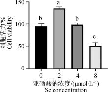
Fig. 1
LCs viability of rams detected by CCK-8 Different letters on the bars indicate significant difference (P < 0.05), the same letter indicates no significant difference (P>0.05), the same as below"
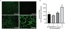
Fig. 2
ROS levels in LCs after treated with different concentrations of Se A. Cell image under the fluorescence microscope after DCFH-DA staining; B. The analysis of the fluorescence intensity of DCFH-DA"
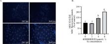
Fig. 3
Autophagy levels in LCs after treated with different concentrations of Se A. LCs image under the fluorescence microscope after MDC staining; B. The analysis of the fluorescence intensity of MDC"
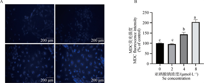
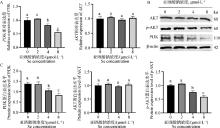
Fig. 4
Excessive Se inhibit the PI3K/AKT signaling pathway in LCs A. qRT-PCR was used to detect mRNA levels of genes; B. Western blot results of proteins; C. Gray value analysis"
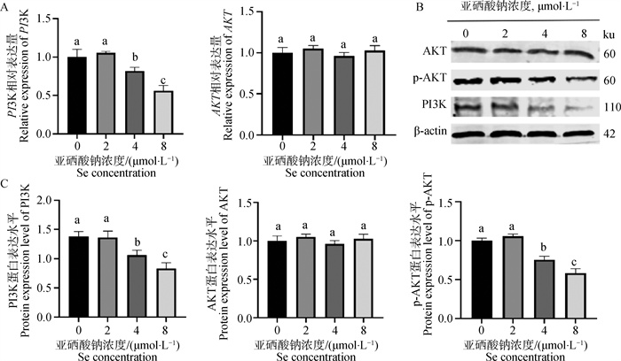

Fig. 5
Effects of AKT activator SC79 on excessive Se-induced autophagy gene expression in LCs A. The viability of cells was assessed with CCK-8 at various concentrations of the SC79 pre-treatment; B. The effect of SC79 (15 mg·L-1) treatment on the expression of p-AKT in cells was detected by Western blot; C. qRT-PCR was used to detect mRNA levels of autophagy genes; D. Gray value analysis; E. Western blot results of proteins"
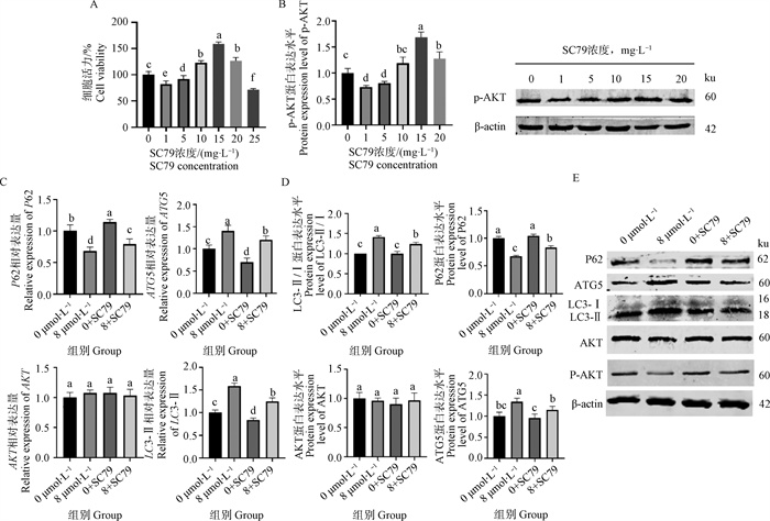

Fig. 6
Regulation of FoxO1 levels, activity, and translocation by Se and AKT activator SC79 in LCs A.B. Cells were treated with Se for 18 h. C.D. Pretreatment with SC79 for 1 h; protein levels of FoxO1 and FoxO1 phosphorylation were analyzed by Western blot and mRNA levels of FoxO1 were tested by qRT-PCR. E, F. Western blot results of proteins"
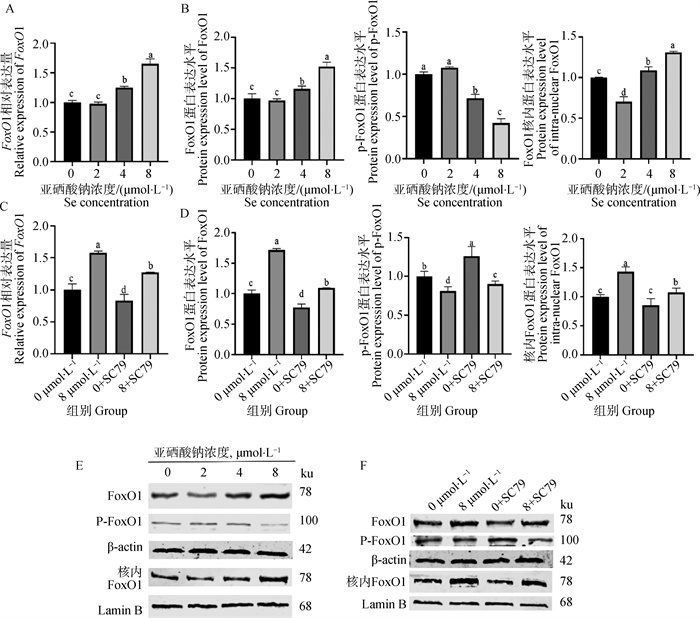
Fig. 7
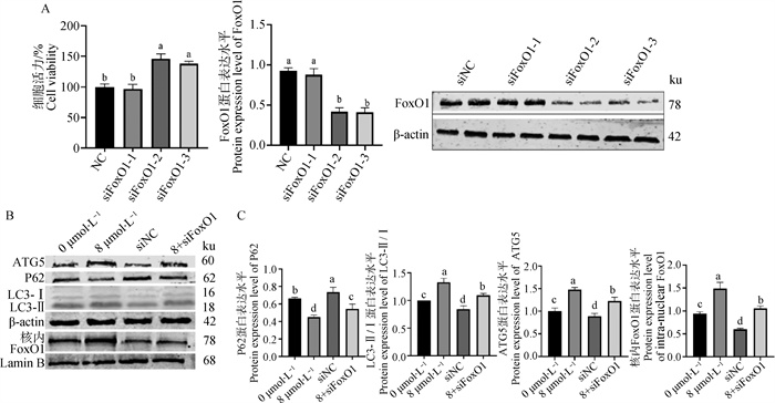
Effect of siFoxO1 on autophagy proteins expression A. Screening and validation of siRNA of FoxO1, LCs were transfected with NC or siFoxO1 for 6 h before being treated with excessive Se for 18 h; B. Western blot results of proteins; C. Gray value analysis"
| 1 |
CALIKA,EMAMIN K,WHITEM B,et al.Influence of dietary vitamin E and selenium supplementation on broilers subjected to heat stress, part Ⅰ: growth performance, body composition and intestinal nutrient transporters[J].Poult Sci,2022,101(6):101857.
doi: 10.1016/j.psj.2022.101857 |
| 2 |
DALIAA M,LOHT C,SAZILIA Q,et al.The effect of dietary bacterial organic selenium on growth performance, antioxidant capacity, and Selenoproteins gene expression in broiler chickens[J].Bmc Vet Res,2017,13(1):254.
doi: 10.1186/s12917-017-1159-4 |
| 3 |
黄靓,胡聪,孙久鹏,等.生物活性硒对不同品种育肥猪生长性能、组织硒含量、抗氧化能力和肉品质的影响[J].动物营养学报,2023,35(10):6301-6317.
doi: 10.12418/CJAN2023.579 |
|
HUANGL,HUC,SUNJ P,et al.Effects of bioactive Selenium on growth performance, tissue Selenium content, antioxidant ability and meat quality of different breeds of finishing pigs[J].Chinese Journal of Animal Nutrition,2023,35(10):6301-6317.
doi: 10.12418/CJAN2023.579 |
|
| 4 | LIC,LIUX,LIJ,et al.Selenomethionine inhibited HADV-induced apoptosis mediated by ROS through the JAK-STAT3 signaling pathway[J].Nutrients,2024,16(12):2969. |
| 5 |
LIUS,YAOS,YANGH,et al.Autophagy: regulator of cell death[J].Cell Death Dis,2023,14(10):648.
doi: 10.1038/s41419-023-06154-8 |
| 6 |
YAMAMOTOH,MATSUIT.Molecular mechanisms of macroautophagy, microautophagy, and chaperone-mediated autophagy[J].Nippon Med Sch,2024,91(1):2-9.
doi: 10.1272/jnms.JNMS.2024_91-102 |
| 7 | ZHANGC,ZHANGH L,LIUS L,et al.P62/SQSTM1 mediates the autophagy-lysosome degradation of CDK2 protein undergoing PI3Kα/AKT T308 inhibition[J].Biochem Bioph Res Co,2022,62(7):11-15. |
| 8 |
HOCHFELDW E,LEES,RUBINSZTEIND C.Therapeutic induction of autophagy to modulate neurodegenerative disease progression[J].Acta Pharmacol Sin,2013,34(5):600-604.
doi: 10.1038/aps.2012.189 |
| 9 |
GLAVIANOA,FOOA S C,LAMH Y,et al.PI3K/AKT/mTOR signaling transduction pathway and targeted therapies in cancer[J].Mol Cancer,2023,22(1):138.
doi: 10.1186/s12943-023-01827-6 |
| 10 | FANGY,OUS,WUT,et al.Lycopene alleviates oxidative stress via the PI3K/Akt/Nrf2pathway in a cell model of Alzheimer's disease[J].Peer J,2020,8(6):9308. |
| 11 |
DE FELICIM,KLINGERF G.PI3K/PTEN/AKT signaling pathways in germ cell development and their involvement in germ cell tumors and ovarian dysfunctions[J].Int J Mol Sci,2021,22(18):9838.
doi: 10.3390/ijms22189838 |
| 12 |
SANZ-CASTILLOB,HURTADOB,VARA-CIRUELOSD,et al.The MASTL/PP2A cell cycle kinase-phosphatase module restrains PI3K-Akt activity in an mTORC1-dependent manner[J].Embo J,2023,42(2):110833.
doi: 10.15252/embj.2022110833 |
| 13 |
ZHUY,WUF,HUJ,et al.LDHA deficiency inhibits trophoblast proliferation via the PI3K/AKT/FoxO1/CyclinD1 signaling pathway in unexplained recurrent spontaneous abortion[J].FASEB J,2023,37(2):22744.
doi: 10.1096/fj.202201219RR |
| 14 |
LUOM,SUZ,GAOH,et al.Cirsiliol induces autophagy and mitochondrial apoptosis through the AKT/FoxO1 axis and influences methotrexate resistance in osteosarcoma[J].Transl Med,2023,21(1):907.
doi: 10.1186/s12967-023-04682-7 |
| 15 |
ZIRKINB R,PAPADOPOULOSV.Leydig cells: formation, function, and regulation[J].Biol Reprod,2018,99(1):101-111.
doi: 10.1093/biolre/ioy059 |
| 16 | YUANF,BAIK,HOUY,et al.Small Molecule cocktails promote fibroblast-to-Leydig-like cell conversion for hypogonadism therapy[J].Pharmaceutics,2023,15(10):56-72. |
| 17 | 王晓蕾. MiR-200a/Nrf2信号通路在硒调控绵羊睾丸间质细胞增殖和凋亡的作用机制[D]. 晋中: 山西农业大学, 2022. |
| WANG X L. Mechanism of MiR-200a/Nrf2 pathway in selenium regulating the proliferation and apoptosis of Leydig cells in sheep [D]. Jinzhong: Shanxi Agricultural University, 2022. (in Chinese) | |
| 18 | 双燕, 李航, 杨振鸿. 城巴断裂带高硒背景区土壤元素地球化学特征[C]. 杭州: 中国矿物岩石地球化学学会第17届学术年会论文摘要集, 2019: 918-919. |
| SHUANG Y, LI H, YANG Z H. Geochemical characteristics of soil elements in the high selenium background area of Chengba fault zone [C]. Hangzhou: Abstract Collection of the 17th Annual Conference of Mineralogical, Petrogeochemical Society of China, 2019: 918-919. (in Chinese) | |
| 19 |
ZHUK,YANGS,LIT,et al.Advances in the study of the mechanism by which Selenium and Selenoproteins boost immunity to prevent food allergies[J].Nutrients,2022,14(15):3133.
doi: 10.3390/nu14153133 |
| 20 | 董小雨,苑景达,付绍印,等.微量元素硒对羊生长发育及繁殖性能的调控作用[J].中国畜牧杂志,2025,61(4):55-60. |
| DONGX Y,YUANJ D,FUS Y,et al.Effects of Selenium on growth and reproductive performance of sheep[J].Chinese Journal of Animal Husbandry and Veterinary Medicine,2025,61(4):55-60. | |
| 21 |
WANGS,LIUX,LEIL,et al.Selenium deficiency induces apoptosis, mitochondrial dynamic imbalance, and inflammatory responses in calf liver[J].Biol Trace Elem Res,2022,200(11):4678-4689.
doi: 10.1007/s12011-021-03059-5 |
| 22 |
ZHANGY,LIUJ,LIX,et al.Dietary selenium excess affected spermatogenesis via DNA damage and telomere-related cell senescence and apoptosis in mice[J].Food Chem Toxicol,2023,171,113556.
doi: 10.1016/j.fct.2022.113556 |
| 23 | 李万栋,李光梅,景建武,等.微量元素硒在反刍动物生产中的应用研究进展[J].中国畜牧杂志,2022,58(11):44-51. |
| LIW D,LIG M,JINGJ W,et al.Effects of Selenium on growth and reproductive performance of sheep[J].Chinese Journal of Animal Husbandry and Veterinary Medicine,2022,58(11):44-51. | |
| 24 | SHIL,SONGR,YAOX,et al.Effects of selenium on the proliferation, apoptosis and testosterone production of sheep Leydig cells in vitro[J].Theriogenology,2017,9(3):24-32. |
| 25 | XUZ J,LIUM,NIUQ J,et al.Both selenium deficiency and excess impair male reproductive system via inducing oxidative stress-activated PI3K/AKT-mediated apoptosis and cell proliferation signaling in testis of mice[J].Free Radical Bio Med,2023,19(7):15-22. |
| 26 |
UDDINM H,RITUJ R,PUTNALAS K,et al.Selenium toxicity in fishes: A current perspective[J].Chemosphere,2024,364,143214.
doi: 10.1016/j.chemosphere.2024.143214 |
| 27 | WANGM,WANGY,WANGS,et al.Selenium alleviates cadmium-induced oxidative stress, endoplasmic reticulum stress and programmed necrosis in chicken testes[J].Sci Total Environ,2023,86(3):160601. |
| 28 |
RENY,WANGR,WENGS,et al.Multifaceted role of redox pattern in the tumor immune microenvironment regarding autophagy and apoptosis[J].Mol Cancer,2023,22(1):130-136.
doi: 10.1186/s12943-023-01831-w |
| 29 |
WANGY,LVJ,LIUG,et al.ZnO NPs impair the viability and function of porcine granulosa cells through autophagy regulated by ROS production[J].Antioxidants-Basel,2024,13(11):1295.
doi: 10.3390/antiox13111295 |
| 30 | LUOD,HEF,LIUJ,et al.Pseudolaric acid B suppresses NSCLC progression through the ROS/AMPK/mTOR/autophagy signalling pathway[J].Biomed Pharmacother,2024,17(5):116614. |
| 31 |
MEIL,CHENX,WEIF,et al.Tethering ATG16L1 or LC3 induces targeted autophagic degradation of protein aggregates and mitochondria[J].Autophagy,2023,19(11):2997-3013.
doi: 10.1080/15548627.2023.2234797 |
| 32 |
ZHONGZ,UMEMURAA,SANCHEZ-LOPEZE,et al.NF-κB Restricts Inflammasome activation via elimination of damaged mitochondria[J].Cell,2016,164(5):896-910.
doi: 10.1016/j.cell.2015.12.057 |
| 33 |
ZHUH,ZHONGY,CHENR,et al.ATG5 knockdown attenuates ischemia-reperfusion injury by reducing excessive autophagy-induced ferroptosis[J].Transl Stroke Res,2024,15(1):153-164.
doi: 10.1007/s12975-022-01118-0 |
| 34 |
LIY,CHENH,LIAOJ,et al.Long-term copper exposure promotes apoptosis and autophagy by inducing oxidative stress in pig testis[J].Environ Sci Pollut Res Int,2021,28(39):55140-55153.
doi: 10.1007/s11356-021-14853-y |
| 35 |
YUDUSHKINI.Getting the Akt together: guiding intracellular Akt ativity by PI3K[J].Biomolecules,2019,9(2):67.
doi: 10.3390/biom9020067 |
| 36 |
KMAL,BARUAHT J.The interplay of ROS and the PI3K/Akt pathway in autophagy regulation[J].Biotechnol Appl Biochem,2022,69(1):248-264.
doi: 10.1002/bab.2104 |
| 37 | SOLINASG,BECATTINNIB.PI3K and AKT at the Interface of signaling and metabolism[J].Curr Top Microbiol,2022,43(6):311-336. |
| 38 | WANGQ,ZHANGX,WANGJ,et al.Effect of high selenium on insulin signaling pathway PI3K-AKT-mTOR in L02 cells[J].Wei Sheng Yan Jiu,2024,53(1):77-87. |
| 39 |
LIS T,CHENN N,QIAOY B,et al.SC79 rescues osteoblasts from dexamethasone though activating Akt-Nrf2 signaling[J].Biochem Bioph Res Co,2016,479(1):54-60.
doi: 10.1016/j.bbrc.2016.09.027 |
| 40 |
LUOH,HUANGQ,HUANGD,et al.HABP2 encapsulated by peripheral Blood-Derived exosomes suppresses astrocyte autophagy to exacerbate neuroinflammatory injury in Mice with ischemic stroke[J].ACS Chem Neurosci,2023,14(12):2347-2361.
doi: 10.1021/acschemneuro.3c00089 |
| 41 | DUY,HUANGF,GUANL,et al.Role of PI3K/Akt/mTOR pathway-mediated macrophage auto-phagy in affecting the phenotype transformation of lung fibroblasts induced by silica dust exposure[J].Zhong Nan Da Xue Xue Bao Yi Xue Ban,2023,48(8):1152-1162. |
| 42 |
LIX,BAIC,WANGH,et al.LncRNA MEG3 regulates autophagy and pyroptosis via FoxO1 in pancreatic β-cells[J].Cell Signal,2022,92,110247.
doi: 10.1016/j.cellsig.2022.110247 |
| 43 |
ZHANGY,WANGM,TANGL,et al.FoxO1 silencing in Atp7b (-/-) neural stem cells attenuates high copper-induced apoptosis via regulation of autophagy[J].J Neurochem,2024,168(9):2762-2774.
doi: 10.1111/jnc.16136 |
| 44 |
IOANNILLIL,CICCARONEF,CIRIOLOM R.Adipose tissue and FoxO1: bridging physiology and mechanisms[J].Cells,2020,9(4):849.
doi: 10.3390/cells9040849 |
| 45 | LEEH,LEEJ.Anti-diabetic effect of hydroxybenzoic acid derivatives in free fatty acid-induced HepG2 cells via miR-1271/IRS1/PI3K/AKT/FOXO1 pathway[J].J Food Biochem,2021,45(12):13993. |
| 46 | MOEINIFARDM,HASSANZ M,FALLAHIANF,et al.Britannin induces apoptosis through AKT-FoxO1 pathway in human pancreatic cancer cells[J].Biomed Pharmacother,2017,9(4):1101-1110. |
| 47 | 陈宇. lncRNA FPMAL竞争抑制FoxO1磷酸化提高奶牛妊娠早期子宫容受性的分子机制研究[D]. 武汉: 华中农业大学, 2023. |
| CHEN Y. Molecular mechanism of lncRNA FPMAL promoting uterinereceptivity in early pregnancy of dairy cows by competitiveinhibition of FoxO1 phosphorylation [D]. Wuhan: Huazhong Agricultural University, 2023. (in Chinese) |
| [1] | LI Yu, BIE Zhiwen, CHEN Shuai, LI Bingzhi, HOU Jinxing, REN Kerun, DENG Yanzhuo, WU Qiang, HU Jianhong. Research Progress on Cryopreservation of Livestock Semen [J]. Acta Veterinaria et Zootechnica Sinica, 2025, 56(8): 3591-3600. |
| [2] | YU Shulong, MAO Nannan, WANG Yunlong, ZHANG Yiran, WANG Yuanyuan, ZHOU Rongyan, ZANG Sumin, XIE Hui. Research Progress on Pigeon Sex Identification [J]. Acta Veterinaria et Zootechnica Sinica, 2025, 56(8): 3601-3609. |
| [3] | YANG Yuxiang, WANG Pengpeng, LI Bin, XIE Liuwei, XIU Fuxiao, LIU Chengwu. Research Progress on the Application and Mechanism of Probiotics in Canine Intestinal Diseases [J]. Acta Veterinaria et Zootechnica Sinica, 2025, 56(8): 3610-3620. |
| [4] | MAO Qianqian, ZHANG Yan, ZHOU Xiangying, SHAN Cuiyan, GUO Chaoqun, LU Tinghuan, WANG Li. Application of Nanotechnology and CRISPR Gene Diagnostics in Precision Detection of Livestock Parasitic Diseases [J]. Acta Veterinaria et Zootechnica Sinica, 2025, 56(8): 3621-3630. |
| [5] | GAO Boquan, WANG Xiumin, HAN Bing, TAO Hui, WANG Zhenlong, WANG Jinquan. Research Progress on Laccase Degradation of Mycotoxins [J]. Acta Veterinaria et Zootechnica Sinica, 2025, 56(8): 3650-3657. |
| [6] | LI Xiaodie, PAN Shiqin, WANG Lu, CHENG Zhentao, OU Deyuan, SONG Xuqin, YANG Jian. Research Progress on the Anti-inflammatory Mechanism of Traditional Chinese Veterinary Medicine based on Network Pharmacology [J]. Acta Veterinaria et Zootechnica Sinica, 2025, 56(8): 3701-3721. |
| [7] | CHI Shunshun, WU Dan, WANG Nan, WANG Wanjie, NIE Yuxin, MU Yulian, LIU Zhiguo, ZHU Zhendong, LI Kui. Establishment and Application of A Detection Method for MSTN Gene-Edited Pigs Based on RPA-CRISPR/Cas12a [J]. Acta Veterinaria et Zootechnica Sinica, 2025, 56(8): 3734-3748. |
| [8] | FAN Jing, LI Wei, ZHU Yan, Wudubala , SHI Jiahui, Husile , WU Jianghong. Study on Rumen Morphological Changes and Gene Expression Differences in Hu Sheep at Different Developmental Stages [J]. Acta Veterinaria et Zootechnica Sinica, 2025, 56(8): 3773-3786. |
| [9] | REN Qianzi, ZHANG Baizhong, WANG Zhenqing, WANG Xianglin, GONG Ying, HU Renke, PU Yabin, SU Peng, LI Yefang, MA Yuehui, LI Haobang, JIANG Lin. Genetic Evolutionary Analysis of Wuxue Goat Based on Whole Genome Resequencing [J]. Acta Veterinaria et Zootechnica Sinica, 2025, 56(8): 3787-3801. |
| [10] | WAN Qiongfei, SHI Shanshan, GUO Ruonan, Lü Hang, HU Debao, GUO Yiwen, ZHANG Linlin, DING Xiangbin, GUO Hong, LI Xin. Screening and Functional Analysis of Key lncRNAs of Bovine Embryonic Muscle Development [J]. Acta Veterinaria et Zootechnica Sinica, 2025, 56(8): 3802-3812. |
| [11] | ZHANG Yang, WANG Zhongfa, LI Minjuan, HE Yunan, GUAN Weijun. Cultivation and Identification of Tenogenic Mesenchymal Stem Cells for Sports-Related Injury Therapy in vitro [J]. Acta Veterinaria et Zootechnica Sinica, 2025, 56(8): 3813-3825. |
| [12] | YUAN Yue, ZHOU Jianxu, LUO Xiaolin, GUAN Jiuqiang, AN Tianwu, ZHAO Hongwen, BAI Qin, REN Zili, ZHANG Xiangfei, ZHAO Yanling. Effect of Rumen-Protected Fat on Growth Performance, Serum Biochemistry and Slaughter Performance of Fattening Yaks [J]. Acta Veterinaria et Zootechnica Sinica, 2025, 56(8): 3849-3860. |
| [13] | BAO Xiaoping, CUI Junwei, ZHAO Yulong, GUO Cheng, CHEN Ming, BI Yanliang. Effects of Different Milking Time after Delivery on Colostrum Quality and the Impact of Different Quality Colostrum on Passive Immune Transfer in Newborn Claves [J]. Acta Veterinaria et Zootechnica Sinica, 2025, 56(8): 3861-3871. |
| [14] | WANG Linwei, WANG Jing, HAN Saibo, LI Hanchuan, WANG Panpan, GUO Gang, JIANG Linshu. Meta-Analysis of the Effects of Essential Oils on Serum Immune and Biochemical Indicators in Suckling Calves [J]. Acta Veterinaria et Zootechnica Sinica, 2025, 56(8): 3872-3892. |
| [15] | CHEN Liu, XIANG Shengrui, YUN Tao, NI Zheng, HUA Jionggang, ZHU Yinchu, ZHANG Cun, YE Weicheng. Construction and Evaluation of Immune Protection Capacity of a Recombinant Duck Enteritis Virus Deleting UL35 Gene [J]. Acta Veterinaria et Zootechnica Sinica, 2025, 56(8): 3933-3941. |
| Viewed | ||||||
|
Full text |
|
|||||
|
Abstract |
|
|||||