


Acta Veterinaria et Zootechnica Sinica ›› 2025, Vol. 56 ›› Issue (3): 1252-1263.doi: 10.11843/j.issn.0366-6964.2025.03.025
• Animal Biotechnology and Reproduction • Previous Articles Next Articles
YAN Yan1,2( ), LIU Yanchen2(), WANG Zhongfa2, LI Minjuan2, HE Yunan2, GUAN Weijun2,*(), JIANG Yunliang1,*()
Received:2024-06-26
Online:2025-03-23
Published:2025-04-02
Contact:
GUAN Weijun, JIANG Yunliang
E-mail:17853651215@163.com;401979899@qq.com;guanweijun@caas.cn;zhaojy@sdau.edu.cn
CLC Number:
YAN Yan, LIU Yanchen, WANG Zhongfa, LI Minjuan, HE Yunan, GUAN Weijun, JIANG Yunliang. Isolation, Culture and Differentiation Potential of Mesenchymal Stem Cells of Yolk Sacs from Rhode Island Red Chicken[J]. Acta Veterinaria et Zootechnica Sinica, 2025, 56(3): 1252-1263.
Table 1
Primers used for RT-PCR"
| 基因 Gene | 引物序列(5'→3') Primer sequence | 产物长度/bp Size | 退火温度/℃ Tm |
| GAPDH | F: AGGTGCTGAGTATGTTGTG R: CGCTGGGATGATGTTCTG | 358 | 56.8 |
| CD29 | F: TGAATGACACGCAGGAAGATGGAAG R: AGGCAGGGCTGTTAGGTTCTCC | 219 | 59.3 |
| CD44 | F: CCTCCTGGCACTGGCATTGATC R: CACTCCACTCTTCATGTCACCATCC | 271 | 58.9 |
| CD73 | F: GGCATCGTTGGCTACACTACACAG R: GCTTGTACCACTGGCACCTTCC | 326 | 54.2 |
| CD90 | F: ACCAAGGACAACAGGAAGCACATC R: GGTGTTCTGGATCAAGAGGCTGAAG | 258 | 57.9 |
| CD166 | F: ACCAGCAGTGCAATGGACAGTTAC R: GCAACCAGCAGAAGACCGACTAC | 294 | 58.5 |
| CD34 | F: GCCACTCGCATCCAGAGAACAC R: GTCAGCATCCCCTTCGCATATCG | 326 | 61.5 |
| CD45 | F: GTCATGGTTACTCGCTGTGAGGAG R: CGTCGGAGTTTGAGAAGGAGATGTG | 263 | 58.7 |
| LPL | F: GTGACCAAGGTAGACCAGCCATTC R: TCGCCTGACTTCACTCTGACTCTC | 248 | 54.3 |
| PPAR-γ | F: CGAATGCCACAAGCGGAGAAGG R: CACTGCCTCCACAGAGCGAAAC | 330 | 59.4 |
| ACAN | F: CCTCAGAGACCTACGATGT R: TGTGGTGCTGGTAGATGG | 258 | 57.8 |
| SOX9 | F: GGAGGCTGCTGAATGAGA R: CTGCTGATGCCGTAGGTA | 550 | 60.4 |
| OPN | F: GGTAAGGATGGTCGCAATGGTCTC R: GGGTCAGCAGGGTCTCAATTTGG | 256 | 59.1 |
| COLⅠ | F: AGCATCAGTGGAGGTAGTT R: CAGGCATTCCAGGCATTC | 355 | 58.1 |
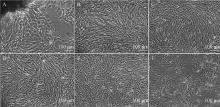
Fig. 1
Cell status of cYS-MSCs(scar bar=100 μm) A-F. Cell morphology of primary (P0), first generation (P1), third generation (P3), 8th generation (P8), 13th generation (P13), 18th generation (P18) cYS-MSCs"


Fig. 2
Fluorescent antibody test results for cell surface antigens (scar bar=50 μm)"
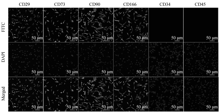
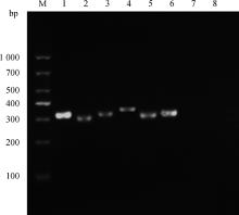
Fig. 3
PCR results of cell surface marker genes M. DL1000 DNA Marker; 1. GAPDH; 2. CD29; 3. CD44; 4. CD73; 5. CD90; 6. CD166; 7. CD34; 8. CD45"


Fig. 4
Growth curves and population doubling time of 3rd, 9th and 15th generation cYS-MSCs A. Growth curves of 3rd generation (P3), 9th generation (P9) and 15th generation (P15) cYS-MSCs; B. Population doubling time of P3, P9 and P15 cYS-MSCs. ***. P < 0.001, the same as below"


Fig. 5
Clonal group morphology and clonal formation rate of cYS-MSCs A, B, C. The clone morphology of P3, P9, P15 cYS-MSCs(scar bar=100 μm); D. Clone formation rate of P3, P9 and P15 cYS-MSCs"

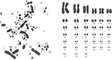
Fig. 6
Karyotype analysis of cYS-MSCs Chromosome 1-4 and chromosome 6-39 are autosome, chromosome 5 is sex chromosome, zz"


Fig. 7
Lipogenic differentiation of cYS-MSCs A. cYS-MSCs before induction(scar bar=100 μm); B. Induction group (before staining) (scar bar=25 μm); C. Induction group was stained by Oil Red O Staining Kit (scar bar=10 μm); D. LPL and PPAR-γ expression: M. DL1000 DNA Marker; 1. GAPDH; 2. LPL; 3. PPAR-γ"


Fig. 8
Chondrogenic differentiation of cYS-MSCs(scar bar=100 μm) A. cYS-MSCs before induction; B. Induction group (before staining); C. Induction group was stained by Alcian Blue; D. ACAN and SOX9 expression: M. DL1000 DNA Marker; 1. GAPDH; 2. ACAN; 3. SOX9"
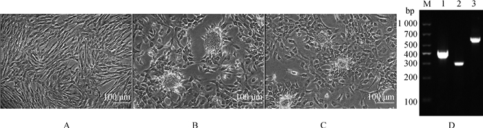

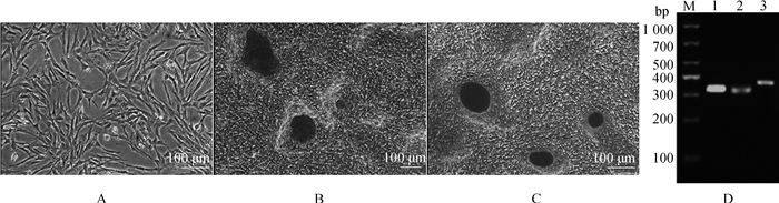
Fig. 9
Osteogenic differentiation of cYS-MSCs(scar bar=100 μm) A. cYS-MSCs before induction; B. Induction group (before staining); C. Induction group was stained by Alizarin Red S; D. OPN and COLⅠ expression; M. DL1000 DNA Marker; 1. GAPDH; 2. OPN; 3. COLⅠ"
| 1 |
WONG E A , UNI Z . Centennial Review: the chicken yolk sac is a multifunctional organ[J]. Poult Sci, 2021, 100 (3): 100821.
doi: 10.1016/j.psj.2020.11.004 |
| 2 |
MIAO X M , WU T , PAN H Y , et al. Integrative analysis of the ovarian metabolome and transcriptome of the Yaoshan chicken and its improved hybrids[J]. Front Genet, 2024, 15, 1416283.
doi: 10.3389/fgene.2024.1416283 |
| 3 |
STARCK J M , STEWART J R , BLACKBURN D G . Phylogeny and evolutionary history of the amniote egg[J]. J Morphol, 2021, 282 (7): 1080- 1122.
doi: 10.1002/jmor.21380 |
| 4 | SHIBATA M , IWASAWA A , YAYOTA M . Gluconeogenesis in the Yolk sac membrane: enzyme activity, gene expression, and metabolites during layer chicken development[J]. J Poult Sci, 2023, 60 (2): 2023020. |
| 5 |
ORNOY A , MILLER R K . Yolk sac development, function and role in rodent pregnancy[J]. Birth Defects Res, 2023, 115 (14): 1243- 1254.
doi: 10.1002/bdr2.2172 |
| 6 | PANDE O , MAKARAM N , SWAMINATHAN R . Effect of extra-abdominal vein varix on the stress distribution in umbilical cord: a simulation study[J]. Annu Int Conf IEEE Eng Med Biol Soc, 2023, 2023, 1- 4. |
| 7 |
ARUTYUNYAN I , ELCHANINOV A , MAKAROV A , et al. Umbilical cord as prospective source for mesenchymal stem cell-based therapy[J]. Stem Cells Int, 2016, 2016, 6901286.
doi: 10.1155/2016/6901286 |
| 8 | FARKHAD N K , SEDAGHAT A , REIHANI H , et al. Specific clinical and immunological changes following mesenchymal stem cell transplantation in COVID-19-induced acute respiratory distress syndrome patients: a phase-Ⅰ clinical trial[J]. Iran J Allergy Asthma Immunol, 2022, 21 (6): 687- 703. |
| 9 |
LEE J H , PARK J , LEE J W . Therapeutic use of mesenchymal stem cell-derived extracellular vesicles in acute lung injury[J]. Transfusion, 2019, 59 (S1): 876- 883.
doi: 10.1111/trf.14838 |
| 10 |
SHI Y F , HU G Z , SU J J , et al. Mesenchymal stem cells: a new strategy for immunosuppression and tissue repair[J]. Cell Res, 2010, 20 (5): 510- 518.
doi: 10.1038/cr.2010.44 |
| 11 |
MARÍN-LLERA J C , GARCÍA-GARCÍA D , GARAY-PACHECO E , et al. Commitment of human mesenchymal stromal cells to skeletal lineages is independent of their morphogenetic capacity[J]. World J Stem Cells, 2023, 15 (7): 701- 712.
doi: 10.4252/wjsc.v15.i7.701 |
| 12 |
BIANCO P , CAO X , FRENETTE P S , et al. The meaning, the sense and the significance: translating the science of mesenchymal stem cells into medicine[J]. Nat Med, 2013, 19 (1): 35- 42.
doi: 10.1038/nm.3028 |
| 13 |
DEWHURST-TRIGG R , HOPKINSON J , RICHARDSON S , et al. Mesenchymal stromal cells and their secretory products reduce the inflammatory crosstalk between islets and endothelial cells[J]. Endocrine, 2024, 87, 94- 105.
doi: 10.1007/s12020-024-03975-1 |
| 14 |
FERNANDES K J L , MCKENZIE I A , MILL P , et al. A dermal niche for multipotent adult skin-derived precursor cells[J]. Nat Cell Biol, 2004, 6 (11): 1082- 1093.
doi: 10.1038/ncb1181 |
| 15 | JIANG Y H , JAHAGIRDAR B N , REINHARDT R L , et al. RETRACTED ARTICLE: pluripotency of mesenchymal stem cells derived from adult marrow[J]. Nature, 2002, 418 (6893): 41- 49. |
| 16 | MAHMOUD N N , HAMAD S , SHRAIM S . Inflammation-modulating biomedical interventions for diabetic wound healing: an overview of preclinical and clinical studies[J]. ACS Omega, 2024, 9 (45): 44860- 44875. |
| 17 | SCHWARTZ R E , REYES M , KOODIE L , et al. Multipotent adult progenitor cells from bone marrow differentiate into functional hepatocyte-like cells[J]. J Clin Invest, 2002, 109 (10): 1291- 1302. |
| 18 | NASKOU M C , COCHRAN A , DARZENTA N , et al. The characteristics and function of small extracellular vesicles derived from human bone marrow and umbilical cord mesenchymal stromal cells are influenced by cell culture conditions[J]. Stem Cells Dev, 2024, 33 (5-6): 117- 127. |
| 19 | SHAN Y L , ZHANG M Y , TAO E X , et al. Pharmacokinetic characteristics of mesenchymal stem cells in translational challenges[J]. Sig Transduct Target Ther, 2024, 9 (1): 242. |
| 20 | SCHWARTZ R E , REYES M , KOODIE L , et al. Multipotent adult progenitor cells from bone marrow differentiate into functional hepatocyte-like cells[J]. J Clin Invest, 2002, 109 (10): 1291- 1302. |
| 21 | JUNG H , JUNG Y , SEO J , et al. Roles of extracellular vesicles from mesenchymal stem cells in regeneration[J]. Mol Cells, 2024, 47 (12): 100151. |
| 22 | KLAYMOOK S , TIRAWANCHAI N , WICHITWIENGRAT S , et al. MSC secretome from amniotic fluid halts IL-1β and TNF-α inflammation via the ERK/MAPK pathway, promoting cartilage regeneration in OA in vitro[J]. J Stem Cells Regen Med, 2024, 20 (1): 3- 13. |
| 23 | KINNAIRD T , STABILE E , BURNETT M S , et al. Local delivery of marrow-derived stromal cells augments collateral perfusion through paracrine mechanisms[J]. Circulation, 2004, 109 (12): 1543- 1549. |
| 24 | SIONOV R V , AHDUT-HACOHEN R . A supportive role of mesenchymal stem cells on insulin-producing langerhans islets with a specific emphasis on the secretome[J]. Biomedicines, 2023, 11 (9): 2558. |
| 25 | ABDELHAKEEM F , MADKOUR F A . Descriptive embryological insights of the colorectum of quail embryos with concern to its functional morphology[J]. BMC Vet Res, 2024, 20 (1): 508. |
| 26 | NIU H X , SONG H N , GUAN Y H , et al. Chicken bone marrow mesenchymal stem cells improve lung and distal organ injury[J]. Sci Rep, 2021, 11 (1): 17937. |
| 27 | DOMINICI M , LE BLANC K , MUELLER I , et al. Minimal criteria for defining multipotent mesenchymal stromal cells.The International Society for Cellular Therapy position statement[J]. Cytotherapy, 2006, 8 (4): 315- 317. |
| 28 | CHEN Y , SHAO J Z , XIANG L X , et al. Mesenchymal stem cells: a promising candidate in regenerative medicine[J]. Int J Biochem Cell Biol, 2008, 40 (5): 815- 820. |
| 29 | JAISWAL N , HAYNESWORTH S E , CAPLAN A I , et al. Osteogenic differentiation of purified, culture-expanded human mesenchymal stem cells in vitro[J]. J Cell Biochem, 1997, 64 (2): 295- 312. |
| 30 |
纪洪兵, 宋哈楠, 赵诗宇, 等. 牛羊水间充质干细胞对小鼠酒精性肝病的影响[J]. 畜牧兽医学报, 2021, 52 (10): 2905- 2914.
doi: 10.11843/j.issn.0366-6964.2021.010.021 |
|
JI H B , SONG H N , ZHAO S Y , et al. The effect of bovine amniotic fluid mesenchymal stem cells on alcoholic liver disease in mice[J]. Acta Veterinaria et Zootechnica Sinica, 2021, 52 (10): 2905- 2914.
doi: 10.11843/j.issn.0366-6964.2021.010.021 |
|
| 31 | BRAMBELL F W R . The passive immunity of the young mammal[J]. Biol Rev, 1958, 33 (4): 488- 531. |
| 32 | GAO Y H , BAI C Y , WANG K F , et al. All-trans retinoic acid promotes nerve cell differentiation of yolk sac-derived mesenchymal stem cells[J]. Appl Biochem Biotechnol, 2014, 174 (2): 682- 692. |
| 33 | LV H J , WANG T , ZHAI S K , et al. Dynamic transcriptome changes during osteogenic differentiation of bone marrow-derived mesenchymal stem cells isolated from chicken[J]. Front Cell Dev Biol, 2022, 10, 940248. |
| 34 | 李双星, 朴丰源, 戚媛, 等. 罗曼鹤鸡骨髓间充质干细胞的分离培养和鉴定[J]. 山东医药, 2014, 54 (11): 1- 4. |
| LI S X , PIAO F Y , QI Y , et al. Isolation, cultivation and identification of chicken bone marrow mesenchymal stem cells[J]. Shandong Medical Journal, 2014, 54 (11): 1- 4. | |
| 35 | WANG X Y , LAN Y , HE W Y , et al. Identification of mesenchymal stem cells in aorta-gonad-mesonephros and yolk sac of human embryos[J]. Blood, 2008, 111 (4): 2436- 2443. |
| 36 | BYDLOWSKI S P , DEBES A A , MASELLI L M F , et al. Características biológicas das células-tronco mesenquimais[J]. Rev Bras Hematol Hemoter, 2009, 31 (S1): 25- 35. |
| 37 | LI C C , LIU Y C , DENG M X , et al. Comparison of the therapeutic effects of mesenchymal stem cells derived from human dental pulp (DP), adipose tissue (AD), placental amniotic membrane (PM), and umbilical cord (UC) on postmenopausal osteoporosis[J]. Front Pharmacol, 2024, 15, 1349199. |
| 38 | DEREN J J , PADYKULA H A , WILSON T H . Development of structure and function in the mammalian yolk sac: Ⅲ.The development of amino acid transport by rabbit yolk sac[J]. Dev Biol, 1966, 13 (3): 370- 384. |
| 39 | ANKRUM J A , ONG J F , KARP J M . Mesenchymal stem cells: immune evasive, not immune privileged[J]. Nat Biotechnol, 2014, 32 (3): 252- 260. |
| 40 | GAO Y H , BAI C Y , WANG K F , et al. All-trans retinoic acid promotes nerve cell differentiation of yolk sac-derived mesenchymal stem cells[J]. Appl Biochem Biotechnol, 2014, 174 (2): 682- 692. |
| [1] | WANG Zhuo, ZHAO Yuwei, TU Yan, DIAO Qiyu, CUI Kai. Research Progress on Biological Characteristics of β-defensins and Their Roles in Regulating Intestinal Barrier in Animals [J]. Acta Veterinaria et Zootechnica Sinica, 2025, 56(3): 995-1005. |
| [2] | HE Haiyang, MA Baohua, PENG Sha. Research Progress on the Role and Mechanism of Mesenchymal Stem Cell-derived Exosomes in Animal Acute Renal Injury [J]. Acta Veterinaria et Zootechnica Sinica, 2025, 56(1): 115-125. |
| [3] | Wei LIU, Jiayi MA, Haoyu GENG, Tian XIE, Sunan MIAO, Zongjie LIAO, Shizhong GENG. Isolation, Identification and Characterization of a Broad Spectrum Salmonella Phage [J]. Acta Veterinaria et Zootechnica Sinica, 2024, 55(9): 4061-4068. |
| [4] | Huanqin ZHENG, Xiaomin JIANG, Hong YUE, Baoyan WANG, Yang LIU, Xingxiao ZHANG, Jianlong ZHANG, Hongwei ZHU. Isolation, Identification and Partial Biological Characteristics Analysis of Feline Herpesvirus-1 [J]. Acta Veterinaria et Zootechnica Sinica, 2024, 55(7): 3040-3048. |
| [5] | Yingguang LÜ, Guangming JIAO, Jinfang SANG, Zhipeng KOU, Tao LIU, Yue WANG, Xiangyu LU, Chenxi PIAO, Yajun MA, Jiantao ZHANG, Hongbin WANG. The Effect of Adipose Mesenchymal Stem Cells on the Healing Process of Autologous Skin Transplantation in Bama Miniature Pigs [J]. Acta Veterinaria et Zootechnica Sinica, 2024, 55(7): 3193-3204. |
| [6] | Mingde ZHU, Yijing CHEN, Pengxiu DAI, Yihua ZHANG, Xinke ZHANG. Study on the Hnf1b, Pdx1, Ngn3 and Pax4/Nkx6.1 Reprograms Canine Adipose-derived MSCs to Differentiate into IPCs [J]. Acta Veterinaria et Zootechnica Sinica, 2024, 55(7): 3205-3212. |
| [7] | GAO Jie, LI Xiaocheng, MU Yang, ZHANG Hui, WEI Rong, LI Jie. Biological Characteristics and Immune Effect Evaluation of Outer Membrane Vesicles of Capsular Type B Pasteurella multocida [J]. Acta Veterinaria et Zootechnica Sinica, 2024, 55(5): 2168-2175. |
| [8] | SU Chuanchen, JIAO Jingya, WANG Yongshuai, LUO Pengna, CHANG Xinghai, HUANG Yanqun. Effect of Introducing 25% Rhode Island Red Chicken Blood on Related Indexes of Henan Fighting Chickens [J]. Acta Veterinaria et Zootechnica Sinica, 2024, 55(3): 1007-1018. |
| [9] | TAN Ning, LI Balun, HAN Miao, LI Chenchen, JING Yuanxiang, KOU Zheng, LI Na, PENG Sha, ZHAO Xianjun, HUA Jinlian. Evaluation of Therapeutic Effect of Mitoquionl Mesylate Pretreated Adipose Derived Mesenchymal Stem Cells on Canine Diabetes Mellitus [J]. Acta Veterinaria et Zootechnica Sinica, 2024, 55(3): 1328-1344. |
| [10] | LIU Yanchen, ZHOU Shiying, ZHANG Yang, GAO Yang, GUAN Weijun. Isolation, Culture and Biological Characteristics Study of Holstein Bovine Lung Stem Cells [J]. Acta Veterinaria et Zootechnica Sinica, 2024, 55(2): 540-551. |
| [11] | ZHOU Zhiyu, DU Jige, XIN Ruolan, ZHANG Jiawen, PAN Chenfan, YIN Chunsheng, CHEN Xiaoyun, ZHU Zhen. Isolation and Identification of Three Lumpy Skin Disease Viruses in China and Their GPCR Gene Analysis [J]. Acta Veterinaria et Zootechnica Sinica, 2024, 55(12): 5620-5630. |
| [12] | ZHANG Gaofeng, WEI Jiayang, FENG Helong, LI Li, ZENG Zhe, TIAN Guangming, NIE Renfeng, LUO Qingping, WEN Guoyuan, WEI Hongbo, SHANG Yu. Effects of Biomineralization on the Biological Characteristics and Immunogenicity of the LaSota Strain of Newcastle Disease Virus [J]. Acta Veterinaria et Zootechnica Sinica, 2024, 55(12): 5663-5671. |
| [13] | Menglei CAI, Dongxu ZHAO, Zhenggang ZHANG, Donghai LIU, Tingting JIANG, Shixuan SU, Xuemin YAN, Xiaoyang XUE, Guolin CUI. The Effect of GreA Protein on the Biological Characteristics and Pathogenicity of Salmonella Enteritidis [J]. Acta Veterinaria et Zootechnica Sinica, 2024, 55(11): 5173-5182. |
| [14] | TIAN Qihui, ZHANG Liang, LONG Yali. Study on the Effect of Astragalus on Proliferation of Bone Marrow Mesenchymal Stem Cells in Anoxic Microenvironment based on PI3K-AKT Signal Pathway [J]. Acta Veterinaria et Zootechnica Sinica, 2024, 55(1): 346-354. |
| [15] | MA Yajun, JIAO Zhihui, LIU Xiaoning, LU Xiangyu, LIU Tao, WANG Yue, PIAO Chenxi, WANG Hongbin. Effects of Adipose-derived Mesenchymal Stem Cells on Pyroptosis of Miniature Pigs with Hepatic Ischemia-Reperfusion Combined with Hepatectomy [J]. Acta Veterinaria et Zootechnica Sinica, 2024, 55(1): 355-364. |
| Viewed | ||||||
|
Full text |
|
|||||
|
Abstract |
|
|||||