


畜牧兽医学报 ›› 2025, Vol. 56 ›› Issue (11): 5635-5648.doi: 10.11843/j.issn.0366-6964.2025.11.023
李志康1( ), 周珂珂2, 陈兆国2, 米荣升2, 黄燕2, 朱琪2, 龚海燕2,*(), 刘伟1,*()
收稿日期:2024-12-19
出版日期:2025-11-23
发布日期:2025-11-27
通讯作者:
龚海燕,刘伟
E-mail:huaerL@stu.hunau.edu.cn;gonghaiyan@shvri.ac.cn;weiliupro@163.com
作者简介:李志康(2000-),男,湖南衡阳人,硕士生,主要从事预防兽医学研究,E-mail: huaerL@stu.hunau.edu.cn
基金资助:
LI Zhikang1(), ZHOU Keke2, CHEN Zhaoguo2, MI Rongsheng2, HUANG Yan2, ZHU Qi2, GONG Haiyan2,*(), LIU Wei1,*()
Received:2024-12-19
Online:2025-11-23
Published:2025-11-27
Contact:
GONG Haiyan, LIU Wei
E-mail:huaerL@stu.hunau.edu.cn;gonghaiyan@shvri.ac.cn;weiliupro@163.com
摘要:
巴贝斯虫是一类由蜱传播的寄生于人或其他动物红细胞内的人兽共患病病原。感染后可引起宿主发热、贫血及血红蛋白尿等临床症状,甚至可导致死亡,严重威胁着人类尤其是无脾、恶性肿瘤、免疫抑制患者的健康。前期的流行病调查发现巴贝斯虫在不同家养动物中的感染率存在明显差异,但是其具体致病机制尚不十分明确。本研究以田鼠巴贝斯虫为研究对象,比较其在常见不同实验动物中的感染能力、血液指数变化和免疫反应,为进一步揭示其致病机制提供依据。以鼠、兔和羊等动物为感染模型,经人工接种田鼠巴贝斯虫后,对0~36 d感染期的染虫率、血液指标及血清中特异性抗体水平进行动态监测。结果表明,小鼠感染后第4天时染虫率达到峰值(62.9%),拷贝数达6.05×10⁷ copies·μL-1,而新西兰兔和羊红细胞染虫率接近于0。血液学监测显示,感染早中期(0~20 d),小鼠血液中红细胞数(RBC)、血红蛋白浓度(HGB)、红细胞压积(HCT)等显著降低。感染早期(0~8 d),血小板数目(PLT)和血小板压积(PCT)较对照组明显降低,此后逐渐恢复正常值。ELISA检测显示,小鼠感染后抗田鼠巴贝斯虫分泌抗原1(BmSA1)抗体持续上升,到第16天达平台期并在后期维持较高水平,而新西兰兔在接种后抗体水平迅速上升,第8天达到平台期并维持较高水平,但绵羊的抗体水平较低。上述结果表明,鼠易感田鼠巴贝斯虫,且血液参数明显异常,而兔和羊不易感,这种特异性可能与寄生虫对宿主细胞识别位点的遗传多样性有关。兔和羊两种非易感动物的抗体水平的差异可能暗示两个物种在抗巴贝斯虫的机制上有本质的区别。本研究为进一步研究巴贝斯虫与宿主的互作机制及致病机理、疫苗与药物筛选奠定了良好的基础。
中图分类号:
李志康, 周珂珂, 陈兆国, 米荣升, 黄燕, 朱琪, 龚海燕, 刘伟. 田鼠巴贝斯虫对鼠、兔和羊的感染性分析[J]. 畜牧兽医学报, 2025, 56(11): 5635-5648.
LI Zhikang, ZHOU Keke, CHEN Zhaoguo, MI Rongsheng, HUANG Yan, ZHU Qi, GONG Haiyan, LIU Wei. The Dynamic Analysis on the Infectivity of Babesia microti in Rats, Rabbits and Sheep[J]. Acta Veterinaria et Zootechnica Sinica, 2025, 56(11): 5635-5648.
表 1
巢式PCR和荧光定量PCR引物序列"
| 引物Primer | 序列(5′→3′)Sequence |
| Bab-1-F | AATTACCCAATCCTGACACAGG |
| Bab-1-R | TTTCGCAGTAGTTCGTCTTTAACA |
| Bab-2-F | GACACAGGGAGGTAGTGACAAGA |
| Bab-2-R | CCCAACTGCTCCTATTAACCATTAC |
| Bm-RF | CAGGGAGGTAGTGACAAGAAATAACA |
| Bm-RR | GGTTTAGATTCCCATCATTCCAAT |
| Bm-Probe | 6FAM-TACAGGGCTTAAAGTCTMGBNFQ[ |
| BmSA1-F | ATGGTGTCATTCAAACCAAC |
| BmSA1-R | TTAGAATAGAAACATAGCGA |

图 1
接种后对三种动物血液中田鼠巴贝斯虫的检测 A~C. 小鼠(A)、兔(B)和羊(C)接种巴贝斯虫后的血涂片(姬姆萨染色,×1 000);D. 镜检反应的染虫率变化曲线;E. Q-PCR检测反应的靶基因拷贝数变化曲线;F~H. 接种后不同时期,小鼠(F)、兔(G)和绵羊(H)血液DNA样本中田鼠巴贝斯虫的巢式PCR检测(M. DNA相对分子质量标准; 1. 阳性对照;2~10. 感染后第4~36天,间隔4 d;11. 阴性对照;12. ddH2O)"
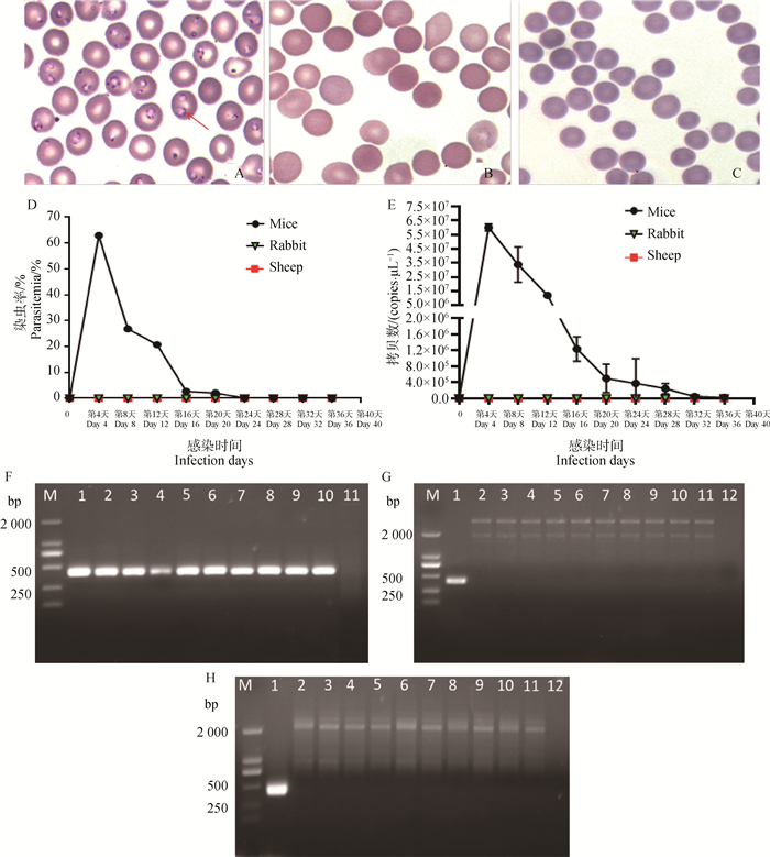

图 2
接种巴贝斯虫后,小鼠(A)、兔(B)和羊(C)体内抗BmSA1抗体水平的变化 Control. 对照组;Infection. 感染组"
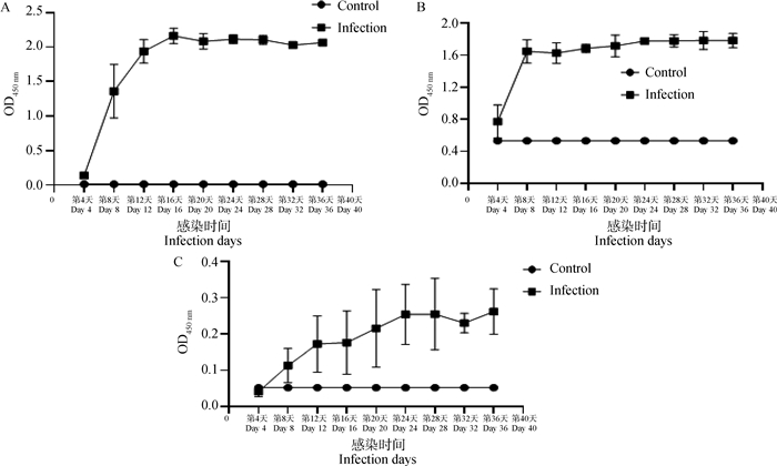
图 3
小鼠(A)、兔和羊(B)接种巴贝斯虫后的体重变化曲线"
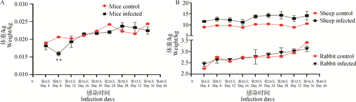
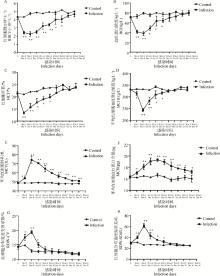
图 4
小鼠红细胞相关参数变化曲线 A. 红细胞数目(RBC);B. 血红蛋白浓度(HGB);C. 红细胞压积(HCT);D. 平均红细胞血红蛋白浓度(MCHC);E. 平均红细胞体积(MCV);F. 平均红细胞血红蛋白含量(MCH);G. 红细胞分布宽度变异系数(RDW-CV);H. 红细胞分布宽度标准差(RDW-SD)。Control. 对照组;Infection. 感染组;Student’s T-test: *. P<0.05; **. P<0.01"
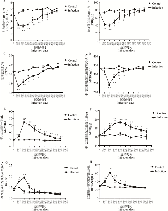
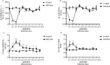
图 5
B.microti感染小鼠血小板参数曲线 A. 血小板数(PLT);B. 血小板压积(PCT%);C. 平均血小板体积(MPV);D. 血小板分布宽度(PDW)。Control. 对照组;Infection. 感染组;Student’s T-test: *. P<0.05; **. P<0.01"
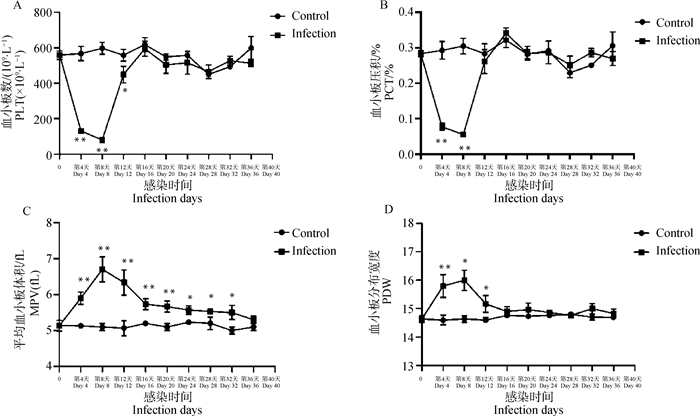
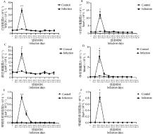
图 6
小鼠白细胞参数变化曲线 A. 白细胞数目(WBC);B. 中性粒细胞数目(Neu);C. 淋巴细胞数目(Lym);D. 单核细胞数目(Mon);E.嗜酸性粒细胞数目(Eos);F. 嗜碱性粒细胞数目(Bas)。Control. 对照组;Infection. 感染组;Student’s T-test: *. P<0.05; **. P<0.01"

| 1 |
HERSH M H , TIBBETTS M , STRAUSS M , et al. Reservoir competence of wildlife host species for Babesia microti[J]. Emerg Infect Dis, 2012, 18 (12): 1951- 1957.
doi: 10.3201/eid1812.111392 |
| 2 |
KRAUSE P J . Human babesiosis[J]. Int J Parasitol, 2019, 49 (2): 165- 174.
doi: 10.1016/j.ijpara.2018.11.007 |
| 3 |
BLOCH E M , KUMAR S , KRAUSE P J . Persistence of Babesia microti infection in humans[J]. Pathogens, 2019, 8 (3): 102.
doi: 10.3390/pathogens8030102 |
| 4 |
HILDEBRANDT A , GRAY J S , HUNFELD K P . Human babesiosis in Europe: what clinicians need to know[J]. Infection, 2013, 41 (6): 1057- 1072.
doi: 10.1007/s15010-013-0526-8 |
| 5 |
KIM J Y , CHO S H , JOO H N , et al. First case of human babesiosis in Korea: detection and characterization of a novel type of Babesia sp. (KO1) similar to ovine babesia[J]. J Clin Microbiol, 2007, 45 (6): 2084- 2087.
doi: 10.1128/JCM.01334-06 |
| 6 |
WESTBLADE L F , SIMON M S , MATHISON B A , et al. Babesia microti: from mice to ticks to an increasing number of highly susceptible humans[J]. J Clin Microbiol, 2017, 55 (10): 2903- 2912.
doi: 10.1128/JCM.00504-17 |
| 7 | 周霞, 王慧, 薛靖波, 等. 国内外巴贝虫病流行现状与研究进展[J]. 中国血吸虫病防治杂志, 2019, 31 (1): 63- 70. |
| ZHOU X , WANG H , XUE J B , et al. Epidemic and research progress of babesiosis[J]. Chinese Journal of Schistosomiasis Control, 2019, 31 (1): 63- 70. | |
| 8 |
ANTUNES S , ROSA C , COUTO J , et al. Deciphering Babesia-vector interactions[J]. Front Cell Infect Microbiol, 2017, 7, 429.
doi: 10.3389/fcimb.2017.00429 |
| 9 |
WEI C Y , WANG X M , WANG Z S , et al. High prevalence of Babesia microti in small mammals in Beijing[J]. Infect Dis Poverty, 2020, 9 (1): 155.
doi: 10.1186/s40249-020-00775-3 |
| 10 |
VANNIER E , KRAUSE P J . Human babesiosis[J]. N Engl J Med, 2012, 366 (25): 2397- 2407.
doi: 10.1056/NEJMra1202018 |
| 11 |
HILDEBRANDT A , HUNFELD K P . Human babesiosis-a rare but potentially dangerous zoonosis[J]. Dtsch Med Wochenschr, 2014, 139 (18): 957- 962.
doi: 10.1055/s-0034-1369936 |
| 12 |
CAI Y C , WU F , HU W , et al. Molecular characterization of Babesia microti seroreactive antigen 5-1-1 and development of rapid detection methods for anti-B. microti antibodies in serum[J]. Acta Trop, 2018, 185, 371- 379.
doi: 10.1016/j.actatropica.2018.03.020 |
| 13 |
ALVAREZ J A , ROJAS C , FIGUEROA J V . An Overview of Current Knowledge on in vitro Babesia cultivation for production of live attenuated vaccines for bovine babesiosis in Mexico[J]. Front Vet Sci, 2020, 7, 364.
doi: 10.3389/fvets.2020.00364 |
| 14 |
STANDFAST N F , BOCK R E , WIECEK M M , et al. Overcoming constraints to meeting increased demand for Babesia bigemina vaccine in Australia[J]. Vet Parasitol, 2003, 115 (3): 213- 222.
doi: 10.1016/S0304-4017(03)00223-1 |
| 15 |
BANETH G . Antiprotozoal treatment of canine babesiosis[J]. Vet Parasitol, 2018, 254, 58- 63.
doi: 10.1016/j.vetpar.2018.03.001 |
| 16 |
GIMENEZ A M , FRANçOSO K S , ERSCHING J , et al. A recombinant multi-antigen vaccine formulation containing Babesia bovis merozoite surface antigens MSA-2a(1), MSA-2b and MSA-2c elicits invasion-inhibitory antibodies and IFN-γ producing cells[J]. Parasit Vectors, 2016, 9 (1): 577.
doi: 10.1186/s13071-016-1862-1 |
| 17 |
BASTOS R G , ALZAN H F , RATHINASAMY V A , et al. Harnessing Mycobacterium bovis BCG trained immunity to control human and bovine babesiosis[J]. Vaccines (Basel), 2022, 10 (1): 123.
doi: 10.3390/vaccines10010123 |
| 18 |
AL-NAZAL H , LOW L M , KUMAR S , et al. A vaccine for human babesiosis: prospects and feasibility[J]. Trends Parasitol, 2022, 38 (10): 904- 918.
doi: 10.1016/j.pt.2022.07.005 |
| 19 | NGUYEN T T H , LEE J S , SHIM H . Construction of rabbit immune antibody libraries[J]. Methods Mol Biol, 2023, 2702, 93- 106. |
| 20 |
FÖLDES K , ALIGHOLIPOUR FARZANI T , ERGüNAY K , et al. Differential growth characteristics of Crimean-Congo hemorrhagic fever virus in kidney cells of human and bovine origin[J]. Viruses, 2020, 12 (6): 685.
doi: 10.3390/v12060685 |
| 21 |
BEUGNET F , MOREAU Y . Babesiosis[J]. Rev Sci Tech, 2015, 34 (2): 627- 639.
doi: 10.20506/rst.34.2.2385 |
| 22 |
KUMAR B , MAHARANA B R , THAKRE B , et al. 18S rRNA gene-based piroplasmid PCR: An assay for rapid and precise molecular screening of Theileria and Babesia species in animals[J]. Acta Parasitol, 2022, 67 (4): 1697- 1707.
doi: 10.1007/s11686-022-00625-2 |
| 23 | 陈倩, 王静, 侯咏, 等. 鼠巴贝斯虫TaqMan探针荧光定量PCR方法的建立及应用[J]. 中国国境卫生检疫杂志, 2014, 37 (4): 221- 225. |
| CHEN Q , WANG J , HOU Y , et al. Establishment and application of a TaqMan probe fluorescence quantitative PCR method for Babesia microti[J]. Chinese Journal of Frontier Health and Quarantine, 2014, 37 (4): 221- 225. | |
| 24 |
TEAL A E , HABURA A , ENNIS J , et al. A new real-time PCR assay for improved detection of the parasite Babesia microti[J]. J Clin Microbiol, 2012, 50 (3): 903- 908.
doi: 10.1128/JCM.05848-11 |
| 25 | 孙明. 牛巴贝斯虫TaqMan荧光定量PCR检测方法的建立[J]. 中国动物检疫, 2020, 37 (10): 99-103, 20. |
| SUN M . Establishment of a TaqMan fluorescent quantitative PCR method for detection of Babesia bovis[J]. China Animal Health Inspection, 2020, 37 (10): 99-103, 20. | |
| 26 |
CAI Y C , YANG C L , SONG P , et al. The protective effects of BMSA1 and BMSA5-1-1 proteins against Babesia microti infection[J]. Parasites Hosts Dis, 2024, 62 (1): 53- 63.
doi: 10.3347/PHD.23077 |
| 27 | 魏金龙, 周勇志, 龚海燕, 等. 田鼠巴贝斯虫感染对宿主凝血系统的影响[J]. 中国兽医科学, 2015, 45 (4): 390- 394. |
| WEI J L , ZHOU Y Z , GONG H Y , et al. Effects of Babesia microti infection on host coagulation system[J]. Chinese Veterinary Science, 2015, 45 (4): 390- 394. | |
| 28 | 张世杰, 米荣升, 张晓丽, 等. 感染微小隐孢子虫绵羊的血常规及血清抗体动态变化[J]. 中国动物传染病学报, 2022, 30 (4): 163- 175. |
| ZHANG S J , MI R S , ZHANG X L , et al. Dynamic changes in blood routine and serum antibodies of sheep infected with Cryptosporidium parvum[J]. Chinese Journal of Animal Infectious Diseases, 2022, 30 (4): 163- 175. | |
| 29 | 李月勤, 曹红梅, 张利卫, 等. 犬巴贝斯虫病的研究概况[J]. 现代牧业, 2019, 3 (4): 44- 46. |
| LI Y Q , CAO H M , ZHANG L W , et al. Research overview of canine babesiosis[J]. Modern Animal Husbandry, 2019, 3 (4): 44- 46. | |
| 30 |
CARIUS H J , LITTLE T J , EBERT D . Genetic variation in a host-parasite association: potential for coevolution and frequency-dependent selection[J]. Evolution, 2001, 55 (6): 1136- 1145.
doi: 10.1111/j.0014-3820.2001.tb00633.x |
| 31 |
FRANK S A . Specific and non-specific defense against parasitic attack[J]. J Theor Biol, 2000, 202 (4): 283- 304.
doi: 10.1006/jtbi.1999.1054 |
| 32 |
HAMILTON W D , AXELROD R , TANESE R . Sexual reproduction as an adaptation to resist parasites (a review)[J]. Proc Natl Acad Sci U S A, 1990, 87 (9): 3566- 3573.
doi: 10.1073/pnas.87.9.3566 |
| 33 |
JIGGINS F M , KIM K W . Contrasting evolutionary patterns in Drosophila immune receptors[J]. J Mol Evol, 2006, 63 (6): 769- 780.
doi: 10.1007/s00239-006-0005-2 |
| 34 | 王婧, 周绪正, 李冰, 等. 抗巴贝斯虫药物的研究进展[J]. 中国畜牧兽医, 2013, 40 (4): 206- 211. |
| WANG J , ZHOU X Z , LI B , et al. Research progress on anti-Babesia drugs[J]. China Animal Husbandry and Veterinary Medicine, 2013, 40 (4): 206- 211. | |
| 35 |
RIZK M A , EL-SAYED S A E , NASSIF M , et al. Assay methods for in vitro and in vivo anti-Babesia drug efficacy testing: Current progress, outlook, and challenges[J]. Vet Parasitol, 2020, 279, 109013.
doi: 10.1016/j.vetpar.2019.109013 |
| 36 | 郭宪国, 黄丽琴. 寄生虫与宿主的协同进化关系[J]. 国际医学寄生虫病杂志, 2009, 36 (1): 49- 54. |
| GUO X G , HUANG L Q . Co-evolution relationship between parasites and hosts[J]. International Journal of Medical Parasitic Diseases, 2009, 36 (1): 49- 54. | |
| 37 |
WEI N , DU Y , LU J , et al. A cysteine protease of Babesia microti and its interaction with tick cystatins[J]. Parasitol Res, 2020, 119 (9): 3013- 3022.
doi: 10.1007/s00436-020-06818-w |
| 38 | 崔筱雨, 隋树丛, 苏炳娟, 等. 血常规检测技术在猪群健康监测中的应用[J]. 黑龙江畜牧兽医, 2018 (2): 86- 88. |
| CUI X Y , SUI S C , SU B J , et al. Application of blood routine detection technology in swine herd health monitoring[J]. Heilongjiang Animal Science and Veterinary Medicine, 2018 (2): 86- 88. | |
| 39 | 王亚东, 韩旭东, 黄晓英. 血常规参数在重症疟疾中的诊断价值[J]. 南通大学学报(医学版), 2016, 36 (5): 464- 467. |
| WANG Y D , HAN X D , HUANG X Y . Diagnostic value of routine blood parameters in severe malaria[J]. Journal of Nantong University (Medical Sciences), 2016, 36 (5): 464- 467. | |
| 40 |
CHAUVIN A , MOREAU E , BONNET S , et al. Babesia and its hosts: adaptation to long-lasting interactions as a way to achieve efficient transmission[J]. Vet Res, 2009, 40 (2): 37.
doi: 10.1051/vetres/2009020 |
| 41 | 陈韶红, 蔡玉春, 陈家旭, 等. 田鼠巴贝虫(Babesia mocroti)的超微结构观察[J]. 中国人兽共患病学报, 2013, 29 (11): 1072- 1075. |
| CHEN S H , CAI Y C , CHEN J X , et al. Ultrastructural observation of Babesia microti[J]. Chinese Journal of Zoonoses, 2013, 29 (11): 1072- 1075. | |
| 42 |
IKE K , KOMATSU T , MURAKAMI T , et al. High susceptibility of Djungarian hamsters (Phodopus sungorus) to the infection with Babesia microti supported by hemodynamics[J]. J Vet Med Sci, 2005, 67 (5): 515- 520.
doi: 10.1292/jvms.67.515 |
| 43 |
NARURKAR R , MAMORSKA-DYGA A , AGARWAL A , et al. Babesiosis-associated immune thrombocytopenia[J]. Stem Cell Investig, 2017, 4, 1.
doi: 10.21037/sci.2017.01.02 |
| 44 |
GODDARD A , LEISEWITZ A L , KRISTENSEN A T , et al. Platelet indices in dogs with Babesia rossi infection[J]. Vet Clin Pathol, 2015, 44 (4): 493- 497.
doi: 10.1111/vcp.12306 |
| 45 |
GODDARD A , LEISEWITZ A L , KRISTENSEN A T , et al. Platelet activation and platelet-leukocyte interaction in dogs naturally infected with Babesia rossi[J]. Vet J, 2015, 205 (3): 387- 392.
doi: 10.1016/j.tvjl.2015.05.008 |
| 46 | AYDOGAN A , AKKUCUK S , ARICA S , et al. The analysis of mean platelet volume and platelet distribution width levels in appendicitis[J]. Indian J Surg, 2015, 77 (Suppl 2): 495- 500. |
| 47 |
SCHWARTZ D , SHARKEY L , ARMSTRONG P J , et al. Platelet volume and plateletcrit in dogs with presumed primary immune-mediated thrombocytopenia[J]. J Vet Intern Med, 2014, 28 (5): 1575- 1579.
doi: 10.1111/jvim.12405 |
| 48 |
VAN ROOYEN L J , HOOIJBERG E H , SCHOEMAN J P , et al. Thromboelastographic platelet mapping in dogs with complicated Babesia rossi infection[J]. Vet Clin Pathol, 2019, 48 (1): 11- 18.
doi: 10.1111/vcp.12689 |
| 49 |
LUO Y , JIA H , TERKAWI M A , et al. Identification and characterization of a novel secreted antigen 1 of Babesia microti and evaluation of its potential use in enzyme-linked immunosorbent assay and immunochromatographic test[J]. Parasitol Int, 2011, 60 (2): 119- 125.
doi: 10.1016/j.parint.2010.11.001 |
| 50 | LI M , AO Y , GUO J , et al. Surface antigen 1 is a crucial secreted protein that mediates Babesia microti invasion into host cells[J]. Front Microbiol, 2019, 10, 3046. |
| 51 |
LEVIN A E , WILLIAMSON P C , BLOCH E M , et al. Serologic screening of United States blood donors for Babesia microti using an investigational enzyme immunoassay[J]. Transfusion, 2016, 56 (7): 1866- 1874.
doi: 10.1111/trf.13618 |
| 52 |
WU J , CAO J , ZHOU Y , et al. Evaluation on Infectivity of Babesia microti to domestic animals and ticks outside the ixodes genus[J]. Front Microbiol, 2017, 8, 1915.
doi: 10.3389/fmicb.2017.01915 |
| 53 |
BLOCH E M , LEVIN A E , WILLIAMSON P C , et al. A prospective evaluation of chronic Babesia microti infection in seroreactive blood donors[J]. Transfusion, 2016, 56 (7): 1875- 1882.
doi: 10.1111/trf.13617 |
| 54 |
AGUILAR-DELFIN I , HOMER M J , WETTSTEIN P J , et al. Innate resistance to Babesia infection is influenced by genetic background and gender[J]. Infect Immun, 2001, 69 (12): 7955- 7958.
doi: 10.1128/IAI.69.12.7955-7958.2001 |
| 55 |
SCHLOSSER J , DäHNERT L , DREMSEK P , et al. Different outcomes of experimental hepatitis E virus infection in diverse mouse strains, wistar rats, and rabbits[J]. Viruses, 2018, 11 (1): 1.
doi: 10.3390/v11010001 |
| 56 |
WHITE N J . Determinants of relapse periodicity in Plasmodium vivax malaria[J]. Malar J, 2011, 10, 297.
doi: 10.1186/1475-2875-10-297 |
| 57 |
KRAUSE P J , GEWURZ B E , HILL D , et al. Persistent and relapsing babesiosis in immunocompromised patients[J]. Clin Infect Dis, 2008, 46 (3): 370- 376.
doi: 10.1086/525852 |
| 58 | HEMMER R M , FERRICK D A , CONRAD P A . Role of T cells and cytokines in fatal and resolving experimental babesiosis: protection in TNFRp55-/- mice infected with the human Babesia WA1 parasite[J]. J Parasitol, 2000, 86 (4): 736- 742. |
| 59 |
CLAWSON M L , PACIORKOWSKI N , RAJAN T V , et al. Cellular immunity, but not gamma interferon, is essential for resolution of Babesia microti infection in BALB/c mice[J]. Infect Immun, 2002, 70 (9): 5304- 5306.
doi: 10.1128/IAI.70.9.5304-5306.2002 |
| 60 |
RAUF U , SULEMAN M , ABID A , et al. Humoral and cell-mediated immune response validation in calves after a live attenuated vaccine of Babesia bigemina[J]. Pathogens, 2020, 9 (11): 936.
doi: 10.3390/pathogens9110936 |
| [1] | 刘峰, 李坤, 章兴赜, 马雪青, 孙普, 李凤娟, 曹轶梅, 白兴文, 付元芳, 袁红, 欧阳一凡, 刘在新, 卢曾军, 李平花. 抗口蹄疫病毒IgA抗体的构建表达及其抗病毒活性的分析[J]. 畜牧兽医学报, 2025, 56(8): 3985-3991. |
| [2] | 李帅鹏, 石正旺, 陈婕, 罗俊聪, 朱昱茜, 石鑫泰, 席韬, 张帆, 何印娣, 郑海学, 张小丽, 田宏. A 型塞内卡病毒抗体检测胶体金免疫层析试纸条的制备及初步应用[J]. 畜牧兽医学报, 2025, 56(8): 4086-4094. |
| [3] | 何印娣, 石正旺, 石鑫泰, 陈婕, 廖焕程, 张帆, 罗俊聪, 朱昱茜, 席韬, 李帅鹏, 王川, 田宏, 郑海学. 羊痘病毒抗体胶体金免疫层析试纸条的研制与初步应用[J]. 畜牧兽医学报, 2025, 56(7): 3368-3377. |
| [4] | 姜艳平, 刘薇, 宫浩阳, 蔡李萌, 李佳璇, 崔文, 周晗, 韩建春, 唐丽杰. IBDV单克隆抗体的制备及双抗体夹心ELISA检测方法的建立[J]. 畜牧兽医学报, 2025, 56(7): 3433-3441. |
| [5] | 王飞燕, 刘超凡, 张亚楠, 周晓甜, 任静, 袁晨, 李潭清, 宋勤叶. 猪IL-15单克隆抗体的制备及双抗体夹心ELISA方法的建立[J]. 畜牧兽医学报, 2025, 56(7): 3442-3452. |
| [6] | 钟梦丹, 吉艳红, 张吉豫, 朱啟超, 冯赛祥, 廖明. 几丁质酶3样蛋白1纳米抗体的筛选与鉴定[J]. 畜牧兽医学报, 2025, 56(7): 3453-3462. |
| [7] | 贾琼, 高帅鹏, 修艳宇, 任泓睿, 张书茵, 杨皓宇, 范瑞文. 基于产气荚膜梭菌β毒素纳米抗体双抗夹心ELISA方法的建立[J]. 畜牧兽医学报, 2025, 56(6): 2906-2916. |
| [8] | 张晓玲, 何兴林, 张梦迪, 李鹏飞, 孙玉梅, 马海龙, 朱红梅, 张梦佳, 李文涛. 猪瘟病毒E2蛋白纳米颗粒的制备及在家兔上的免疫原性分析[J]. 畜牧兽医学报, 2025, 56(5): 2301-2311. |
| [9] | 张宇, 程凡玉, 俞赵荣, 邵颖, 魏宁波, 陈芳芳, 王振宇, 宋祥军, 涂健, 祁克宗. 禽4型腺病毒单克隆抗体的制备、鉴定与初步应用[J]. 畜牧兽医学报, 2025, 56(5): 2364-2371. |
| [10] | 曹丽艳, 孔祥雨, 袁聪, 段月月, 马国祥, 施磊, 张宇, 万颖, 李想通, 王娅婷, 杜煜, 郑海学, 王琦. 猪急性腹泻综合征冠状病毒核衣壳蛋白新型线性B细胞表位的鉴定[J]. 畜牧兽医学报, 2025, 56(4): 1854-1864. |
| [11] | 张越, 茹毅, 郝荣增, 杨锐, 赵陇和, 李亚军, 杨洋, 张荣, 蒋成辉, 郑海学. 非洲猪瘟病毒H108R蛋白的制备及其免疫原性评价[J]. 畜牧兽医学报, 2025, 56(3): 1344-1354. |
| [12] | 马晓莉, 李段, 曾道平, 刘燕玲, 王晓敏, 彭国良, 宋长绪, 王磊, 徐铮. 非洲猪瘟病毒p72蛋白抗体全自动化学发光酶免疫检测方法的建立[J]. 畜牧兽医学报, 2025, 56(3): 1355-1365. |
| [13] | 赵龙, 林静怡, 豆薇, 徐婷萱, 顾庆云, 高海慧, 李生庆, 郭抗抗. 体外对牛冠状病毒复制具有抑制效应藏药的筛选[J]. 畜牧兽医学报, 2025, 56(2): 826-838. |
| [14] | 李璠, 孙海凤, 孙萌, 高雁怩, 孙杨杨, 张路捷, 白娟, 姜平. 猪IL-1β单克隆抗体制备及其抗炎症反应活性[J]. 畜牧兽医学报, 2025, 56(2): 890-899. |
| [15] | 孙心如, 武文清, 罗亚娟, 谢锐, 彭长江, 华琳, 吴斌, 彭忠. 胞内劳森菌MltA蛋白单克隆抗体的制备及阻断ELISA抗体检测方法的建立[J]. 畜牧兽医学报, 2025, 56(11): 5660-5669. |
| 阅读次数 | ||||||
|
全文 |
|
|||||
|
摘要 |
|
|||||