


畜牧兽医学报 ›› 2025, Vol. 56 ›› Issue (3): 1453-1464.doi: 10.11843/j.issn.0366-6964.2025.03.043
王子姣( ), 刘春晓, 李光玉*()
收稿日期:2024-04-24
出版日期:2025-03-23
发布日期:2025-04-02
通讯作者:
李光玉
E-mail:wzjdgg1022@163.com;tcslgy@126.com
作者简介:王子姣(1998-),女,山东青岛人,硕士生,主要从事宠物营养与健康研究,E-mail: wzjdgg1022@163.com
基金资助:
WANG Zijiao(), LIU Chunxiao, LI Guangyu*()
Received:2024-04-24
Online:2025-03-23
Published:2025-04-02
Contact:
LI Guangyu
E-mail:wzjdgg1022@163.com;tcslgy@126.com
摘要:
犬特应性皮炎(canine atopic dermatitis,CAD)是一种复杂的炎症性皮肤病,与皮肤微生物组、免疫和皮肤屏障改变有关。紫花前胡苷(nodakenin,NK)是一种具有抗炎、抗菌、抗氧化等作用的香豆素类糖苷。本研究旨在探究紫花前胡苷对2,4-二硝基甲苯(DNCB)诱导的犬特应性皮炎的干预效果以及皮肤菌群的影响。首先建立DNCB诱导的犬特应性皮炎,给与紫花前胡苷14 d局部涂抹治疗,通过体重指标、血常规检测、血清生化检测、血清免疫球蛋白E(immunoglobulin E,IgE)含量、皮肤病变和瘙痒程度以及皮肤菌群16S rRNA测序结果来评估紫花前胡苷治疗效果。结果显示:与DNCB组相比,经紫花前胡苷治疗后犬的体重明显上升,血常规指标中白细胞总数和中性粒细胞百分比显著提高(P<0.05),而血清生化相关指标无显著差异(P>0.05),血清中IgE含量显著下降(P<0.05),皮肤病变和瘙痒程度均显著改善(P<0.01)。皮肤菌群中的α多样性和β多样性均存在显著差异(P<0.05),在门水平上,紫花前胡苷治疗后显著恢复了厚壁菌门(Firmicutes)的相对丰度(P<0.01)。在属水平上,紫花前胡苷治疗后显著增加了罗姆布茨菌属(Romboutsia)、狭义梭菌属(Clostridium_sensu_stricto_1)和土孢杆菌属(Terrisporobacter)的相对丰度(P<0.01,P<0.05),显著降低了莫拉菌属(Moraxella)和葡萄球菌属(Staphylococcus)相对丰度(P<0.05)。涂抹紫花前胡苷能够有效减轻犬皮损程度、缓解皮肤瘙痒情况、降低血清IgE含量以及调节皮肤菌群失衡,从而改善犬特应性皮炎的症状,为CAD的药物研发提供新思路。
中图分类号:
王子姣, 刘春晓, 李光玉. 紫花前胡苷对犬特应性皮炎的疗效分析[J]. 畜牧兽医学报, 2025, 56(3): 1453-1464.
WANG Zijiao, LIU Chunxiao, LI Guangyu. Analysis of the Efficacy of Nodakenin in Canine Atopic Dermatitis[J]. Acta Veterinaria et Zootechnica Sinica, 2025, 56(3): 1453-1464.
表 1
皮肤视觉宏观病变程度评分表"
| 病变程度 Lesion level | 评分 Score |
| 无明显变化 No significant change | 0 |
| 红斑 Erythema | 1 |
| 红斑、皮肤苔藓化或丘疹 Erythema, mossy skin or pimples | 2 |
| 红斑、皮肤苔藓化和更严重反应 Erythema, mossy skin and more severe reactions | 3 |
表 2
皮肤视觉瘙痒程度评分表"
| 瘙痒程度 Itchiness | 评分 Score |
| 没有瘙痒 No itch | 0 |
| 轻度瘙痒 Mild itch | 1~2 |
| 中度瘙痒 Moderate itch | 3~4 |
| 持续且剧烈瘙痒 Constant and intense itch | 5 |
表 3
各组犬在第0、7、14、21天体重($\bar x \pm s$)"
| 时间 Time | 组别Group | ||
| Ctrl | DNCB | DNCB+NK | |
| 第0天Day 0 | 15.06±1.77 | 16.25±2.78 | 16.86±1.18 |
| 第7天Day 7 | 15.28±1.84 | 15.60±2.60 | 16.44±1.29 |
| 第14天Day 14 | 15.27±1.72 | 15.88±3.01 | 16.46±1.41 |
| 第21天Day 21 | 15.35±2.10 | 15.84±2.52 | 16.60±1.47 |
表 4
各组犬血常规检测结果($\bar x \pm s$)"
| 检测项目 Item | 组别Group | 参考范围 Reference scope | ||
| Ctrl | DNCB | DNCB+NK | ||
| 白细胞总数/(109·L-1) WBC | 16.06±1.27 | 11.89±2.32## | 14.42±1.36* | 6.0~17.0 |
| 淋巴细胞数量/(109·L-1) LYM | 2.13±0.65 | 1.65±1.05 | 1.89±0.20 | 1.0~4.8 |
| 单核细胞数量/(109·L-1) MON | 1.20±0.42 | 0.99±0.29 | 1.21±0.24 | 0.2~1.5 |
| 中性粒细胞数量/(109·L-1) NEU | 9.48±1.12 | 6.40±1.52# | 8.78±1.95 | 3~12 |
| 嗜酸性粒细胞数量/(109·L-1) EOS | 0.95±0.31 | 0.78±0.18 | 0.84±0.38 | 0~0.9 |
| 嗜碱性粒细胞数量/(109·L-1) BAS | 0.07±0.03 | 0.02±0.01 | 0.03±0.02 | 0~0.4 |
| 淋巴细胞百分比/% LYM | 18.26±3.72 | 19.03±3.55 | 17.77±4.97 | 12~30 |
| 单核细胞百分比/% MON | 8.40±1.93 | 9.96±1.88## | 9.78±1.88 | 3~14 |
| 中性粒细胞百分比/% NEU | 74.32±6.27 | 61.02±5.13# | 68.09±6.26* | 60~80 |
| 嗜酸性粒细胞百分比/% EOS | 6.37±0.49 | 6.03±2.07 | 6.56±2.59 | 0~10 |
| 嗜碱性粒细胞百分比/% BAS | 0.38±0.06 | 0.18±0.05 | 0.21±0.08 | 少有 |
| 红细胞总数/(1012·L-1) RBC | 5.83±0.74 | 4.85±1.48 | 5.13±0.63 | 5.5~8.5 |
| 血红蛋白浓度/(g·L-1) HGB | 137.25±32.33 | 130.00±38.57 | 138.75±15.76 | 120~180 |
| 红细胞压积/% HCT | 35.50±7.83 | 33.63±6.74 | 32.08±5.72 | 30~55 |
| 平均红细胞体积/fL MCV | 68.25±1.23 | 68.50±2.15 | 68.80±0.14 | 60~77 |
| 平均红细胞血红蛋白含量/pg MCH | 26.78±0.59 | 27.53±0.90 | 26.73±0.13 | 19.5~26.5 |
| 平均红细胞血红蛋白浓度/(g·L-1) MCHC | 385.25±12.28 | 386.25±5.32 | 391.50±6.45 | 310~390 |
| 红细胞体积分布宽度变异系/% RDWc | 13.48±0.13 | 14.05±0.84 | 13.60±0.50 | ———— |
| 血小板总数/(109·L-1) PLT | 284.67±109.71 | 203.75±51.60 | 240.50±62.78 | 165~500 |
表 5
各组犬血清生化指标检测结果($\bar x \pm s$)"
| 检测项目 Item | 组别Group | ||
| Ctrl | DNCB | DNCB+NK | |
| 丙氨酸氨基转氨酶/[nmol· (min·mL)-1] ALT | 3.41±0.81 | 3.32±1.25 | 3.65±1.09 |
| 天冬氨酸氨基转氨酶/[nmol· (min·mL)-1] AST | 1.58±0.76 | 1.51±1.24 | 1.45±0.77 |
| 肌酐/(μmol·L) -1 CRE | 39.36±4.79 | 36.13±4.79 | 40.88±2.89 |
| 白蛋白/(g·L) -1 ALB | 30.48±3.29 | 33.47±2.05 | 31.81±0.82 |
| 碱性磷酸酶/[μmol· (min·mL)-1] AKP | 0.55±0.06 | 0.28±0.01## | 0.33±0.09 |
| 葡萄糖/(mg·mL) -1 GLU | 61.24±6.20 | 49.41±3.99# | 51.62±5.82 |
| 血中γ-谷氨酰转移酶/(U·L) -1 γ-GT | 7.18±0.48 | 8.65±1.87 | 8.62±1.22 |
| 钙含量/(mmol·L) -1 Ca | 2.57±0.13 | 2.62±0.13 | 2.60±0.04 |

图 1
各组犬血清IgE含量 与DNCB组比较,*. P < 0.05;**. P<0.01"
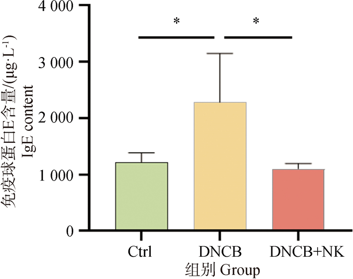

图 2
各组随机一只犬第0、4、8、12、16、23天刺激部位皮肤图片"


图 3
各组犬皮肤病变评分分析图(A)和皮肤瘙痒评分分析图(B) 与DNCB组比较:*.P < 0.05,**.P<0.001,***.P<0.001"
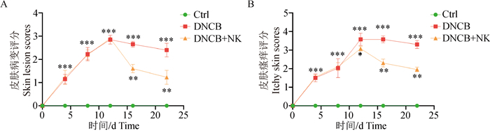

图 4
皮肤菌群OTUs韦恩图分析"


图 5
各组样本的Alpha多样性分析指数 *.P < 0.05; ns. P>0.05"


图 6
Beta多样性主坐标成分分析 *.P < 0.05; ns. P>0.05"
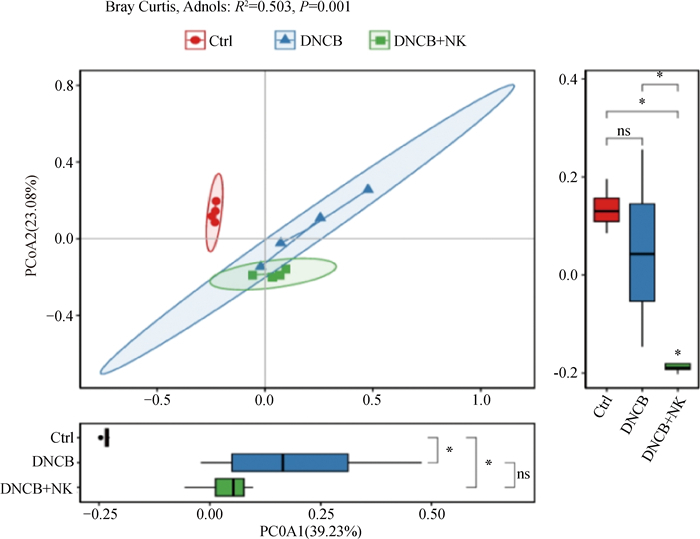

图 7
各组犬皮肤菌群在门水平上的变化 A. 物种组成分析柱状图;B.在门水平上显著差异的皮肤菌群。*.P < 0.05; **.P < 0.01"

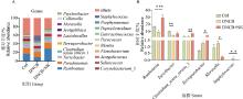
图 8
各组犬皮肤菌群在属水平上的变化 A. 物种组成分析柱状图;B.在属水平上显著差异的皮肤菌群.*.P < 0.05; **.P < 0.01"


图 9
LEfSe分析柱状图和分支图"

| 1 |
SANTORO D , SARIDOMICHELAKIS M , EISENSCHENK M , et al. Update on the skin barrier, cutaneous microbiome and host defence peptides in canine atopic dermatitis[J]. Vet Dermatol, 2024, 35 (1): 5- 14.
doi: 10.1111/vde.13215 |
| 2 | MARSELLA R , DE BENEDETTO A . Atopic dermatitis in animals and people: an update and comparative review[J]. Vet Sci, 2017, 4 (3): 37. |
| 3 | FERNANDES B , ALVES S , SCHMIDT V , et al. Primary prevention of canine atopic dermatitis: breaking the cycle-a narrative review[J]. Vet Sci, 2023, 10 (11): 659. |
| 4 |
GEDON N K Y , MUELLER R S . Atopic dermatitis in cats and dogs: a difficult disease for animals and owners[J]. Clin Transl Allergy, 2018, 8, 41.
doi: 10.1186/s13601-018-0228-5 |
| 5 |
NOLI C . Assessing quality of life for pets with dermatologic disease and their owners[J]. Vet Clin North Am Small Anim Pract, 2019, 49 (1): 83- 93.
doi: 10.1016/j.cvsm.2018.08.008 |
| 6 |
FAVROT C , STEFFAN J , SEEWALD W , et al. A prospective study on the clinical features of chronic canine atopic dermatitis and its diagnosis[J]. Vet Dermatol, 2010, 21 (1): 23- 31.
doi: 10.1111/j.1365-3164.2009.00758.x |
| 7 |
BIZIKOVA P , SANTORO D , MARSELLA R , et al. Review: clinical and histological manifestations of canine atopic dermatitis[J]. Vet Dermatol, 2015, 26 (2): 79- e24.
doi: 10.1111/vde.12196 |
| 8 |
HENSEL P , SANTORO D , FAVROT C , et al. Canine atopic dermatitis: detailed guidelines for diagnosis and allergen identification[J]. BMC Vet Res, 2015, 11, 196.
doi: 10.1186/s12917-015-0515-5 |
| 9 |
MARSELLA R , DOERR K , GONZALES A , et al. Oclacitinib 10 years later: lessons learned and directions for the future[J]. J Am Vet Med Assoc, 2023, 261 (S1): S36- S47.
doi: 10.2460/javma.22.12.0570 |
| 10 |
ROEKEVISCH E , SPULS P I , KUESTER D , et al. Efficacy and safety of systemic treatments for moderate-to-severe atopic dermatitis: a systematic review[J]. J Allergy Clin Immunol, 2014, 133 (2): 429- 438.
doi: 10.1016/j.jaci.2013.07.049 |
| 11 | YU S H , DRUCKER A M , LEBWOHL M , et al. A systematic review of the safety and efficacy of systemic corticosteroids in atopic dermatitis[J]. J Am Acad Dermatol, 2017, 78 (4): 733- 740. e11. |
| 12 |
LIM J Y , LEE J H , YUN D H , et al. Inhibitory effects of nodakenin on inflammation and cell death in lipopolysaccharide-induced liver injury mice[J]. Phytomedicine, 2021, 81, 153411.
doi: 10.1016/j.phymed.2020.153411 |
| 13 |
JIN B R , LEE M , AN H J . Nodakenin represses obesity and its complications via the inhibition of the VLDLR signalling pathway in vivo and in vitro[J]. Cell Prolif, 2021, 54 (8): e13083.
doi: 10.1111/cpr.13083 |
| 14 |
PARK S J , CHA H S , LEE Y H , et al. Effect of nodakenin on atopic dermatitis-like skin lesions[J]. Biosci Biotechnol Biochem, 2014, 78 (9): 1568- 1571.
doi: 10.1080/09168451.2014.923296 |
| 15 | LEE N Y , CHUNG K S , JIN J S , et al. The inhibitory effect of nodakenin on mast-cell-mediated allergic inflammation via downregulation of NF-κB and caspase-1 activation[J]. J Cell Biochem, 2017, 118 (11): 3993- 4001. |
| 16 | 肖晓杰, 丁明星, 齐智利, 等. 添加补骨脂提取物的低敏处方粮对犬皮肤健康的影响[J]. 养殖与饲料, 2022, 21 (9): 18- 24. |
| XIAO X J , DING M X , QI Z L , et al. Effects of hypoallergenic prescription diet containing psoralea on health and disease in dogs[J]. Animals Breeding and Feed, 2022, 21 (9): 18- 24. | |
| 17 | TAO R , LI R Y , WANG R J . Dysbiosis of skin mycobiome in atopic dermatitis[J]. Mycoses, 2022, 65 (3): 285- 293. |
| 18 | CHAI R R , TAI Z G , ZHU Y J , et al. Symbiotic microorganisms: prospects for treating atopic dermatitis[J]. Expert Opin Biol Ther, 2022, 22 (7): 911- 927. |
| 19 | KRAWIEC D R , GAAFAR S M . A comparative study of allergic and primary irritant contact dermatitis with dinitrochlorobenzene (DNCB) in dogs[J]. J Invest Dermatol, 1975, 65 (2): 248- 251. |
| 20 | PARAMASIVAN P , LAI C , PICKARD C , et al. Repeated low-dose skin exposure is an effective sensitizing stimulus, a factor to be taken into account in predicting sensitization risk[J]. Br J Dermatol, 2010, 162 (3): 594- 597. |
| 21 | LIAO Y , CHEN Z , YANG Y K , et al. Antibiotic intervention exacerbated oxidative stress and inflammatory responses in SD rats under hypobaric hypoxia exposure[J]. Free Radic Biol Med, 2023, 209, 70- 83. |
| 22 | 张伟, 胡莉萍, 孙艳, 等. 动物应激的评价指标的探讨[J]. 畜牧兽医科技信息, 2018 (3): 5. |
| ZHANG W , HU L P , SUN Y , et al. Exploration of evaluation indexes of animal stress[J]. Chinese Journal of Animal Husbandry and Veterinary Medicine, 2018 (3): 5. | |
| 23 | CZARNOWICKI T , HE H , KRUEGER J G , et al. Atopic dermatitis endotypes and implications for targeted therapeutics[J]. J Allergy Clin Immunol, 2019, 143 (1): 1- 11. |
| 24 | HUANG K K , LI F , LIU Y Y , et al. Multi-omics analyses reveal interactions between the skin microbiota and skin metabolites in atopic dermatitis[J]. Front Microbiol, 2024, 15, 1349674. |
| 25 | SAVVA M , PAPADOPOULOS N G , GREGORIOU S , et al. Recent advancements in the atopic dermatitis mechanism[J]. Front Biosci (Landmark Ed), 2024, 29 (2): 84. |
| 26 | CHERMPRAPAI S , EDERVEEN T H A , BROERE F , et al. The bacterial and fungal microbiome of the skin of healthy dogs and dogs with atopic dermatitis and the impact of topical antimicrobial therapy, an exploratory study[J]. Vet Microbiol, 2019, 229, 90- 99. |
| 27 | BRADLEY C W , MORRIS D O , RANKIN S C , et al. Longitudinal evaluation of the skin microbiome and association with microenvironment and treatment in canine atopic dermatitis[J]. J Invest Dermatol, 2016, 136 (6): 1182- 1190. |
| 28 | RODRIGUES H A , PATTERSON A P , DIESEL A , et al. The skin microbiome in healthy and allergic dogs[J]. PLoS One, 2014, 9 (1): e83197. |
| 29 | KIM J , KIM B E , GOLEVA E , et al. Alterations of epidermal lipid profiles and skin microbiome in children with atopic dermatitis[J]. Allergy Asthma Immunol Res, 2023, 15 (2): 186- 200. |
| 30 | THOMSEN M , KUNSTNER A , WOHLERS I , et al. A comprehensive analysis of gut and skin microbiota in canine atopic dermatitis in Shiba Inu dogs[J]. Microbiome, 2023, 11 (1): 232. |
| 31 | WANG Y W , HOU J P , TSUI J C , et al. Unique gut microbiome signatures among adult patients with moderate to severe atopic dermatitis in southern Chinese[J]. Int J Mol Sci, 2023, 24 (16): 12856. |
| 32 | SUNDE R B , THORSEN J , KIM M , et al. Bacterial colonisation of the airway in neonates and risk of asthma and allergy until age 18 years[J]. Eur Respir J, 2024, 63 (1): 2300471. |
| 33 | NAKATSUJI T , CHEN T H , NARALA S , et al. Antimicrobials from human skin commensal bacteria protect against Staphylococcus aureus and are deficient in atopic dermatitis[J]. Sci Transl Med, 2017, 9 (378): eaah4680. |
| 34 | IWAMOTO K , MORIWAKI M , MIYAKE R , et al. Staphylococcus aureus in atopic dermatitis: Strain-specific cell wall proteins and skin immunity[J]. Allergol Int, 2019, 68 (3): 309- 315. |
| 35 | HERZ U , SCHNOY N , BORELLI S , et al. A human-SCID mouse model for allergic immune response bacterial superantigen enhances skin inflammation and suppresses IgE production[J]. J Invest Dermatol, 1998, 110 (3): 224- 231. |
| [1] | 靳茹文, 王英杰, 蒋全兴, 刘天强, 邓阳, 罗杰, 赵玲, 叶刚, 施飞, 李英伦, 唐华侨. 加味枳术散对断奶仔兔肠黏膜屏障和抗氧化能力的影响[J]. 畜牧兽医学报, 2024, 55(12): 5825-5838. |
| [2] | 黄麒霖, 刘晨, 景晓涵, 黄丽娜, 潘香逸, 石雄伟, 仇正英, 辛蕊华. 柚皮苷可通过多种途径修复猪胸膜肺炎放线杆菌诱导的小鼠肺上皮屏障损伤[J]. 畜牧兽医学报, 2024, 55(11): 5299-5309. |
| [3] | 冯琦, 刘义钢, 何琴, 李泽龙, 马英才, 易鹏飞, 李娜, 孙亚伟, 陈如龙, 姚刚, 马雪连. 大青叶水提物对牛传染性鼻气管炎病毒的体外增殖抑制活性评价[J]. 畜牧兽医学报, 2024, 55(11): 5287-5298. |
| [4] | 苑庆欣, 刘阔, 包旭华, 高东阳, 李鹤, 宋军, 周志新. 白屈菜红碱抗耐甲氧西林金黄色葡萄球菌作用机制研究[J]. 畜牧兽医学报, 2024, 55(10): 4670-4678. |
| [5] | 文安林, 杨芸芸, 罗永荣, 杨颖, 程振涛, 欧德渊, 文明. 黄连防治鸭病毒性肠炎机制的网络药理学分析及动物试验验证[J]. 畜牧兽医学报, 2024, 55(7): 3225-3233. |
| [6] | 李美艳, 邢晓莹, 李娜, 张椰莉, 乔宏萍, 武晓英. 甘草多糖对LPS诱导小鼠睾丸炎的调节作用[J]. 畜牧兽医学报, 2024, 55(6): 2741-2750. |
| [7] | 陈富斌, 徐国伟, 王磊, 刘琴, 冯海鹏, 张康, 郭志廷, 韩松伟, 刘佳惠, 古雪艳, 张景艳, 李建喜, Huub F. J. Savelkoul. 黄芪多糖对HD11鸡巨噬细胞转录组和代谢组的影响[J]. 畜牧兽医学报, 2024, 55(3): 1290-1301. |
| [8] | 魏苗伊, 吴世海, 杨富琳, 余宸昀, 孙志刚, 刘馨媛, 徐媛媛, 梁冰冰, 李复煌, 孙鸿, 刘晓晔, 董虹. 中药龙胆草抗鸽毛滴虫的临床药效分析[J]. 畜牧兽医学报, 2024, 55(2): 785-796. |
| [9] | 肖乐, 刘峻源, 曾雯玉, 汪芹, 韩雯珏, 刘彦泠, 范誉, 徐雨婷, 杨贝妮, 肖雄, 王自力. 基于微生物组和宿主转录组整合分析香砂六君子汤对ETEC诱导断奶腹泻仔猪回肠损伤的调控机制[J]. 畜牧兽医学报, 2024, 55(2): 797-808. |
| [10] | 薛霖莉, 孙睿, 郝晓静, 曹校瑞, 王海东, 卢嘉茵. 基于小鼠骨骼肌损伤模型分析丹参素对骨骼肌损伤后修复再生的促进作用[J]. 畜牧兽医学报, 2023, 54(12): 5252-5263. |
| [11] | 池幸子, 李耀星, 王慧婷, 严铭恩, 杨诗靖, 杨泊文, 孙晗, 郭世宁, 石达友, 武力, 刘翠. 紫锥菊提取物联合柳氮磺吡啶对湿热泄泻大鼠Th17/Treg免疫失衡的影响[J]. 畜牧兽医学报, 2023, 54(11): 4817-4826. |
| [12] | 张旭梅, 魏玉荣, 许丞惠, 杨彤, 史慧君, 付强, 杨莉. 基于网络药理学和试验验证分析小檗碱治疗鸡沙门菌感染的作用机制[J]. 畜牧兽医学报, 2023, 54(8): 3557-3570. |
| [13] | 巩志国, 赵佳敏, 顾柏臣, 任佩佩, 于琢雅, 白云洁, 刘鑫煜, 王超, 刘博. 基于网络药理学分析党参减轻大肠杆菌感染小鼠急性肺损伤的作用机制[J]. 畜牧兽医学报, 2023, 54(8): 3571-3581. |
| [14] | 潘婵媛, 赵梓轩, 段铭洁, 蒋林树, 童津津. 基于网络药理学预测青蒿缓解奶牛氧化应激的作用机制[J]. 畜牧兽医学报, 2023, 54(3): 1071-1084. |
| [15] | 常颖艳, 赵鸿雁, 胡茂志, 刘宗平. 枸杞酰胺体外对镉致成骨细胞凋亡的保护作用[J]. 畜牧兽医学报, 2023, 54(3): 1273-1280. |
| 阅读次数 | ||||||
|
全文 |
|
|||||
|
摘要 |
|
|||||