


Acta Veterinaria et Zootechnica Sinica ›› 2025, Vol. 56 ›› Issue (1): 404-416.doi: 10.11843/j.issn.0366-6964.2025.01.037
• Basic Veterinary Medicine • Previous Articles Next Articles
JIN Congli1( ), JIA Qiong1, REN Hongrui1, CHI Zhiduan1, BAI Rui1, GUO Xiang3, FAN Ruiwen1,*(), HERRID Muren2,*()
Received:2024-02-26
Online:2025-01-23
Published:2025-01-18
Contact:
FAN Ruiwen, HERRID Muren
E-mail:18339908802@163.com;ruiwenfan@163.com;mherrid@gmail.com
CLC Number:
JIN Congli, JIA Qiong, REN Hongrui, CHI Zhiduan, BAI Rui, GUO Xiang, FAN Ruiwen, HERRID Muren. The Expression of Qa-1b/NKG2A in the Skins of Mongolia Cattle and Preparation and Functional Roles of the Qa-1b Nanobody[J]. Acta Veterinaria et Zootechnica Sinica, 2025, 56(1): 404-416.
Table 1
Primers for qPCR"
| 引物名称 Primer name | 序列(5′→3′) Primer sequence | 扩增产物大小/bp Size of PCR product |
| β-actin-F | TTGCTGACAGGATGCAGAAG | 141 |
| β-actin-R | ACATCTGCTGGAAGGTGGAC | |
| Qa-1b-F | AGTATTGGGAGCGGGAGACT | 572 |
| Qa-1b-R | CACCACAGATGCCCACTTCT | |
| NKG2A-F | GCCCCTGCAAAGATACCGAA | 62 |
| NKG2A-R | TCTGTGGGTTCTAGTCATTGAGG |

Fig. 1
Expression of Qa-1b in skins of Mongolia cattle A. Results of the mRNA expression of Qa-1b in the skin, Y is calf, M is adult samples; B. Results of the protein expression of Qa-1b in the skin, 1-3 is calf, 4-6 is adult cattle; C. Grayscale value analysis; D. The distribution of Qa-1b in the skin tissues was detected by immunohistochemistry (400×), MF was the female adult and YF was the female calf. *.P < 0.05, **.P < 0.01, ***.P < 0.001"


Fig. 2
Expression of NKG2A in skins of Mongolia cattle A. Results of the mRNA expression of NKG2A in the skin, Y is calf, M is adult; B. Results of NKG2A protein expression in the skin samples, 1-3 is calf, 4-6 is adult cattle; C. Grayscale value analysis; D. The distribution of NKG2A in the skin tissues was detected by immunohistochemistry(400×), MF is the female adult and YF is the female calf. *.P < 0.05, **.P < 0.01, ***.P < 0.001"


Fig. 3
Qa-1b/NKG2A interaction in skins by immunoprecipitation"

Table 2
Screening of Qa-1b positive monoclonal colonies by ELISA"
| 阳性克隆编号 No. of positive clones | S/N | 阳性强度 Positive strength |
| Qa-1b-VHH-A-6 | 6.041 | +++ |
| Qa-1b-VHH-A-8 | 4.453 | +++ |
| Qa-1b-VHH-A-9 | 4.706 | +++ |
| Qa-1b-VHH-C-1 | 4.057 | +++ |
| Qa-1b-VHH-C-7 | 3.414 | ++ |
| Qa-1b-VHH-D-4 | 3.511 | ++ |
| Qa-1b-VHH-E-7 | 3.773 | +++ |
| Qa-1b-VHH-F-10 | 4.082 | +++ |

Fig. 4
Amino acid sequence analysis of Qa-1b-VHH-A-6 and nanobody P13. Camel source amino acid sequence of VHH; Qa-1. pET28a-Qa-1b-VHH amino acid sequence"


Fig. 5
Plasmids of pET-28a and Qa-1b-VHH-A-6 were cut by BamH Ⅰand EcoR Ⅰ M1 and M2 were 5 000 and 500 bp marker, respectively; 1 channel was the plasmid of pET-28a; Other channels were product of plasmids cut by BamH Ⅰand EcoR Ⅰ"
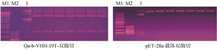

Fig. 6
Alignment between the sequence of pET28a-Qa-1b-VHH recombinant plasmid and Qa-1b-VHH"


Fig. 7
Protein expression levels in the supernatant after IPTG induction M. Protein molecular mass standard; The expression levels from 1 to 7 were induced by IPTG concentrations of 0, 0.2, 0.4, 0.5, 0.6, 0.8 and 1.0 mmol·L-1, respectively"


Fig. 8
Purification of Qa-1b nanobody In the UV absorption peak, Qa-1b nanobody was eluted at 30% imidazole; A. Bands at 30% imidazole were detected by Western blot; B. The results of affinity chromatography by SDS-PAGE (Coomassie blue staining) showed the single band at 30% imidazole"

Table 3
Detection of binding force between Qa-1b nanobody and Qa-1b"
| 组别 Groups | Qa-1b纳米抗体稀释度 Dilution of Qa-1b nanobody | |||||||
| 原液 Stock solution | 1∶2 | 1∶4 | 1∶8 | 1∶16 | 1∶32 | 1∶64 | 1∶128 | |
| 试验组 Experiment group | 1.320 | 0.608 | 0.603 | 0.299 | 0.325 | 0.160 | 0.097 | 0.071 |
| 对照组 Control group | 0.034 | 0.031 | 0.040 | 0.037 | 0.037 | 0.035 | 0.034 | 0.027 |

Fig. 9
The distribution of Qa-1b in canine melanoma tissues was detected by immunohistochemistry (400×)"


Fig. 10
Qa-1b nanobody were used as primary antibodies in Western blotting to detect the expression of Qa-1b in skins samples A. Western blot, 1-3. Calf, 4-6. Adult cattle; B. Grayscale value analysis"

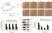
Fig. 11
Effects of Qa-1b nanobody on proliferation and migration of B16 cells A. CCK8 assay was used to detect the inhibitory effect of Qa-1b nanobody on cell proliferation when the concentration of QA-1b nanobody was 120 μg·mL-1; B. Cell scratch assay showed that the addition of Qa-1b nanobody significantly inhibited cell migration; C. The mRNA expressions of MEK1, CDK4, Ras and CyclinD1 were down-regulated by qPCR assay; D, E. Western blot method was used to detect the expression of proliferation-related proteins, and the expression levels of MEK1, CDK4, Ras and Cyclin D1 were reduced, where E was the result of Image J gray analysis. *.P < 0.05, **.P < 0.01, ***.P < 0.001"

| 1 | 张霞, 聂红. Qa-1分子在免疫调节中的研究进展[J]. 现代免疫学, 2010, 30 (2): 159- 163. |
| ZHANG X , NIE H . Progress on Qa-1 molecules in immune regulation[J]. Current Immunology, 2010, 30 (2): 159- 163. | |
| 2 | 曹朔文, 刘丹, 施明. 基于HLA-E功能特点的肿瘤免疫治疗新策略[J]. 中国肿瘤生物治疗杂志, 2022, 29 (5): 383- 390. |
| CAO S W , LIU D , SHI M . Novel strategies of tumor immunotherapy based on the functional features of HLA-E[J]. Chinese Journal of Cancer Biotherapy, 2022, 29 (5): 383- 390. | |
| 3 |
IMANI F , SOLOSKI M J . Heat shock proteins can regulate expression of the Tla region-encoded class Ib molecule Qa-1[J]. Proc Natl Acad Sci U S A, 1991, 88 (23): 10475- 10479.
doi: 10.1073/pnas.88.23.10475 |
| 4 |
VIVIER E , ROMAGNÉ F . Good news, bad news for missing-self recognition by NK cells: autoimmune control but viral evasion[J]. Immunity, 2007, 26 (5): 549- 551.
doi: 10.1016/j.immuni.2007.05.006 |
| 5 |
VIVIER E , RAULET D H , MORETTA A , et al. Innate or adaptive immunity? The example of natural killer cells[J]. Science, 2011, 331 (6013): 44- 49.
doi: 10.1126/science.1198687 |
| 6 |
FEREZ M , KNUDSON C J , LEV A , et al. Viral infection modulates Qa-1b in infected and bystander cells to properly direct NK cell killing[J]. J Exp Med, 2021, 218 (5): e20201782.
doi: 10.1084/jem.20201782 |
| 7 |
RAHBARIZADEH F , AHMADVAND D , SHARIFZADEH Z . Nanobody; an old concept and new vehicle for immunotargeting[J]. Immunol Invest, 2011, 40 (3): 299- 338.
doi: 10.3109/08820139.2010.542228 |
| 8 |
MUYLDERMANS S , ATARHOUCH T , SALDANHA J , et al. Sequence and structure of VH domain from naturally occurring camel heavy chain immunoglobulins lacking light chains[J]. Protein engineering, 1994, 7 (9): 1129- 1135.
doi: 10.1093/protein/7.9.1129 |
| 9 |
VU K B , GHAHROUDI M A , WYNS L , et al. Comparison of llama VH sequences from conventional and heavy chain antibodies[J]. Mol Immunol, 1997, 34 (16-17): 1121- 1131.
doi: 10.1016/S0161-5890(97)00146-6 |
| 10 | OLIVEIRA S , VAN DONGEN G A M S , STIGTER-VAN WALSUM M , et al. Rapid visualization of human tumor xenografts through optical imaging with a near-infrared fluorescent anti-epidermal growth factor receptor nanobody[J]. Mol Imaging, 2012, 11 (1): 33- 46. |
| 11 |
ABULROB A , SPRONG H , VAN BERGEN EN HENEGOUWEN P , et al. The blood-brain barrier transmigrating single domain antibody: mechanisms of transport and antigenic epitopes in human brain endothelial cells[J]. J Neurochem, 2005, 95 (4): 1201- 1214.
doi: 10.1111/j.1471-4159.2005.03463.x |
| 12 | 郭湘. Qa-1b对恶性黑色素瘤生长发展作用的机制及其受体NKG2A纳米抗体的制备与应用[D]. 晋中: 山西农业大学, 2022. |
| GUO X. The mechanism of Qa-1b in malignant melanomaand preparation and application of nanobody of its receptor NKG2A[D]. Jinzhong: Shanxi Agricultural University, 2022. (in Chinese) | |
| 13 |
HSU Y C , LI L S , FUCHS E . Emerging interactions between skin stem cells and their niches[J]. Nat Med, 2014, 20 (8): 847- 856.
doi: 10.1038/nm.3643 |
| 14 |
OVAERE P , LIPPENS S , VANDENABEELE P , et al. The emerging roles of serine protease cascades in the epidermis[J]. Trends Biochem Sci, 2009, 34 (9): 453- 463.
doi: 10.1016/j.tibs.2009.08.001 |
| 15 | 王峰, 田春英, 荣威恒. 蒙古牛改良与肉牛育种规划[J]. 畜牧与饲料科学, 2004, 25 (6): 62- 63. |
| WANG F , TIAN C Y , RONG W H . Mongolian cattle improvement and beef cattle breeding program[J]. Animal Husbandry and Feed Science, 2004, 25 (6): 62- 63. | |
| 16 | 张文兰, 王静瑜, 王筱珊, 等. 阿拉善蒙古牛不同部位肉品质特性研究[J]. 当代畜牧, 2023, (8): 39- 41. |
| ZHANG W L , WANG J Y , WANG X S , et al. Study on the meat quality characteristics of different parts of Alxa Mongolian cattle[J]. Contemporary Animal Husbandry, 2023, (8): 39- 41. | |
| 17 |
齐昱, 邢燕平, 潘静, 等. 基于转录组数据的蒙古牛皮肤组织抗寒相关信号通路及候选基因的筛选[J]. 畜牧兽医学报, 2017, 48 (12): 2301- 2313.
doi: 10.11843/j.issn.0366-6964.2017.12.010 |
|
QI Y , XING Y P , PAN J , et al. Screening of cold tolerance-related signaling pathways and candidate genes in Mongolia cattle skin tissues based on transcriptome data[J]. Acta Veterinaria et Zootechnica Sinica, 2017, 48 (12): 2301- 2313.
doi: 10.11843/j.issn.0366-6964.2017.12.010 |
|
| 18 | 贾琼, 金聪俐, 胡世雄, 等. 丝裂原活化蛋白激酶15纳米抗体的制备及其在B16-F10黑素瘤细胞生长过程中的抑制作用[J]. 中国生物化学与分子生物学报, 2024, 40 (1): 72- 80. |
| JIA Q , JIN C L , HU S X , et al. Preparation of MAPK15 nanobody and its inhibitory effect on the growth of B16-F10 melanoma cells[J]. Chinese Journal of Biochemistry and Molecular Biology, 2024, 40 (1): 72- 80. | |
| 19 |
NGUYEN A V , SOULIKA A M . The dynamics of the skin's immune system[J]. Int J Mol Sci, 2019, 20 (8): 1811.
doi: 10.3390/ijms20081811 |
| 20 | 付国强. 小鼠毛囊免疫系统的产生及周期性改变[J]. 国外医学皮肤性病学分册, 1999, 25 (3): 185. |
| FU G Q . Production and periodic changes of the immune system[J]. International Journal of Dermatology and Venereology, 1999, 25 (3): 185. | |
| 21 |
RÖLLE A , JÄGER D , MOMBURG F . HLA-E peptide repertoire and dimorphism-centerpieces in the adaptive NK cell puzzle?[J]. Front Immunol, 2018, 9, 2410.
doi: 10.3389/fimmu.2018.02410 |
| 22 | 张亮, 陈美凯, 刘小锋, 等. 牛几种常见皮肤病的防治措施[J]. 甘肃畜牧兽医, 2022, 52 (2): 16- 19. |
| ZHANG L , CHEN M K , LIU X F , et al. Prevention and treatment measures for several common skin diseases in cattle[J]. Gansu Animal Husbandry and Veterinary Medicine, 2022, 52 (2): 16- 19. | |
| 23 | 李玮, 胡佳丽, 王凯, 等. 黑色素瘤免疫治疗的研究现状与展望[J]. 中国临床药理学与治疗学, 2021, 26 (9): 1053- 1064. |
| LI W , HU J L , WANG K , et al. Research progress and prospect of immunotherapy in the treatment of melanoma[J]. Chinese Journal of Clinical Pharmacology and Therapeutics, 2021, 26 (9): 1053- 1064. | |
| 24 |
HERNANDEZ B , ADISSU H , WEI B R , et al. Naturally occurring canine melanoma as a predictive comparative oncology model for human mucosal and other triple wild-type melanomas[J]. Int J Mol Sci, 2018, 19 (2): 394.
doi: 10.3390/ijms19020394 |
| 25 | 郑保纹, 徐福南, 钱存忠. 乳牛皮肤恶性黑色素瘤形态学的观察[J]. 中国兽医科技, 1988, (8): 13- 14. |
| ZHENG B W , XU F N , QIAN C Z . Morphology of malignant melanoma in dairy bovine skin[J]. Chinese Journal of Veterinary Science and Technology, 1988, (8): 13- 14. | |
| 26 | 袁荣茂, 景积友. 水牛恶性黑色素瘤病例报告[J]. 甘肃畜牧兽医, 1986, (3): 30. |
| YUAN R M , JING J Y . Case report of malignant melanoma in water buffalo[J]. Gansu Animal Husbandry and Veterinary Medicine, 1986, (3): 30. | |
| 27 | 孙钟琴, 江多轩, 郭宗英. 黄牛恶性黑色素瘤一例[J]. 中国兽医杂志, 1983, (11): 36- 37. |
| SUN Z Q , JIANG D X , GUO Z Y . A case of malignant melanoma in scalpers[J]. Chinese Journal of Veterinary Medicine, 1983, (11): 36- 37. | |
| 28 |
FURGE K A , KIEWLICH D , LE P , et al. Suppression of Ras-mediated tumorigenicity and metastasis through inhibition of the Met receptor tyrosine kinase[J]. Proc Natl Acad Sci U S A, 2001, 98 (19): 10722- 10727.
doi: 10.1073/pnas.191067898 |
| 29 |
SULLIVAN R J , FLAHERTY K . MAP kinase signaling and inhibition in melanoma[J]. Oncogene, 2013, 32 (19): 2373- 2379.
doi: 10.1038/onc.2012.345 |
| 30 | VAKIANI E , SOLIT D B . KRAS and BRAF: drug targets and predictive biomarkers[J]. J Pathol, 2011, 223 (2): 219- 229. |
| 31 | FERRELL J E JR . MAP kinases in mitogenesis and development[J]. Curr Top Dev Biol, 1996, 33, 1- 60. |
| 32 |
MORRISON D K , CUTLER R E . The complexity of Raf-1 regulation[J]. Curr Opin Cell Biol, 1997, 9 (2): 174- 179.
doi: 10.1016/S0955-0674(97)80060-9 |
| 33 |
MONSEL G , ORTONNE N , BAGOT M , et al. c-Kit mutants require hypoxia-inducible factor 1α to transform melanocytes[J]. Oncogene, 2010, 29 (2): 227- 236.
doi: 10.1038/onc.2009.320 |
| 34 | 于吉军, 杨光, 周婷婷, 等. 骆驼来源单域抗体的研究进展[J]. 国际药学研究杂志, 2017, 44 (1): 18- 23. |
| YU J J , YANG G , ZHOU T T , et al. Single domain antibodies from camel: research advances[J]. Journal of International Pharmaceutical Research, 2017, 44 (1): 18- 23. | |
| 35 | MARQUARDT A , MUYLDERMANS S , PRZYBYLSKI M . A synthetic camel anti-lysozyme peptide antibody (peptibody) with flexible loop structure identified by high-resolution affinity mass spectrometry[J]. Chemistry, 2010, 12 (7): 1915- 1923. |
| [1] | SONG Wenyan, ZHANG Hanwen, WU Aodi, ZHANG Liyan, LIU Zhao, YE Tongtong, CHEN Chuangfu, SHENG Jinliang. Screening of Nanobodies against Porcine Reproductive and Respiratory Syndrome Virus GP5 Protein and Exploration of Their Inhibitory Effect on Virus Replication [J]. Acta Veterinaria et Zootechnica Sinica, 2024, 55(1): 258-270. |
| [2] | HU Xiangyun, CAO Yanhong, LÜ Lingyan, LIU Zheng, HUANG Facai, WU Zhuyue, XIAO Zhengzhong. Nanobodies and Their Research Status in Veterinary Field [J]. Acta Veterinaria et Zootechnica Sinica, 2023, 54(8): 3164-3172. |
| [3] | WANG Ying, ZHU Jiahong, ZHAO Jiakai, JI Pinpin, CHEN Xu, ZHANG Lu, LIU Baoyuan, SUN Yani, ZHAO Qin. Screening and Identification of Nanobodies against NP419L Protein of African Swine Fever Virus and Its Preliminary Application of Antibody Detection [J]. Acta Veterinaria et Zootechnica Sinica, 2023, 54(6): 2509-2520. |
| [4] | MA Zhijie, WANG Shikang, ZHANG Weizhong, GUO Weixing, ZHAO Haiming, LEI Chuzhao. Y-chromosome Genomic Diversity and Paternal Origins of Qaidam Cattle and Mongolian Cattle [J]. Acta Veterinaria et Zootechnica Sinica, 2023, 54(3): 1026-1033. |
| [5] | WANG Tianyu, LI Zhiwei, YANG Ting, DONG Linfang, MA Zhiqian, BIANBA Ciren, XIAO Shuqi, LI Shuang. Screening and Identification of Nanobodies against Porcine Epidemic Diarrhea Virus S Protein [J]. Acta Veterinaria et Zootechnica Sinica, 2021, 52(9): 2589-2598. |
| [6] | YANG Lamei, ZHANG Ting, ZHANG Ding. Pathological Diagnosis of a Case of Liver Malignant Melanoma in Broiler Chicken [J]. Acta Veterinaria et Zootechnica Sinica, 2021, 52(1): 268-272. |
| [7] | LI Yuan, LIU Wen-yan, CAI Yong-qiang, ZHANG Li-huan, LI Hong, ZHU Zhi-wei. Analysis of Long Non-coding RNA HOTAIR and Prediction of Its Interaction Molecules in Melanoma Pathogenesis [J]. ACTA VETERINARIA ET ZOOTECHNICA SINICA, 2017, 48(8): 1424-1435. |
| [8] | HE Sheng-fang, FU Xiang-jing, LIU Yang-kun, MU Guo-hui, LIU Hai-jin,WANG Xing-long, DU En-qi, YANG Zeng-qi. Establishing a Method for Highly Efficient Producing Nanometer Antibodies [J]. ACTA VETERINARIA ET ZOOTECHNICA SINICA, 2013, 44(9): 1487-1493. |
| Viewed | ||||||
|
Full text |
|
|||||
|
Abstract |
|
|||||