


Acta Veterinaria et Zootechnica Sinica ›› 2024, Vol. 55 ›› Issue (11): 5035-5049.doi: 10.11843/j.issn.0366-6964.2024.11.021
• Animal Biotechnology and Reproduction • Previous Articles Next Articles
Helin LI1,3( ), Yufen JIANG1,3(), Na CHENG1,3, Yuchen HAN1,3, Xiaoying HUO2,3, Hongding SU3, Yue CHANG1,3, Yuzhu FANG2,3, Pei WANG1,3, Baoyu JIA2,3, Hongjiang WEI2,3,*(), Wenmin CHENG1,3,*()
Received:2024-03-29
Online:2024-11-23
Published:2024-11-30
Contact:
Hongjiang WEI, Wenmin CHENG
E-mail:sonnyspike@163.com;2962181956@qq.com;hongjiangwei@126.com;cheng_8097@163.com
CLC Number:
Helin LI, Yufen JIANG, Na CHENG, Yuchen HAN, Xiaoying HUO, Hongding SU, Yue CHANG, Yuzhu FANG, Pei WANG, Baoyu JIA, Hongjiang WEI, Wenmin CHENG. The Study of Regulatory Effect of Differentially Expressed microRNAs on the Npm2 Expression in Pig Oocytes[J]. Acta Veterinaria et Zootechnica Sinica, 2024, 55(11): 5035-5049.
Table 1
Primers sequence"
| 引物名称 Primer name | 引物序列(5′→3′) Primer sequence | 注释 Exegesis |
| miR-32-RT | GTCGTATCCAGTGCAGGGTCCGAGGTATTCGCACTGGATACGACGCAACT | miR-32反转录引物 |
| miR-32-qPF | AGCCAGCGTATTGCACATTAC | miR-32上游引物 |
| miR-32-qPR | AGTGCAGGGTCCGAGGTATT | miR-32下游引物 |
| miR-150-RT | GTCGTATCCAGTGCAGGGTCCGAGGTATTCGCACTGGATACGACCACTGG | miR-150反转录引物 |
| miR-150-qPF | GCGTCTCCCAACCCTTGTA | miR-150上游引物 |
| miR-150-qPR | AGTGCAGGGTCCGAGGTATT | miR-150下游引物 |
| miR-296-5p-RT | GTCGTATCCAGTGCAGGGTCCGAGGTATTCGCACTGGATACGACACAGGA | miR-296-5p反转录引物 |
| miR-296-5p-qPF | AGAGGGCCCCCCCCAA | miR-296-5p上游引物 |
| miR-296-5p-qPR | AGTGCAGGGTCCGAGGTATT | miR-296-5p下游引物 |
| miR-296-3p-RT | GTCGTATCCAGTGCAGGGTCCGAGGTATTCGCACTGGATACGACGGAAAG | miR-296-3p反转录引物 |
| miR-296-3p-qPF | AACAATAGGGTTGGGCGGA | miR-296-3p上游引物 |
| miR-296-3p-qPR | GTCGTATCCAGTGCAGGGT | miR-296-3p下游引物 |
| U6-qPF | CTCGCTTCGGCAGCACA | U6上游引物 |
| U6-qPR&RT | AACGCTTCACGAATTTGCGT | U6下游引物/反转录引物 |
| NPM2-qPF | GCTTAGCACGATTTGCCTGG | Npm2上游引物 |
| NPM2-qPR | CCACTGAGGAACACAGGTCC | Npm2下游引物 |
| GAPDH-qPF | AGGGCATCCTGGGCTACACT | GAPDH上游引物 |
| GAPDH-qPR | TCCACCACCCTGTTGCTGTAG | GAPDH下游引物 |
Table 2
miRNAs qPCR reaction system"
| 试剂 Reagent | 使用量/μL Usage amount | 终浓度 Final concentration |
| 2×miRNA qPCR master mix | 10 | 1× |
| Forward Primer(10 μmol·L-1) | 0.5 | 0.25 μmol·L-1 |
| Reverse Primer(10 μmol·L-1) | 0.5 | 0.25 μmol·L-1 |
| DNA模板Template DNA | 1~2 | |
| ROX Reference Dye(L)/(H) | 1 | |
| 无酶水RNase-free water | up to 20 |
Table 3
Genes qPCR reaction system"
| 试剂 Reagent | 使用量/μL Usage amount | 终浓度 Final concentration |
| TB Green Premix Ex Taq II(Tli RNaseH Plus)(2×) | 12.5 | 1× |
| PCR Forward Primer(10 μmol·L-1) | 1 | 0.4 μmol·L-1 |
| PCR Reverse Primer(10 μmol·L-1) | 1 | 0.4 μmol·L-1 |
| DNA模板Template DNA(< 100 ng)*2 | 2 | |
| 无酶水RNase-free water | 8.5 | |
| 总量Total | 25 |

Fig. 1
pmiRGLO plasmid profile"

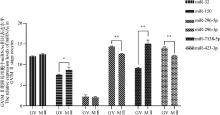
Fig. 2
Quantitative validation of differentially expressed miRNAs *.P < 0.05; **.P < 0.01; ***.P < 0.001. The same as below"


Fig. 3
Dual luciferase test results A. miR-32 mimic+Npm2 3′UTR wild-type and mutant relative luciferase intensity; B. miR-150 mimic+Npm2 3′UTR wild-type and mutant relative luciferase intensity; C. miR-296-5p mimic+Npm2 3′UTR wild-type and mutant relative luciferase intensity; D. miR-296-3p mimic+Npm2 3′UTR wild-type and mutant relative luciferase intensity; E. miR-423-3p mimic+Npm2 3′UTR wild-type and mutant relative luciferase intensity; F. miR-7138-5p mimic+Npm2 3′UTR wild-type and mutant relative luciferase intensity.NC. Control; mimic. miRNA mimic; NPM2-WT. Wild-type Npm2 3′UTR sequence; NPM2-MT. Mutant Npm2 3′UTR sequence. ns.P>0.05, the same as below"


Fig. 4
The effect of adding different concentration miRNAs mimic/inhibitor on the oocytes maturation rate and developmental ability of parthenogenetic activated embryos A. Added miR-32 mimic; B. Added miR-32 inhibitor; C. Added miR-150 mimic; D. Added miR-150 inhibitor; E. Added miR-296-5p mimic; F. Added miR-296-5p inhibitor; G. Added miR-296-3p mimic; H. Added miR-296-3p inhibitor; I. Added miR-423-3p mimic; J. Added miR-423-3p inhibitor; K. Added miR-7138-5p mimic; L. Added miR-7138-5p inhibitor"


Fig. 5
The effect of miRNA mimic/inhibitor addition on the activity of MⅡ oocytes cortical granules A. Addition of optimal concentration miR-32 mimic/inhibitor; B. Addition of optimal concentration miR-150 mimic/inhibitor; C. Addition of optimal concentration miR-296-5p mimic/inhibitor; D. Addition of optimal concentration miR-296-3p mimic/inhibitor; E. Addition of optimal concentration miR-423-3p mimic/inhibitor; F. Addition of optimal concentration miR-7138-5p mimic/inhibitor; G. Fluorescence intensity of cortical granules in MⅡ stage oocytes. CG PNA-FITC. Peanut agglutinin labeled with green fluorescence (granulosa cells); Nucleus. Cell nucleus (including polar body); Merge. Combined layers"


Fig. 6
The effect of miRNAs mimic/inhibitor addition on the expression level of Npm2 mRNA in in vitro matured oocytes A. Relative expression level of Npm2 in GV/MⅡ stage oocytes; B. Relative expression level of Npm2 in MⅡ stage oocytes after adding miRNA mimics/inhibitors"

| 1 | READER K L , STANTON J A L , JUENGEL J L .The role of oocyte organelles in determining developmental competence[J].Biology (Basel),2017,6(3):35. |
| 2 |
MILLS A D , LASKEY R A , BLACK P , et al.An acidic protein which assembles nucleosomes in vitro is the most abundant protein in Xenopus oocyte nuclei[J].J Mol Biol,1980,139(3):561-568.
doi: 10.1016/0022-2836(80)90148-5 |
| 3 | 姚顺发, 许庆龙, 张君正, 等.精细胞中促进组蛋白-鱼精蛋白替换的分子机制研究进展[J].中国畜牧杂志,2024,60(7):60-65. |
| YAO S F , XU Q L , ZHANG J Z , et al.Research progress on the molecular mechanism of promoting histone-protamine replacement in spermatids[J].Chinese Journal of Animal Science,2024,60(7):60-65. | |
| 4 |
FREHLICK L J , EIRÍN-LÓPEZ J M , AUSIÓ J .New insights into the nucleophosmin/nucleoplasmin family of nuclear chaperones[J].BioEssays,2007,29(1):49-59.
doi: 10.1002/bies.20512 |
| 5 |
LASKEY R A , HONDA B M , MILLS A D , et al.Nucleosomes are assembled by an acidic protein which binds histones and transfers them to DNA[J].Nature,1978,275(5679):416-420.
doi: 10.1038/275416a0 |
| 6 |
ONIKUBO T , NICKLAY J J , XING L , et al.Developmentally regulated post-translational modification of nucleoplasmin controls histone sequestration and deposition[J].Cell Rep,2015,10(10):1735-1748.
doi: 10.1016/j.celrep.2015.02.038 |
| 7 |
OKUWAKI M , SUMI A , HISAOKA M , et al.Function of homo- and hetero-oligomers of human nucleoplasmin/ nucleophosmin family proteins NPM1, NPM2 and NPM3 during sperm chromatin remodeling[J].Nucleic Acids Res,2012,40(11):4861-4878.
doi: 10.1093/nar/gks162 |
| 8 |
TAMADA H , VAN THUAN N , REED P , et al.Chromatin decondensation and nuclear reprogramming by nucleoplasmin[J].Mol Cell Biol,2006,26(4):1259-1271.
doi: 10.1128/MCB.26.4.1259-1271.2006 |
| 9 | 张宸艺博, 余彤, 任斌斌, 等.动物早期胚胎发育中表观重编程的机制[J].畜牧兽医学报,2023,54(12):4898-4909. |
| ZHANG C Y B , YU T , REN B B , et al.Mechanism of epigenetic reprogramming of early animal embryos[J].Acta Veterinaria et Zootechnica Sinica,2023,54(12):4898-4909. | |
| 10 |
OKUWAKI M , MATSUMOTO K , TSUJIMOTO M , et al.Function of nucleophosmin/B23, a nucleolar acidic protein, as a histone chaperone[J].FEBS Lett,2001,506(3):272-276.
doi: 10.1016/S0014-5793(01)02939-8 |
| 11 |
BURNS K H , VIVEIROS M M , REN Y S , et al.Roles of NPM2 in chromatin and nucleolar organization in oocytes and embryos[J].Science,2003,300(5619):633-636.
doi: 10.1126/science.1081813 |
| 12 |
WEN J , FRIEDMAN J R .miR-122 regulates hepatic lipid metabolism and tumor suppression[J].J Clin Invest,2012,122(8):2773-2776.
doi: 10.1172/JCI63966 |
| 13 |
BRENNECKE J , HIPFNER D R , STARK A , et al.bantam encodes a developmentally regulated microRNA that controls cell proliferation and regulates the proapoptotic gene hid in Drosophila[J].Cell,2003,113(1):25-36.
doi: 10.1016/S0092-8674(03)00231-9 |
| 14 |
SALILEW-WONDIM D , GEBREMEDHN S , HOELKER M , et al.The role of MicroRNAs in mammalian fertility: from gametogenesis to embryo implantation[J].Int J Mol Sci,2020,21(2):585.
doi: 10.3390/ijms21020585 |
| 15 |
ROSENBLUTH E M , SHELTON D N , SPARKS A E T , et al.MicroRNA expression in the human blastocyst[J].Fertil Steril,2013,99(3):855-861.
doi: 10.1016/j.fertnstert.2012.11.001 |
| 16 |
PAULSON E E , FISHMAN E L , MA J , et al.Embryonic microRNAs are essential for bovine preimplantation embryo development[J].Proc Natl Acad Sci U S A,2022,119(45):e2212942119.
doi: 10.1073/pnas.2212942119 |
| 17 |
GIRALDEZ A J , MISHIMA Y , RIHEL J , et al.Zebrafish MiR-430 promotes deadenylation and clearance of maternal mRNAs[J].Science,2006,312(5770):75-79.
doi: 10.1126/science.1122689 |
| 18 |
LV C , YU W X , WANG Y , et al.MiR-21 in extracellular vesicles contributes to the growth of fertilized eggs and embryo development in mice[J].Biosci Rep,2018,38(4):BSR20180036.
doi: 10.1042/BSR20180036 |
| 19 |
WANG Y Z , ZHOU C , MENG F M , et al.Ssc-miR-92b-3p regulates porcine trophoblast cell proliferation and migration via the PFKM gene[J].Int J Mol Sci,2022,23(24):16138.
doi: 10.3390/ijms232416138 |
| 20 |
TANGA B M , FANG X , BANG S , et al.MiRNA-155 inhibition enhances porcine embryo preimplantation developmental competence by upregulating ZEB2 and downregulating ATF4[J].Theriogenology,2022,183,90-97.
doi: 10.1016/j.theriogenology.2022.02.019 |
| 21 |
LINGENFELTER B M , TRIPURANI S K , TEJOMURTULA J , et al.Molecular cloning and expression of bovine nucleoplasmin 2 (NPM2): a maternal effect gene regulated by miR-181a[J].Reprod Biol Endocrinol,2011,9,40.
doi: 10.1186/1477-7827-9-40 |
| 22 | 张珂, 严继猛, 刘耀文, 等.猪GV/MⅡ期卵母细胞miRNAs表达谱及Npm2基因相关miRNAs筛选[J].畜牧兽医学报,2021,52(4):996-1009. |
| ZHANG K , YAN J M , LIU Y W , et al.miRNAs expression profile of porcine GV/MⅡ-stage oocytes and screening of miRNAs related to Npm2 gene[J].Acta Veterinaria et Zootechnica Sinica,2021,52(4):996-1009. | |
| 23 | KOBAYASHI H .Imprinting genes associated with endometriosis[J].EXCLI J,2014,13,252-264. |
| 24 |
LEE J Y , AHN E H , KIM J O , et al.Associations between microRNA (miR-25, miR-32, miR-125, and miR-222) polymorphisms and recurrent implantation failure in Korean women[J].Hum Genomics,2019,13(1):68.
doi: 10.1186/s40246-019-0246-y |
| 25 |
GUO J , LU W F , LIANG S , et al.Peroxisome proliferator-activated receptor δ improves porcine blastocyst hatching via the regulation of fatty acid oxidation[J].Theriogenology,2017,90,266-275.
doi: 10.1016/j.theriogenology.2016.11.018 |
| 26 | ZOU G , LIU T , GUO L H , et al.MicroRNA-32 silences WWP2 expression to maintain the pluripotency of human amniotic epithelial stem cells and β islet-like cell differentiation[J].Int J Mol Med,2018,41(4):1983-1991. |
| 27 |
REVEL A , ACHACHE H , STEVENS J , et al.MicroRNAs are associated with human embryo implantation defects[J].Hum Reprod,2011,26(10):2830-2840.
doi: 10.1093/humrep/der255 |
| 28 |
ABD EL NABY W S , HAGOS T H , HOSSAIN M M , et al.Expression analysis of regulatory microRNAs in bovine cumulus oocyte complex and preimplantation embryos[J].Zygote,2013,21(1):31-51.
doi: 10.1017/S0967199411000566 |
| 29 |
LIN Y C , KUO M W , YU J , et al.c-Myb is an evolutionary conserved miR-150 target and miR-150/c-Myb interaction is important for embryonic development[J].Mol Biol Evol,2008,25(10):2189-2198.
doi: 10.1093/molbev/msn165 |
| 30 |
WANG R K , LI G F , ZHUANG G Y , et al.Overexpression of microRNA-423-3p indicates poor prognosis and promotes cell proliferation, migration, and invasion of lung cancer[J].Diagn Pathol,2019,14(1):53.
doi: 10.1186/s13000-019-0831-3 |
| 31 |
BIGNOTTI E , CALZA S , TASSI R A , et al.Identification of stably expressed reference small non-coding RNAs for microRNA quantification in high-grade serous ovarian carcinoma tissues[J].J Cell Mol Med,2016,20(12):2341-2348.
doi: 10.1111/jcmm.12927 |
| 32 |
RAO M , ZENG Z Y , TANG L , et al.Next-generation sequencing-based microRNA profiling of mice testis subjected to transient heat stress[J].Oncotarget,2017,8(67):111672-111682.
doi: 10.18632/oncotarget.22900 |
| 33 |
TOKUMOTO T , TOKUMOTO M , OSHIMA T , et al.Characterization of multiple membrane progestin receptor (mPR) subtypes from the goldfish ovary and their roles in the induction of oocyte maturation[J].Gen Comp Endocrinol,2012,177(1):168-176.
doi: 10.1016/j.ygcen.2012.03.005 |
| 34 |
SCHNEIDER A , MATKOVICH S J , VICTORIA B , et al.Changes of ovarian microRNA profile in long-living ames dwarf mice during aging[J].PLoS One,2017,12(1):e0169213.
doi: 10.1371/journal.pone.0169213 |
| 35 |
CAPRA E , LAZZARI B , RUSSO M , et al.Seasonal effects on miRNA and transcriptomic profile of oocytes and follicular cells in buffalo (Bubalus bubalis)[J].Sci Rep,2020,10(1):13557.
doi: 10.1038/s41598-020-70546-5 |
| 36 | GALOCZOVA M , COATES P , VOJTESEK B .STAT3, stem cells, cancer stem cells and p63[J].Cell Mol Biol Lett,2018,23,12. |
| [1] | Jingjing TIAN, Xiaoqing WANG, Mianyan LI, Hailing WANG, Qitian WU, Lixian WANG, Longchao ZHANG, Fuping ZHAO. Analysis of the Whole Genome Run of Homozygosity (ROH) and Selection Signal in Beijing Black Pigs [J]. Acta Veterinaria et Zootechnica Sinica, 2024, 55(9): 3833-3842. |
| [2] | Dong CHEN, Wenxuan ZHOU, Zhenjian ZHAO, Qi SHEN, Yang YU, Shengdi CUI, Junge WANG, Ziyang CHEN, Shixin YU, Jiamiao CHEN, Xiangfeng WANG, Pingxian WU, Zongyi GUO, Jinyong WANG, Guoqing TANG. Development of a Pig Intramuscular Fat Content and Eye Muscle Area Measurement System Based on Computer Vision Technology [J]. Acta Veterinaria et Zootechnica Sinica, 2024, 55(9): 3843-3852. |
| [3] | Baigao YANG, Xi LONG, Liang ZHANG, Jiehuan XU, Jianjun DAI, Xueming ZHAO, Hongmei PAN. Exploring the Effect of Vitrification on Gene Expression in Porcine Parthenogenetic Blastocysts by Smart-seq2 [J]. Acta Veterinaria et Zootechnica Sinica, 2024, 55(9): 3936-3946. |
| [4] | Cong REN, Hu ZHANG, Yuming WANG, Jingjing XIE, Renna SA, Feng ZHAO. Study on the Accuracy and Additivity of Effective Energy in Feed for Growing Pigs Predicted by Simulated Digestion Method [J]. Acta Veterinaria et Zootechnica Sinica, 2024, 55(9): 3988-4000. |
| [5] | Shan ZHANG, Dahu LIU, Baojing LIU, Lin LIANG, Ruiying LIANG, Xinming TANG, Xusheng QIU, Chan DING, Jiabo DING, Shaohua HOU. Isolation, Identification and Pathogenicity Analysis of a Pigeon Paramyxovirus-1 Strain [J]. Acta Veterinaria et Zootechnica Sinica, 2024, 55(9): 4051-4060. |
| [6] | Xiaojuan LIANG, Yushuang LI, Zhou FU, Duo TANG, Yingying LI, Shouwei WANG. Isolation, Culture and Adipogenic Differentiation of Pigeon Preadipocytes [J]. Acta Veterinaria et Zootechnica Sinica, 2024, 55(8): 3482-3492. |
| [7] | Jing CHEN, Xuebei WU, Dongzhi MIAO, Chi ZHANG, Zhenyu GUO, Ying WANG. Comparative Analysis of Transcriptome of Pigeon Follicles at Early Stage of Laying Interval Reveals Genes Related to Follicular Development [J]. Acta Veterinaria et Zootechnica Sinica, 2024, 55(8): 3503-3515. |
| [8] | Yue LI, Changchun ZHANG, Guangyu LIU, Mengyuan GAO, Chaojun FU, Jiabao XING, Sijia XU, Qiyuan KUANG, Jing LIU, Xiaopeng GAO, Heng WANG, Lang GONG, Guihong ZHANG, Yankuo SUN. Application and Analysis of Meta-transcriptomics Sequencing Technology in the Diagnosis of Viral Diarrhea Diseases in Piglets [J]. Acta Veterinaria et Zootechnica Sinica, 2024, 55(8): 3579-3589. |
| [9] | Tengfei DOU, Jiahao WU, Ziyi WU, Liyao BAI, Xinjian LI, Xuelei HAN, Ruimin QIAO, Kejun WANG, Feng YANG, Yining WANG, Xiuling LI. Application Progress of Genomic Selection and Mating Allocation Techniques in Pig Breeding [J]. Acta Veterinaria et Zootechnica Sinica, 2024, 55(7): 2795-2808. |
| [10] | Yaxuan MENG, Yan LIU, Jing WANG, Guoshun CHEN, Tao FENG. Research Progress in the Effect of Oxidative Stress on Ovarian Function in Female Livestock [J]. Acta Veterinaria et Zootechnica Sinica, 2024, 55(7): 2825-2835. |
| [11] | Xiaojuan LIANG, Yushuang LI, Yingying LI, Shouwei WANG. Isolation, Culture and Adipogenic Differentiation of Beijing Black Pig Preadipocytes [J]. Acta Veterinaria et Zootechnica Sinica, 2024, 55(7): 2877-2889. |
| [12] | Weizhe LIU, Chenggang LUO, Rong YUAN, Yijie LIAO, Yimin WEN, Ying SUN, Enbo YU, Sanjie CAO, Xiaobo HUANG. Isolation and Identification of a Highly Pathogenic Strain of Porcine Epidemic Diarrhea Virus [J]. Acta Veterinaria et Zootechnica Sinica, 2024, 55(7): 3049-3063. |
| [13] | Yingguang LÜ, Guangming JIAO, Jinfang SANG, Zhipeng KOU, Tao LIU, Yue WANG, Xiangyu LU, Chenxi PIAO, Yajun MA, Jiantao ZHANG, Hongbin WANG. The Effect of Adipose Mesenchymal Stem Cells on the Healing Process of Autologous Skin Transplantation in Bama Miniature Pigs [J]. Acta Veterinaria et Zootechnica Sinica, 2024, 55(7): 3193-3204. |
| [14] | Bin LIU, Yan LIU, Chen ZHENG, Tao FENG. Effects of Glucosamine on Growth Performance, Antioxidant Capacity, and Immune Function in Weaned Piglets [J]. Acta Veterinaria et Zootechnica Sinica, 2024, 55(7): 3246-3254. |
| [15] | Tiantian YAN, Jianliang WU, Zhaojun WANG, Li XU, Qingli MENG, Meiyu SU, Hanqiao LI, Guoying HUANG, Chao WANG, Jiaqi LIN. Genetic Parameter Estimation and Genetic Progress Analysis of Reproductive Traits in French Large White Pigs [J]. Acta Veterinaria et Zootechnica Sinica, 2024, 55(6): 2388-2396. |
| Viewed | ||||||
|
Full text |
|
|||||
|
Abstract |
|
|||||