


Acta Veterinaria et Zootechnica Sinica ›› 2026, Vol. 57 ›› Issue (1): 401-413.doi: 10.11843/j.issn.0366-6964.2026.01.035
• PREVENTIVE VETERINARY MEDICINE • Previous Articles Next Articles
LI Meng1,2, SU Siyuan1,2, LI Zipan1,2, XU Yuntiao1,2, HUANG Zibei1,2, LUO Jianya1,2, ZHAO Yiheng1,2, GUO Xin1,2, BIAN Hongkai1,2, PAN Xinyu1,2, LIU Wenbo1,2( )
)
Received:2024-10-25
Online:2026-01-23
Published:2026-01-26
Contact:
LIU Wenbo
E-mail:lwb@yzu.edu.cn
CLC Number:
LI Meng, SU Siyuan, LI Zipan, XU Yuntiao, HUANG Zibei, LUO Jianya, ZHAO Yiheng, GUO Xin, BIAN Hongkai, PAN Xinyu, LIU Wenbo. Preparation and Application of Cat PD-L1 Monoclonal Antibody and Feline Derived Single Chain Antibody[J]. Acta Veterinaria et Zootechnica Sinica, 2026, 57(1): 401-413.

Fig.1
Construction of PET-MPD-L1 and antigen preparationA. Feline PD-L1 gene: M. 200 bp marker; 1. Feline PD-L1 gene. B. Double digestion identification of PET-MPD-L1: M. 1000 bp marker; 1. PET-MPD-L1 digestion result. C. PET-MPD-L1 protein expression: M. Protein molecular weight marker; 1. Lysate supernatant of induced PET-32a; 2. Lysate precipitate of induced PET-32a; 3. Lysate supernatant of uninduced PET-32a; 4. Lysate precipitate of uninduced PET-32a; 5. Lysate precipitate of uninduced PET-MPD-L1; 6. Lysate supernatant of uninduced PET-MPD-L1; 7. Lysate precipitate of induced PET-MPD-L1; 8. Lysate supernatant of induced PET-MPD-L1. D. PET-MPD-L1 protein purification: M. Protein molecular weight marker; 1. Bacterial lysate; 2. Flow-through; 3-9. Wash fractions; 10-12. Elution fractions"
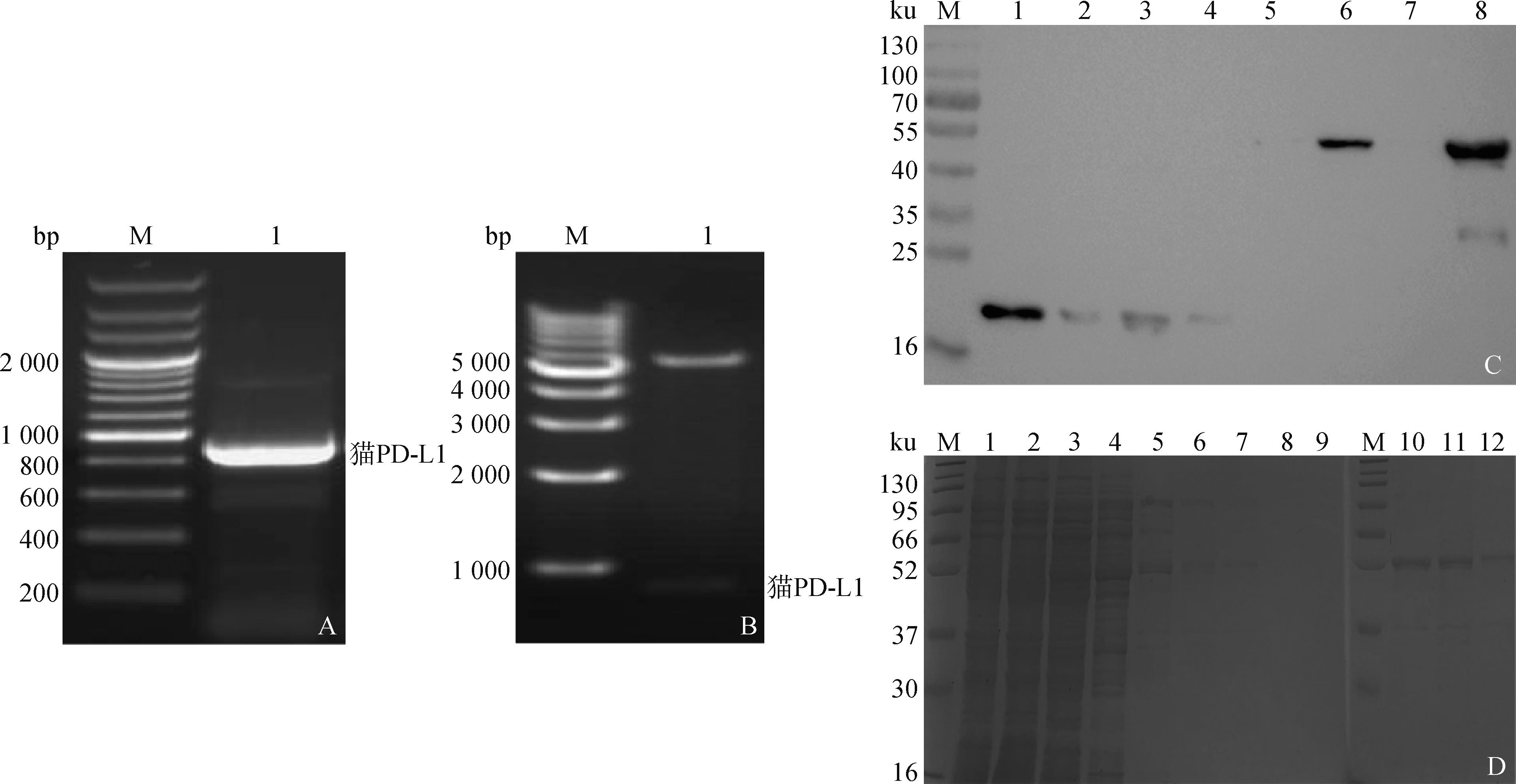

Fig.2
Results of SDS-PAGE validation antibody purificationM. Protein molecular weight marker; 1. Preliminary extraction of ascites; 2. Flowing fluid; 3. Washing solution; 4. Purification of antibodies"
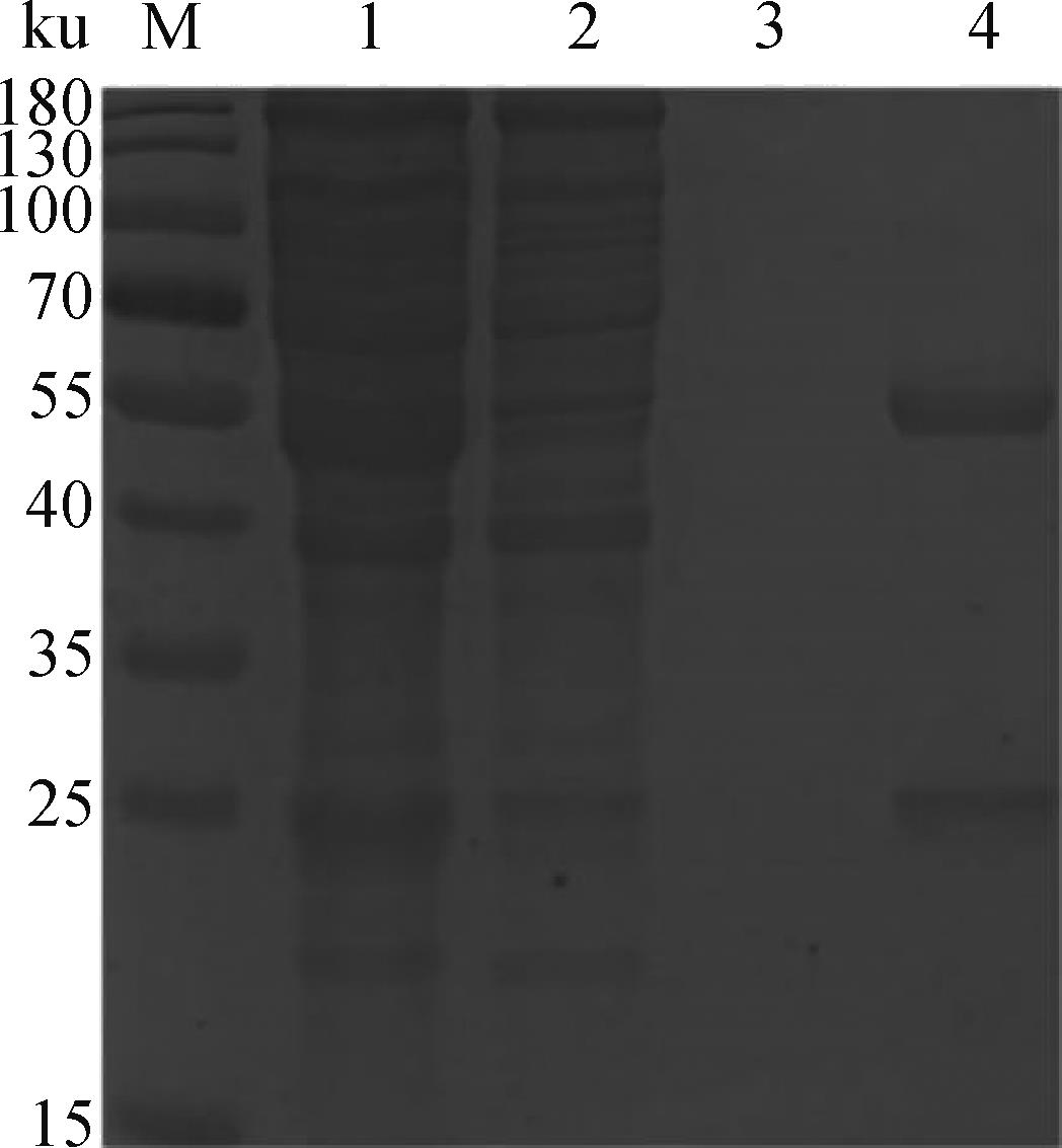
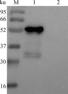
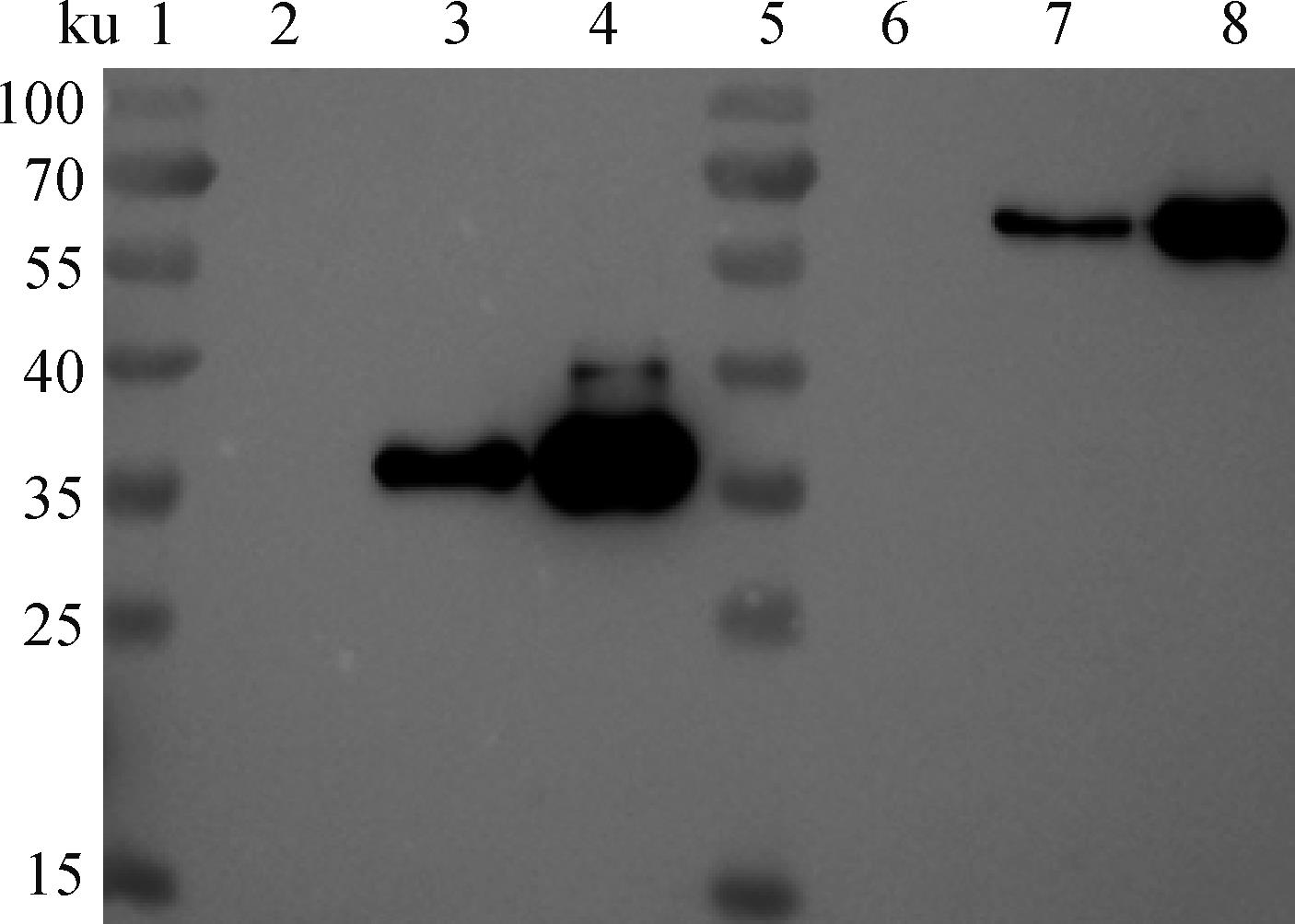
Fig.3
Western blot result of McAb reacted with feline PD-L1M. Protein molecular weight marker; 1. PET-MPD-L1 protein; 2. PET-32a"
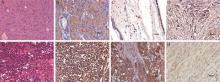
Fig.4
Identification of canine and feline tumor tissues using HE staining and IHC with 2A5A2F4D7A. Canine perianal epithelial tumor, HE staining (100×); B. Canine perianal epithelial tumor, IHC; C. Canine pilomatricoma, IHC; D. Canine mammary fibroadenoma, IHC; E. Feline lymphoma, HE staining; F. Feline lymphoma, (IHC); G. Canine mammary carcinoma, IHC; H. Peritumoral tissue of canine mammary carcinoma, IHC. Arrows point to the positive tumor cells"


Fig.5
Antibody gene amplificationA. Antibody heavy chain gene amplification: M1. 100 bp marker; M2. 200 bp marker; 1-16. The first to the 16th pairs of heavy chain gene amplification. B. Antibody light chain gene amplification: M1. 100 bp marker; 1-19. The first to the 19th pairs of light chain gene amplification"

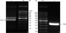
Fig.6
Amplification of antibody light and heavy chain genesA. Antibody heavy chain gene amplification: M. 100 bp marker; 1. The 5th pair of heavy chain gene amplification. B. Antibody light chain gene amplification: M. 100 bp marker; 1. The 8th pair of light chain gene amplification"

Fig.7
Antibody gene alignmentA. Comparison of antibody gene amplification using the 5th pair of light chain primers; B. Comparison of antibody gene amplification using the 8th pair of light chain primers"

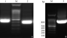
Fig.8
Amplification of VH5 and VK8 and splicing of their scFvA. Antibody light and heavy chain gene amplification: M. 100 bp marker; 1. VH5 Linker; 2. VK8 Linker. B. ScFv splicing: M. 100 bp marker;3. scFv"

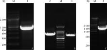
Fig.9
Assembly of the feline-derived chimeric single-chain antibody geneA. Amplification of the feline constant region antibody gene: M. 200 bp marker; 1. Feline constant region antibody gene. B. Amplification of scFv and feline IgG-Fc genes: M. 100 bp marker; 2. scFv; 3. Feline IgG-Fc. C. Feline-derived chimeric single-chain antibody gene: M. 200 bp marker; 4. scFv-fFc"


Fig.10
Rescue of recombinant baculoviruses rBV-scFv and rBV-scFv-fFcA. Double enzyme digestion production of rBacmid-scFv: 1. 1 kb marker; 2. 200 bp marker; 3. Double enzyme digestion of rBacmid-scFv. B. Double enzyme digestion production of rBacmid-scFv-fFc: 1. 1kb marker; 2. 200 bp marker; 3. Double enzyme digestion of rBacmid-scFv-fFc. C. PCR production of rBacmid-scFv and rBacmid-scFv-fFc using M13 primers and specific primers: 1, 4. 1 kb marker; 2. Identification of rBacmid-scFv using scFv primers; 3, 6. Identification of rBacmid-scFv and rBacmid-scFv-fFc using M13 primers; 5. Identification of rBacmid-scFv-fFc using scFv-fFc primers. D. PCR production of P1-P3 generations of recombinant baculovirus rBV-scFv and rBV-scFv-fFc using M13 primers and specific primers: 1, 11. 1 kb marker; 2. 200 bp marker; 3-5. Identification of P1-P3 generations of rBV-scFv using scFv primers; 7-9, 16-18. Identification of P1-P3 generations of rBV-scFv and rBV-scFv-fFc using M13 primers; 6, 10, 15, 19. Non-infected controls; 12-14. Identification of P1-P3 generations of rBV-scFv-fFc using scFv-fFc primers"
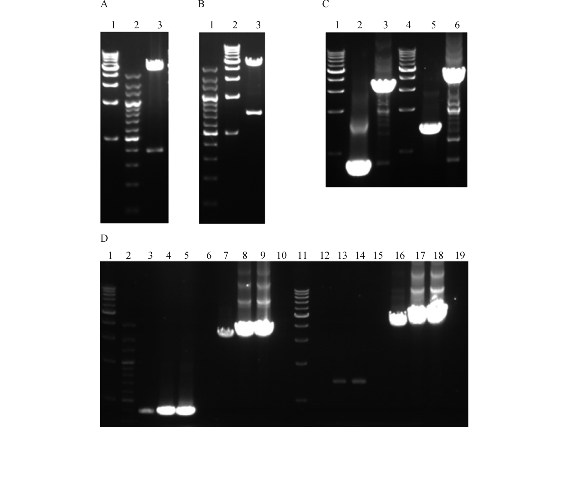
Fig.11
Western blot analysis of scFv and scFv-fFc expression1,5. Marker; 2-4 and 6-8: Represent the P3 generation rBV-scFv- and rBV-scFv-fFc-infected SF9 cell culture medium, cell lysate supernatant, and cell lysate pellet, respectively"
| [1] | 黄琳娜.广东省犬猫肿瘤流行病学调查及加味柴胡疏肝散治疗恶性乳腺肿瘤的临床疗效[D].广州:华南农业大学,2023. |
| HUANG L N.Epidemiological investigation of canine and cat tumors in Guangdong province and clinical efficacy of modified chaihu shugan powder in the treatment of malignant breast tumors[D].Guangzhou:South China Agricultural University,2023.(in Chinese) | |
| [2] | BLACKWOOD L.Cats with cancer:where to start [J].J Feline Med Surg,2013,15(5):366-377. |
| [3] | BRØNDEN L B,NIELSEN S S,TOFT N,et al.Data from the danish veterinary cancer registry on the occurrence and distribution of neoplasms in dogs in denmark [J].Vet Rec,2010,166(19):586-590. |
| [4] | 罗盈滢.犬猫肿瘤性疾病的临床诊疗方法[J].湖北畜牧兽医,2017,38(5):23-24. |
| LUO Y Y.Clinical diagnosis and treatment methods for canine and feline tumor diseases[J].Hubei Animal Husbandry and Veterinary Medicine,2017,38(5):23-24.(in Chinese) | |
| [5] | HE X,XU C.Immune checkpoint signaling and cancer immunotherapy [J].Cell Res,2020,30(8):660-669. |
| [6] | DONG H,STROME S E,SALOMAO D R,et al.Tumor-associated B7-H1 promotes T-cell apoptosis:A potential mechanism of immune evasion [J].Nat Med,2002,8(8):793-800. |
| [7] | SZNOL M,CHEN L.Antagonist antibodies to PD-1 and B7-H1 (PD-L1) in the treatment of advanced human cancer [J].Clin Cancer Res,2013,19(5):1021-1034. |
| [8] | WONG R M,SCOTLAND R R,LAU R L,et al.Programmed death-1 blockade enhances expansion and functional capacity of human melanoma antigen-specific CTLs [J].Int Immunol,2007,19(10):1223-1234. |
| [9] | THOMPSON R H,KUNTZ S M,LEIBOVICH B C,et al.Tumor B7-H1 is associated with poor prognosis in renal cell carcinoma patients with long-term follow-up [J].Cancer Res,2006,66(7):3381-3385. |
| [10] | COY J,CALDWELL A,CHOW L,et al.PD‐1 expression by canine T cells and functional effects of PD‐1 blockade [J].Vet Comp Oncol,2017,15(4):1487-1502. |
| [11] | NEMOTO Y,SHOSU K,OKUDA M,et al.Development and characterization of monoclonal antibodies against canine PD-1 and PD-L1 [J].Vet Immunol Immunopathol,2018,198:19-25. |
| [12] | MAEKAWA N,KONNAI S,IKEBUCHI R,et al.Expression of PD-L1 on canine tumor cells and enhancement of IFN-γ production from tumor-infiltrating cells by PD-L1 blockade [J].PLoS One,2014,9(6):e98415. |
| [13] | NISHIBORI S,KANEKO M K,NAKAGAWA T,et al.Development of anti-feline PD-1 antibody and its functional analysis [J].Sci Rep,2023,13(1):6420. |
| [14] | IGASE M,NEMOTO Y,ITAMOTO K,et al.A pilot clinical study of the therapeutic antibody against canine PD-1 for advanced spontaneous cancers in dogs [J].Sci Rep,2020,10(1):18311. |
| [15] | MAEKAWA N,KONNAI S,NISHIMURA M,et al.PD-L1 immunohistochemistry for canine cancers and clinical benefit of anti-PD-L1 antibody in dogs with pulmonary metastatic oral malignant melanoma [J].NPJ Precis Oncol,2021,5(1):10. |
| [16] | MAEKAWA N,KONNAI S,TAKAGI S,et al.A canine chimeric monoclonal antibody targeting PD-L1 and its clinical efficacy in canine oral malignant melanoma or undifferentiated sarcoma [J].Sci Rep,2017,7(1):8951. |
| [17] | IGASE M,INANAGA S,TANI K,et al.Long‐term survival of dogs with stage 4 oral malignant melanoma treated with anti‐canine PD‐1 therapeutic antibody:A follow‐up case report [J].Vet Comp Oncol,2022,20(4):901-905. |
| [18] | BLAZEK D,CELER V.The production and application of single-chain antibody fragments [J].Folia Microbiol (Praha),2003,48(5):687-698. |
| [19] | 任 锐,王艳伟,吕雪峰.单链抗体在兽医上的应用前景 [J].兽医导刊,2010(12):39-40 |
| REN R,WANG Y W,LÜ X F.Application prospects of single chain antibodies in veterinary medicine [J] Veterinary Guide,2010 (12):39-40.(in Chinese) | |
| [20] | HOLLIGER P,HUDSON P J.Engineered antibody fragments and the rise of single domains [J].Nat Biotechnol,2005,23(9):1126-1136. |
| [21] | WEISSER N E,HALL J C.Applications of single-chain variable fragment antibodies in therapeutics and diagnostics[J].Biotechnol Adv,2009,27(4):502-520. |
| [22] | STONE C A,SPILLER B W,SMITH S A.Engineering therapeutic monoclonal antibodies [J].J Allergy Clin Immunol,2024,153(3):539-548. |
| [23] | 郭 鑫.抗不同物种动物IgG单克隆抗体的制备及初步应用[D].扬州:扬州大学,2023. |
| GUO X.Preparation and preliminary application of monoclonal antibodies against IgG of different species of animals[D].Yangzhou:Yangzhou University,2023.(in Chinese) | |
| [24] | ROHATGI S,GANJU P,SEHGAL D.Systematic design and testing of nested (RT-)PCR primers for specific amplification of mouse rearranged/expressed immunoglobulin variable region genes from small number of B cells[J].J Immunol Methods,2008,339(2):205-219. |
| [25] | YU Y,WANG G,LI Q,et al.Single-chain anti-idiotypic antibody retains its specificity to porcine reproductive and respiratory syndrome virus GP5[J].Immunol Lett,2015,163(1):8-13. |
| [26] | CANNON C.Cats,Cancer and comparative oncology [J].Vet Sci,2015,2(3):111-126. |
| [27] | NISHIBORI S,SAKURAI M,KAGAWA Y,et al.Cross-reactivity of anti-human programmed cell death ligand 1 (PD-L1) monoclonal antibody,clone 28-8 against feline PD-L1[J].J Vet Med Sci,2023,85(6):592-600. |
| [28] | MITTENDORF E A,PHILIPS A V,MERIC-BERNSTAM F,et al.PD-L1 expression in triple-negative breast cancer[J].Cancer Immunol Res,2014,2(4):361-370. |
| [29] | PARSA A T,WALDRON J S,PANNER A,et al.Loss of tumor suppressor PTEN function increases B7-H1 expression and immunoresistance in glioma [J].Nat Med,2007,13(1):84-88. |
| [30] | TOPALIAN S L,HODI F S,BRAHMER J R,et al.Safety,activity,and immune correlates of anti-PD-1 antibody in cancer[J].N Engl J Med,2012,366(26):2443-2454. |
| [31] | ZAK K M,GRUDNIK P,MAGIERA K,et al.Structural biology of the immune checkpoint receptor PD-1 and its ligands PD-L1/PD-L2[J].Structure,2017,25(8):1163-1174. |
| [1] | HUANG Jin, LI Siyuan, MAO Li, CAI Xuhang, XIE Lingling, WANG Fu, ZHOU Hua, LI Jizong, LI Bin. Eukaryotic Expression of Bovine Coronavirus S1 Protein and Establishment and Application of Indirect ELISA [J]. Acta Veterinaria et Zootechnica Sinica, 2024, 55(5): 2050-2060. |
| [2] | WANG Ying, LI Jiakang, ZENG Yue, SHI Kaituo, WEI Liting, PENG Jiajia, LI Qiuyan, CAO Longlong, ZHOU Dengyuan. Development of Monoclonal Antibodies against Feline Calicivirus [J]. Acta Veterinaria et Zootechnica Sinica, 2024, 55(1): 401-405. |
| [3] | QI Yanli, LIU Taoxue, YU Haishen, ZHANG Chao, LU Weifei, WANG Jiang, CHU Beibei, ZHANG Gaiping. Preparation of the Monoclonal Antibody against the African Swine Fever Virus p54 Protein and Identification of the Antigenic Epitope [J]. Acta Veterinaria et Zootechnica Sinica, 2023, 54(1): 281-292. |
| [4] | CAI Weimin, LI Wenjing, WANG Lele, SU-DING Zeyang, ZHU Yu, LIU Dandan, XU Jinjun, TAO Jianping. Preparation and Characterization of Monoclonal Antibodies against the Gametocyte Antigen EnGAM22 of Eimeria necatrix [J]. Acta Veterinaria et Zootechnica Sinica, 2022, 53(3): 875-882. |
| [5] | GUO Ruizhen, SU Bingqian, WANG Qi, WANG Yi, YU Pengwei, MENG Jiejie, QI Yanli, YANG Guoyu, CHU Beibei. Preparation of the Monoclonal Antibody against the Serotype 4 Fowl Adenovirus Fiber2 Protein and Identification of the Antigenic Epitope [J]. Acta Veterinaria et Zootechnica Sinica, 2021, 52(7): 2000-2012. |
| [6] | WANG Lijuan, SHI Zhengwang, YANG Bo, MA Yuan, LUO Juncong, WAN Ying, SONG Rui, TIAN Hong, ZHENG Haixue. Construction, Expression and Activity Identification of Anti-ASFV Single-Chain Antibody [J]. Acta Veterinaria et Zootechnica Sinica, 2021, 52(5): 1328-1336. |
| [7] | ZHAO Fei-fei;ZHAO Qin;HU Shou-bin;CHEN Fu-yong;XIAO Yi-hong;ZHOU En-min. Preparation and Characterization of Monoclonal Antibodies Specific for Capsid Protein of Swine Hepatitis E Virus Genotype Ⅳ [J]. ACTA VETERINARIA ET ZOOTECHNICA SINICA, 2012, 43(6): 950-955. |
| [8] | LI Yanxin;MA Guiping;YANG Hanchun;LIU Quanguo;SHI Xiju;LI Bingling. Expression of a Chimera of Bovine Prion Protein and Yeast Ure2p Prion Domain in Escherichia coli and Preparation of Monoclonal Antibodies Against Prion Protein [J]. ACTA VETERINARIA ET ZOOTECHNICA SINICA, 2012, 43(1): 105-111. |
| [9] | WU Xiang-dong;QU Yue;CHEN Nan-hui;WANG Ping;HE Hou-jun. Construction and Screening of ScFv Phage Display Library of the Bursin Antiidiotype Antibody from Chicken Immunized with Anti-Bursin McAb [J]. ACTA VETERINARIA ET ZOOTECHNICA SINICA, 2008, 39(4): 488-493. |
| [10] | SHAO Jian-jun;MIAO Xiang-yang;ZHU Rui-liang;DING Shu-yan;CHEN Yong-fu. Construction of Naive Mouse Phagemid Single Chain Antibody Library [J]. ACTA VETERINARIA ET ZOOTECHNICA SINICA, 2005, 36(1): 98-99. |
| Viewed | ||||||
|
Full text |
|
|||||
|
Abstract |
|
|||||