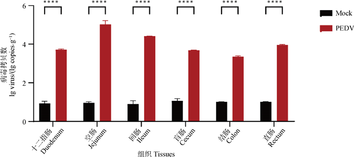
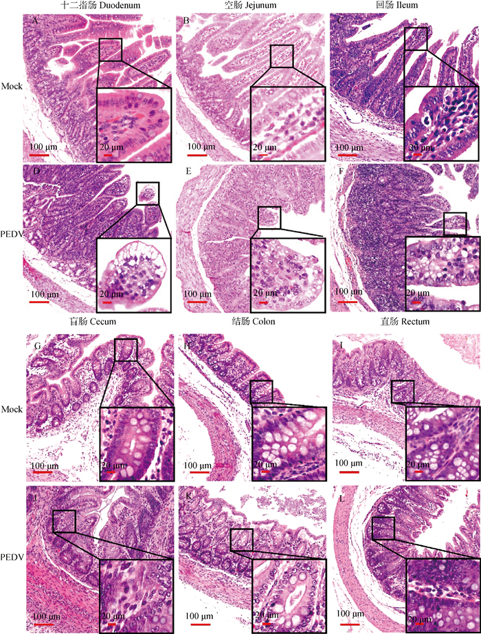
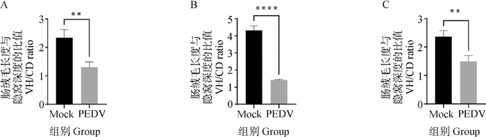
| 1 |
KOCHERHANS R , BRIDGEN A , ACKERMANN M , et al. Completion of the porcine epidemic diarrhoea coronavirus (PEDV) genome sequence[J]. Virus Gene, 2001, 23 (2): 137- 144.
doi: 10.1023/A:1011831902219
|
| 2 |
LI W , LI H , LIU Y , et al. New variants of porcine epidemic diarrhea virus, China, 2011[J]. Emerg Infect Dis, 2012, 18 (8): 1350- 1353.
doi: 10.3201/eid1803.120002
|
| 3 |
高改林, 王晨星. 猪流行性腹泻流行趋势与防控[J]. 北方牧业, 2023 (19): 34.
|
|
GAO G L , WANG C X . Epidemic trend and prevention of porcine epidemic diarrhea[J]. Northern Animal Husbandry, 2023 (19): 34.
|
| 4 |
JAYARAMAN B , NYACHOTI C M . Husbandry practices and gut health outcomes in weaned piglets: A review[J]. Anim Nutr, 2017, 3 (3): 205- 211.
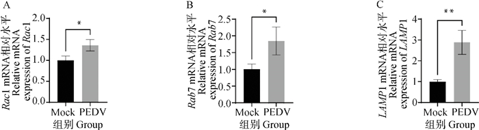
doi: 10.1016/j.aninu.2017.06.002
|
| 5 |
ZHENG X , ZHU D , XIANG Q , et al. Ginsenoside Rb1 inhibits porcine epidemic diarrhea virus replication through suppressing S1 protein mediated the MAPK/ERK pathway and reducing apoptosis[J]. Int J Biol Macromol, 2025, Pt2, 140937.
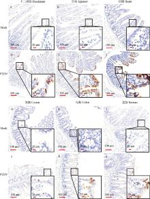
|
| 6 |
阚先进. 猪流行性腹泻病毒诱导细胞自噬的分子机制[D]. 扬州: 扬州大学, 2018.
|
|
KAN X J. Mechanisms of porcine epidemic diarrhea virus-induced autophagy[D]. Yangzhou: Yangzhou University, 2018. (in Chinese)
|
| 7 |
闫清源, 周书亭, 谢洋洋, 等. 猪流行性腹泻病毒感染Vero细胞的细胞病理学研究[J]. 上海交通大学学报(农业科学版), 2018, 36 (2): 39- 42.
|
|
YAN Q Y , ZHOU S T , XIE Y Y , et al. Cytopathology study on porcine epidemic diarrhea virus (PEDV) infected vero cells[J]. Journal of Shanghai Jiaotong University (Agricultural Science), 2018, 36 (2): 39- 42.
|
| 8 |
蔡红兵, 刘金坤, 范钦, 等. 巨泡式死亡: 一种新的细胞死亡方式[J]. 南方医科大学学报, 2013, 33 (12): 1844- 1847.
|
|
CAI H B , LIU J K , FAN Q , et al. Methuosis-a novel type of cell death[J]. Journal of Southern Medical University, 2013, 33 (12): 1844- 1847.
|
| 9 |
RITTER M , BRESGEN N , KERSCHBAUM H H . From pinocytosis to methuosis-fluid consumption as a risk factor for cell death[J]. Front Cell Dev Biol, 2021, 9, 651982.
doi: 10.3389/fcell.2021.651982
|
| 10 |
OVERMEYER J H , KAUL A , JOHNSON E E , et al. Active ras triggers death in glioblastoma cells through hyperstimulation of macropinocytosis[J]. Mol Cancer Res, 2008, 6 (6): 965- 977.
doi: 10.1158/1541-7786.MCR-07-2036
|
| 11 |
BHANOT H , YOUNG A M , OVERMEYER J H , et al. Induction of nonapoptotic cell death by activated ras requires inverse regulation of Rac1 and Arf6[J]. Mol Cancer Res, 2010, 8 (10): 1358- 1374.
doi: 10.1158/1541-7786.MCR-10-0090
|
| 12 |
OVERMEYER J H , YOUNG A M , BHANOT H , et al. A chalcone-related small molecule that induces methuosis, a novel form of non-apoptotic cell death, in glioblastoma cells[J]. Mol Cancer, 2011, 10 (1): 1- 17.
doi: 10.1186/1476-4598-10-1
|
| 13 |
BHATTACHARYA S S , JIN L , ROY D , et al. Abstract 2658: PFKFB3 inhibition reprograms malignant pleural mesothelioma to glycolytic stress-induced macropinocytosis and ER stress as independent binary adaptive responses[J]. Cell Death Dis, 2019, 10 (10): 725.
doi: 10.1038/s41419-019-1916-3
|
| 14 |
HAN Q F , LI W J , HU K S , et al. Exosome biogenesis: machinery, regulation, and therapeutic implications in cancer[J]. Mol Cancer, 2022, 21 (1): 207.
doi: 10.1186/s12943-022-01671-0
|
| 15 |
LUO Y , GUAN B , DENG X , et al. Methuosis inducer SGI-1027 cooperates with everolimus to promote apoptosis and pyroptosis by triggering lysosomal membrane permeability in renal cancer[J]. Adv Sci (Weinh), 2024, 11 (38): e2404693.
doi: 10.1002/advs.202404693
|
| 16 |
MULLIGAN R J , MAGAJ M M , DIGILIO L , et al. Collapse of late endosomal pH elicits a rapid Rab7 response via the V-ATPase and RILP[J]. J Cell Sci, 2024, 137 (9): jcs261765.
doi: 10.1242/jcs.261765
|
| 17 |
ZHANG Y H , LI H X , CHEN X M , et al. Genetic characteristics and pathogenicity of a novel porcine epidemic diarrhea virus with a naturally occurring truncated ORF3 Gene[J]. Viruses, 2022, 14 (3): 487.
doi: 10.3390/v14030487
|
| 18 |
李洪炫, 滑宇鹏, 彭琳颖, 等. 猪流行性腹泻病毒和猪圆环病毒4型双重SYBR GreenⅠ实时荧光定量PCR方法的建立[J]. 中国兽医学报, 2023, 43 (1): 23- 28.
|
|
LI H X , HUA Y P , PENG L Y , et al. Development of a SYBR Green Ⅰ-based real-time PCR assay for detection of virus and porcine circovirus 4[J]. Chinese Journal of Veterinary Science, 2023, 43 (1): 23- 28.
|
| 19 |
倪艳秀, 林继煌, 何孔旺, 等. 猪流行性腹泻研究概况[J]. 畜牧与兽医, 2001 (1): 38- 40.
|
|
NI Y X , LIN J H , HE K W , et al. Research overview of porcine epidemic diarrhea[J]. Animal Husbandry & Veterinary Medicine, 2001 (1): 38- 40.
|
| 20 |
卓秀萍, 朱玲, 乔小改, 等. 人工感染猪流行性腹泻病毒的哺乳仔猪的病理学观察[J]. 中国兽医科学, 2015, 45 (2): 202- 207.
|
|
ZHUO X P , ZHU L , QIAO X G , et al. Pathological observation of suckling piglets infected with porcine epidemic diarrhea virus artificially[J]. Chinese Veterinary Science, 2015, 45 (2): 202- 207.
|
| 21 |
JUNG K , MIYAZAKI A , SAIF L J . Immunohistochemical detection of the vomiting-inducing monoamine neurotransmitter serotonin and enterochromaffin cells in the intestines of conventional or gnotobiotic (Gn) pigs infected with porcine epidemic diarrhea virus (PEDV) and serum cytokine responses of Gn pigs to acute PEDV infection[J]. Res Vet Sci, 2018, 119, 99- 108.
doi: 10.1016/j.rvsc.2018.06.009
|
| 22 |
JUNG K , SAIF L J . Goblet cell depletion in small intestinal villous and crypt epithelium of conventional nursing and weaned pigs infected with porcine epidemic diarrhea virus[J]. Res Vet Sci, 2017, 110, 12- 15.
doi: 10.1016/j.rvsc.2016.10.009
|
| 23 |
肖静, 董斌, 王俊棋, 等. 细胞骨架与细胞膜损伤介导药物引起心肌细胞巨泡式死亡的作用与机制[C]. 厦门: 中国毒理学会第九次全国青年科技大会暨第二届生物技术药物毒理与安全评价委员会学术会议论文集, 2023: 46-47.
|
|
XIAO J, DONG B, WANG J Q, et al. The role and mechanism of cytoskeleton and cell membrane damage mediating drug induced cardiomyocyte methuosis[C]. Xiamen: Proceedings of the 9th National Youth Science and Technology Conference of the Chinese Society of Toxicology and the 2nd Academic Conference of the Committee for Toxicology and Safety Evaluation of Biotechnology Drugs, 2023: 46-47.
|
| 24 |
LUISONI S , SUOMALAINEN M , BOUCKE K , et al. Co-option of membrane wounding enables virus penetration into cells[J]. Cell Host Microbe, 2015, 18 (1): 75- 85.
doi: 10.1016/j.chom.2015.06.006
|


 ), 著龙祥1,2(