


畜牧兽医学报 ›› 2025, Vol. 56 ›› Issue (9): 4267-4278.doi: 10.11843/j.issn.0366-6964.2025.09.012
余时隆( ), 岳潇雨, 栾悦, 王勤*()
收稿日期:2024-08-26
出版日期:2025-09-23
发布日期:2025-09-30
通讯作者:
王勤
E-mail:slyu@cau.edu.cn;wangqin@cau.edu.cn
作者简介:余时隆(2001-),男,江西都昌人,硕士生,主要从事动物遗传育种与繁殖研究,E-mail:slyu@cau.edu.cn
基金资助:
YU Shilong(), YUE Xiaoyu, LUAN Yue, WANG Qin*()
Received:2024-08-26
Online:2025-09-23
Published:2025-09-30
Contact:
WANG Qin
E-mail:slyu@cau.edu.cn;wangqin@cau.edu.cn
摘要:
皮肤伤口愈合是一个涉及炎症、组织再生和细胞外基质重塑的复杂生理过程,其失调可能导致疤痕增生或慢性溃疡,影响皮肤功能。本文综述了动物皮肤伤口愈合的分子机制,探讨了包括新型药物开发、改善药物传输、调控激素及外源信号、基因治疗和组织工程法在内的多种修复策略。这些方法通过改善药物传输、调控炎症反应、促进血管及组织再生,从而有效提升了伤口愈合效果。本研究旨在为未来动物皮肤伤口愈合提供新的视角,指出了潜在的研究思路和方向,以期开发出更高效、安全的治疗策略,改善动物的健康和福利。
中图分类号:
余时隆, 岳潇雨, 栾悦, 王勤. 动物皮肤伤口愈合的分子机制与创新治疗策略[J]. 畜牧兽医学报, 2025, 56(9): 4267-4278.
YU Shilong, YUE Xiaoyu, LUAN Yue, WANG Qin. Molecular Mechanisms and Innovative Therapeutic Strategies for Cutaneous Wound Healing in Animals[J]. Acta Veterinaria et Zootechnica Sinica, 2025, 56(9): 4267-4278.
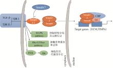
图 1
TGF-β信号转导通路"

表 1
非编码RNA影响伤口愈合的通路"
| 非编码RNA Non-coding RNA | 靶向目标 Targeting | 靶基因 Target gene | 作用(通路) Role (pathway) | 参考文献 Reference |
| circRNA_101238 | miR-138-5p | CDK-6 | 促进瘢痕成纤维细胞凋亡Promoting apoptosis in scar fibroblasts | Yang等[ |
| circCOL5A1 | miR-7-5p | Epac1 | 通过PI3K/Akt信号通路促进瘢痕疙瘩增殖Promoting keloid proliferation through the PI3K/Akt signalling pathway | Lv等[ |
| circRNA-MYLK | miR-29a | VEGFA | TGF-β信号通路,NF-κB信号通路和β联蛋白途径加快 创面修复 TGF-β signalling pathway, NF-κB signalling pathway and β-linked protein pathway accelerate wound repair | Zhong等[ |
| circRNA_010567 | miR-141 | TGF-β1 | 促进胶原生成 Promotes collagen production | Zhou和Yu[ |
| circ-Amotl1 | miR-17-5p | STAT3、 DNMT3A | 增加细胞黏附、迁移、增殖、存活和伤口修复 Increases cell adhesion, migration, proliferation, survival and wound repair | Yang等[ |
| LncRNA FOXD2-AS1 | miR-185-5p | ROCK2 | 促进角质形成细胞迁移和增殖 Promote migration and proliferation of keratinocytes | Chang等[ |
| HOTAIR | miR-126 | VEGF | 促进血管生成和伤口愈合Promotes angiogenesis and wound healing | Born等[ Jiang等[ |
| LncRNA HULC | miR-663b | TLR4 | 抑制皮肤成纤维细胞的增殖、侵袭和ECM合成Inhibition of proliferation, invasion and ECM synthesis in skin fibroblasts | Liu等[ |
| MALAT1 | miR-124 | 激活Wnt/β-连环蛋白通路帮助伤口愈合 Activation of the Wnt/β-Cyclin pathway helps wound healing | He等[ | |
| SCARNA2 | miR-143-3p | ADD3 | 促进体外伤口愈合Promote wound healing in vitro | Xiong等[ |
| H19 | NLRP3炎症小体途径NLRP3 inflammatory vesicle pathway | Yang等[ |
| 1 |
VILEIKYTE L . Stress and wound healing[J]. Clin Dermatol, 2007, 25 (1): 49- 55.
doi: 10.1016/j.clindermatol.2006.09.005 |
| 2 | SHAW T J , MARATIN P . Wound repair at a glance[J]. J Cell Sci, 2009, Pt 18 (122): 3209- 3213. |
| 3 |
MARTIN P , LEIBOVICH S J . Inflammatory cells during wound repair: The good, the bad and the ugly[J]. Trends Cell Biol, 2005, 15 (11): 599- 607.
doi: 10.1016/j.tcb.2005.09.002 |
| 4 | DARBY I A , LAVERDET B , BONTE F , et al. Fibroblasts and myofibroblasts in wound healing[J]. Clin Cosmet Investig Dermatol, 2014, 7, 301- 311. |
| 5 | BERMAN B , MADERAL A , RAPHAEL B . Keloids and hypertrophic scars: Pathophysiology, classification, and treatment[J]. Dermatol Surg, 2017, 43 (Suppl 1): S3- S18. |
| 6 | TSAI C H , OGAWA R . Keloid research: current status and future directions[J]. Scars Burn Heal, 2019, 5, 1007974669. |
| 7 |
LIMANDJAJA G C , NIESSEN F B , SCHEPER R J , et al. Hypertrophic scars and keloids: Overview of the evidence and practical guide for differentiating between these abnormal scars[J]. Exp Dermatol, 2021, 30 (1): 146- 161.
doi: 10.1111/exd.14121 |
| 8 | 刘梦琪. 慢性伤口患者疾病心理社会适应水平现状及影响因素研究[D]. 芜湖: 皖南医学院, 2023. |
| LIU M Q. A study on the current situation of the level of psychosocial adaptation to illness in patients with chronic wounds and the factors affecting it[D]. Wuhu: Wannan Medical College, 2023. (in Chinese) | |
| 9 | NAVARRO-ALVAREZ N , GONCALVES B , ANDREWS A R , et al. A CFA-induced model of inflammatory skin disease in miniature swine[J]. Int J Inflam, 2018, 2018, 6916920. |
| 10 | 雷映文. 母水牛伤口感染治疗[J]. 四川畜牧兽医, 2014, 41 (2): 56. |
| LEI Y W . Treatment of wound infection in female buffaloes[J]. Sichuan Animal & Veterinary Sciences, 2014, 41 (2): 56. | |
| 11 | 余良政, 陈建材, 周华波, 等. 一例犬严重伤口感染病例的诊治[J]. 云南畜牧兽医, 2021 (4): 16- 19. |
| YU Z L , CHEN J C , ZHOU H B , et al. Diagnosis and treatment of a case of severe wound infection in a dog[J]. Yunnan Journal of Animal Science and Veterinary Medicine, 2021 (4): 16- 19. | |
| 12 |
MCGOWAN C . Clinical pathology in the racing horse: the role of clinical pathology in assessing fitness and performance in the racehorse[J]. Vet Clin North Am Equine Pract, 2008, 24 (2): 405- 421.
doi: 10.1016/j.cveq.2008.03.001 |
| 13 |
CHANG H Y , SNEDDON J B , ALIZADEH A A , et al. Gene expression signature of fibroblast serum response predicts human cancer progression: similarities between tumors and wounds[J]. PLoS Biol, 2004, 2 (2): E7.
doi: 10.1371/journal.pbio.0020007 |
| 14 |
BEANES S R , DANG C , SOO C , et al. Skin repair and scar formation: the central role of TGF-beta[J]. Expert Rev Mol Med, 2003, 5 (8): 1- 22.
doi: 10.1017/S1462399403005817 |
| 15 |
YANG L , CHAN T , DEMARE J , et al. Healing of burn wounds in transgenic mice overexpressing transforming growth factor-beta 1 in the epidermis[J]. Am J Pathol, 2001, 159 (6): 2147- 2157.
doi: 10.1016/S0002-9440(10)63066-0 |
| 16 |
HEYDEMANN A . The super super-healing MRL mouse strain[J]. Front Biol (Beijing), 2012, 7 (6): 522- 538.
doi: 10.1007/s11515-012-1192-4 |
| 17 |
DERYNCK R , ZHANG Y E . Smad-dependent and Smad-independent pathways in TGF-beta family signalling[J]. Nature, 2003, 425 (6958): 577- 584.
doi: 10.1038/nature02006 |
| 18 |
WANG X J , HAN G , OWENS P , et al. Role of TGF beta-mediated inflammation in cutaneous wound healing[J]. J Investig Dermatol Symp Proc, 2006, 11 (1): 112- 117.
doi: 10.1038/sj.jidsymp.5650004 |
| 19 | 王佩茹, 韩佳彤, 魏茗蕾, 等. TGFβ通路介导的低剂量5-氨基酮戊酸光动力疗法重塑光老化皮肤真皮胶原[J]. 中国激光医学杂志, 2018, 27 (2): 72. |
| WANG P R , HAN J T , WEI M L , et al. TGFβ pathway-mediated low-dose 5-aminolevulinic acid photodynamic therapy for remodelling of dermal collagen in photodamaged skin[J]. Chinese Journal of Laser Medicine & Surgery, 2018, 27 (2): 72. | |
| 20 |
FERRARI G , COOK B D , TERUSHKIN V , et al. Transforming growth factor-beta 1 (TGF-beta1) induces angiogenesis through vascular endothelial growth factor (VEGF)-mediated apoptosis[J]. J Cell Physiol, 2009, 219 (2): 449- 458.
doi: 10.1002/jcp.21706 |
| 21 |
POHLERS D , BRENMOEHL J , LOFFLER I , et al. TGF-beta and fibrosis in different organs-molecular pathway imprints[J]. Biochim Biophys Acta, 2009, 1792 (8): 746- 756.
doi: 10.1016/j.bbadis.2009.06.004 |
| 22 |
BULLARD K M , CASS D L , BANDA M J , et al. Transforming growth factor beta-1 decreases interstitial collagenase in healing human fetal skin[J]. J Pediatr Surg, 1997, 32 (7): 1023- 1027.
doi: 10.1016/S0022-3468(97)90391-2 |
| 23 |
FERGUSON M W , O'KANE S . Scar-free healing: from embryonic mechanisms to adult therapeutic intervention[J]. Philos Trans R Soc Lond B Biol Sci, 2004, 359 (1445): 839- 850.
doi: 10.1098/rstb.2004.1475 |
| 24 |
SULLIVAN K M , LORENZ H P , MEULI M , et al. A model of scarless human fetal wound repair is deficient in transforming growth factor beta[J]. J Pediatr Surg, 1995, 30 (2): 198- 203.
doi: 10.1016/0022-3468(95)90560-X |
| 25 | YANG D , LI M , DU N . Effects of the circ_101238/miR-138-5p/CDK6 axis on proliferation and apoptosis keloid fibroblasts[J]. Exp Ther Med, 2020, 20 (3): 1995- 2002. |
| 26 |
LV W , LIU S , ZHANG Q , et al. Circular RNA circCOL5A1 sponges the miR-7-5p/Epac1 axis to promote the progression of keloids through regulating PI3K/Akt signaling pathway[J]. Front Cell Dev Biol, 2021, 9, 626027.
doi: 10.3389/fcell.2021.626027 |
| 27 |
ZHONG Z , HUANG M , LV M , et al. Circular RNA MYLK as a competing endogenous RNA promotes bladder cancer progression through modulating VEGFA/VEGFR2 signaling pathway[J]. Cancer Lett, 2017, 403, 305- 317.
doi: 10.1016/j.canlet.2017.06.027 |
| 28 |
ZHOU B , YU J W . A novel identified circular RNA, circRNA_010567, promotes myocardial fibrosis via suppressing miR-141 by targeting TGF-beta1[J]. Biochem Biophys Res Commun, 2017, 487 (4): 769- 775.
doi: 10.1016/j.bbrc.2017.04.044 |
| 29 |
YANG Z G , AWAN F M , DU W W , et al. The Circular RNA interacts with STAT3, increasing its nuclear translocation and wound repair by modulating Dnmt3a and miR-17 function[J]. Mol Ther, 2017, 25 (9): 2062- 2074.
doi: 10.1016/j.ymthe.2017.05.022 |
| 30 |
CHANG H , CHEN J , DING K , et al. Highly-expressed lncRNA FOXD2-AS1 in adipose mesenchymal stem cell derived exosomes affects HaCaT cells via regulating miR-185-5p/ROCK2 axis[J]. Adipocyte, 2023, 12 (1): 2173513.
doi: 10.1080/21623945.2023.2173513 |
| 31 |
BORN L J , CHANG K H , SHOURESHI P , et al. HOTAIR-loaded mesenchymal stem/stromal cell extracellular vesicles enhance angiogenesis and wound healing[J]. Adv Healthc Mater, 2022, 11 (5): e2002070.
doi: 10.1002/adhm.202002070 |
| 32 |
JIANG B , TANG Y , WANG H , et al. Down-regulation of long non-coding RNA HOTAIR promotes angiogenesis via regulating miR-126/SCEL pathways in burn wound healing[J]. Cell Death Dis, 2020, 11 (1): 61.
doi: 10.1038/s41419-020-2247-0 |
| 33 | LIU Y , QI X , ZHOU Y . Long non-coding RNA HULC regulates TLR4 expression by acting as ceRNA to attract miR-663b in skin fibroblasts of pediatric burns[J]. Am J Transl Res, 2021, 13 (4): 2499- 2510. |
| 34 |
HE L , ZHU C , JIA J , et al. ADSC-Exos containing MALAT1 promotes wound healing by targeting miR-124 through activating Wnt/beta-catenin pathway[J]. Biosci Rep, 2020, 40 (5): BSR20192549.
doi: 10.1042/BSR20192549 |
| 35 |
XIONG H , REN S , CHEN J , et al. Knockdown of long noncoding RNA SAN rejuvenates aged adipose-derived stem cells via miR-143-3p/ADD3 axis[J]. Stem Cell Res Ther, 2023, 14 (1): 213.
doi: 10.1186/s13287-023-03441-1 |
| 36 | YANG H , ZHANG Y , DU Z , et al. Hair follicle mesenchymal stem cell exosomal lncRNA H19 inhibited NLRP3 pyroptosis to promote diabetic mouse skin wound healing[J]. Aging (Albany NY), 2023, 15 (3): 791- 809. |
| 37 |
RAZIYEVA K , KIM Y , ZHARKINBEKOV Z , et al. Immunology of acute and chronic wound healing[J]. Biomolecules, 2021, 11 (5): 700.
doi: 10.3390/biom11050700 |
| 38 | 崔允美, 崔晶, 李今子. 皮肤无瘢痕愈合的研究现状[J]. 中国皮肤性病学杂志, 2023, 37 (9): 988- 993. |
| CUI Y M , CUI J , LI J Z . Current status of research on scarless healing of the skin[J]. The Chinese Journal of Dermatovenereology, 2023, 37 (9): 988- 993. | |
| 39 |
ZHAO M , SONG B , PU J , et al. Electrical signals control wound healing through phosphatidylinositol-3-OH kinase-gamma and PTEN[J]. Nature, 2006, 442 (7101): 457- 460.
doi: 10.1038/nature04925 |
| 40 |
RAJAGOPALAN K , SELVAN C J , CHELLADURAI K S , et al. Understanding the molecular mechanism of regeneration through apoptosis-induced compensatory proliferation studies-updates and future aspects[J]. Apoptosis, 2024, 29 (9-10): 1399- 1414.
doi: 10.1007/s10495-024-01958-1 |
| 41 | 海洋. 软刚度抑制人成纤维细胞增殖和分化的关键基因特征研究[D]. 长春: 吉林大学, 2023. |
| HAI Y. Characterisation of key genes involved in soft stiffness inhibition of human fibroblast proliferation and differentiation[D]. Changchun: Jilin University, 2023. (in Chinese) | |
| 42 | 尚瑾, 秦盼月, 杨兴鑫, 等. 基于皮肤"微环境污染"的人参皂苷Rg3修复糖尿病皮肤损伤的作用机理研究[J]. 世界科学技术-中医药现代化, 2022, 24 (2): 582- 590. |
| SHANG J , QIN P Y , YANG X X , et al. Study on the mechanism of ginsenoside Rg3 in repairing diabetic skindamage based on skin "micro-environmental pollution"[J]. Modernization of Traditional Chinese Medicine and Materia Medica-World Science and Technology, 2022, 24 (2): 582- 590. | |
| 43 | XU X , YANG W H , MIAO Z W , et al. Modified hongyu decoction promotes wound healing by activating the VEGF/PI3K/Akt signaling pathway[J]. Acta Biochim Pol, 2023, 70 (4): 843- 853. |
| 44 |
CHAK K F , HSIAO C Y , CHEN T Y . A study of the effect of shiunko, a traditional chinese herbal medicine, on fibroblasts and its implication on wound healing processes[J]. Adv Wound Care (New Rochelle), 2013, 2 (8): 448- 455.
doi: 10.1089/wound.2012.0368 |
| 45 |
ANIS A , SHARSHAR A , HANBALLY S E , et al. A novel organic composite accelerates wound healing: Experimental and clinical study in equine[J]. J Equine Vet Sci, 2021, 99, 103406.
doi: 10.1016/j.jevs.2021.103406 |
| 46 | 陈淼. 灵芝孢子粉对犬皮肤创伤愈合研究[D]. 长春: 吉林农业大学, 2022. |
| CHEN M. Study of ganoderma lucidum spore powder on wound healing of dog skin[D]. Changchun: Jilin Agricutural University, 2022. (in Chinese) | |
| 47 | 朴雪. 马勃促进犬皮肤创面愈合的效果观察[D]. 哈尔滨: 东北农业大学, 2023. |
| PIAO X. The effect of lasiosphaera calvatia on promoting skin wound healing in canines[D]. Harbin: Northeast Agricultural University, 2023. (in Chinese) | |
| 48 | GUPTA R , KAUR T , SHARMA S . Transfersomes for escalating effectiveness of drugs via transdermal and topical administration: A review[J]. Pharm Biosci J, 2022, 9- 18. |
| 49 | 梁舒韵, 赵莉娜, 邓清月, 等. 罗非鱼皮抗氧化肽联合维生素C对皮肤损伤的愈合作用[J]. 现代食品科技, 2023, 39 (6): 10- 17. |
| LIANG S Y , ZHAO L N , DENG Q Y , et al. Synergistic healing of skin injuries by tilapia skin antioxidant peptide and vitamin C[J]. Modern Food Science and Technology, 2023, 39 (6): 10- 17. | |
| 50 |
BIYASHEV D , ONAY U V , DALAL P , et al. A novel treatment for skin repair using a combination of spironolactone and vitamin D3[J]. Ann N Y Acad Sci, 2020, 1480 (1): 170- 182.
doi: 10.1111/nyas.14485 |
| 51 |
ZHANG S , TAN H , CHENG X , et al. Autologous platelet-rich fibrin enhances skin wound healing in a feline trauma model[J]. BMC Vet Res, 2024, 20 (1): 504.
doi: 10.1186/s12917-024-04358-4 |
| 52 | 廖雪玉. 犬富血小板纤维蛋白对猫创口恢复的初步研究[D]. 武汉: 华中农业大学, 2024. |
| LIAO X Y. Canine platelet-rich fibrin on wound recovery in cats: A preliminary study[D]. Wuhan: Huazhong Agricultural University, 2024. (in Chinese) | |
| 53 | 贺丝雨. 富血小板纤维蛋白(PRF)促进兔急性伤口愈合的实验研究[D]. 南充: 川北医学院, 2023. |
| HE S Y. Experimental study on Platelet-rich fibrin(PRF)promoting the healing of acute wounds in rabbits[D]. Nanchong: North Sichuan Medical College, 2023. (in Chinese) | |
| 54 | 肖梓腾, 王婷禹, 张雯雯, 等. 外泌体与皮肤创伤的修复[J]. 中国组织工程研究, 2024, 28 (19): 3104- 3110. |
| XIAO Z T , WANG T Y , ZHANG W W , et al. Exosomes and skin wound healing[J]. Chinese Journal of Tissue Engineering Research, 2024, 28 (19): 3104- 3110. | |
| 55 | 张慧敏. 犬脂肪间充质干细胞外泌体对犬皮肤损伤治疗作用探究[D]. 杨凌: 西北农林科技大学, 2022. |
| ZHANG H M. The therapeutic effect of canine adipose mesenchymal stem cell-derived exosomes on canine skin injury[D]. Yangling: Northwest A&F University, 2022. (in Chinese) | |
| 56 |
GONG C , WU Q , WANG Y , et al. A biodegradable hydrogel system containing curcumin encapsulated in micelles for cutaneous wound healing[J]. Biomaterials, 2013, 34 (27): 6377- 6387.
doi: 10.1016/j.biomaterials.2013.05.005 |
| 57 | 许真真, 焦红军, 任明. 回阳生肌脂质体凝胶治疗糖尿病足溃疡大鼠的作用及其对TGF-β1/Smad3通路的调控机制[J]. 湖北中医杂志, 2024, 46 (7): 7- 11. |
| XU Z Z , JIAO H J , REN M . Effect of Huiyangshengji liposome gel on diabetes foot ulcer rats and its regulating mechanism on TGF- 1/Smad3 pathway[J]. Hubei Journal of Traditional Chinese Medicine, 2024, 46 (7): 7- 11. | |
| 58 | 周杰. 负载CpG ODNs水凝胶的制备及其用于伤口修复的研究[D]. 无锡: 江南大学, 2023. |
| ZHOU J. Preparation of CpG ODNs-loaded hydrogel for wound repair[D]. Wuxi: Jiangnan University, 2023. (in Chinese) | |
| 59 |
ALENABI A , BEHFAR M , MALEKINEJAD H , et al. Allotransplantation of ascorbic acid-treated fibroblasts improves healing of excisional cutaneous wound in diabetic rats[J]. Acta Histochem, 2022, 124 (2): 151857.
doi: 10.1016/j.acthis.2022.151857 |
| 60 |
SPARKS H D , SIGAEVA T , TARRAF S , et al. Biomechanics of wound healing in an equine limb model: effect of location and treatment with a peptide-modified collagen-chitosan hydrogel[J]. ACS Biomater Sci Eng, 2021, 7 (1): 265- 278.
doi: 10.1021/acsbiomaterials.0c01431 |
| 61 | 刘倩倩, 陈玉琪, 杨斯琪, 等. 壳聚糖纳米银水凝胶对犬感染创的治疗效果[C]//中国畜牧兽医学会兽医外科学分会第十届会员代表大会暨第24次学术研讨会. 2019: 344. |
| LIU Q Q, CHEN Y Q, YANG S Q, et al. Therapeutic efficacy of chitosan nanosilver hydrogel on canine infected wounds[C]//The 10th General Meeting and 24th Symposium of the Veterinary Surgery Branch of the Chinese Association of Animal Husbandry and Veterinary Science. 2019: 344. (in Chinese) | |
| 62 | 李明聪, 陈文静, 孙明楷, 等. 静电纺羧甲基纤维素伤口敷料的研究进展[J]. 棉纺织技术, 2025, 53 (5): 68- 73. |
| LI M C , CHEN W J , SUN M K , et al. Research progress of wound dressing for electrospinning carboxymethyl cellulose[J]. Cotton Textile Technology, 2025, 53 (5): 68- 73. | |
| 63 |
GAD E S , HALAWANI E M , ALZAHRANI S . Biosynthesis of silver nano-drug using Juniperus excelsa and its synergistic antibacterial activity against multidrug-resistant bacteria for wound dressing applications[J]. 3 Biotech, 2021, 11 (6): 255.
doi: 10.1007/s13205-021-02782-z |
| 64 |
ROY A P , JANA S , DAS H , et al. Stimulated full-thickness cutaneous wound healing with bioactive dressings of zinc and cobalt ion-doped bioactive glass-coated eggshell membranes in a diabetic rabbit model[J]. ACS Biomater Sci Eng, 2024, 10 (7): 4510- 4524.
doi: 10.1021/acsbiomaterials.4c00691 |
| 65 | 岳圆. 静电纺黄芪多糖及黄芪甲苷脂质体纳米纤维膜促糖尿病创面愈合的效果研究[D]. 成都: 成都中医药大学, 2022. |
| YUE Y. Effect of electrospun astragalus polysaccharide andastragaloside IV liposome nanofiber membrane on promoting diabetic wound healing[D]. Chengdu: Chengdu University of TCM, 2022. | |
| 66 |
CHRISTIAN L M , GRAHAM J E , PADGETT D A , et al. Stress and wound healing[J]. Neuroimmunomodulation, 2006, 13 (5-6): 337- 346.
doi: 10.1159/000104862 |
| 67 |
MARUCHA P T , KIECOLT-GLASER J K , FAVAGEHI M . Mucosal wound healing is impaired by examination stress[J]. Psychosom Med, 1998, 60 (3): 362- 365.
doi: 10.1097/00006842-199805000-00025 |
| 68 |
LEAUTE-LABREZE C , DUMAS D L R E , HUBICHE T , et al. Propranolol for severe hemangiomas of infancy[J]. N Engl J Med, 2008, 358 (24): 2649- 2651.
doi: 10.1056/NEJMc0708819 |
| 69 | MOHAMMADI A A , BAKHSHAEEKIA A , ALIBEIGI P , et al. Efficacy of propranolol in wound healing for hospitalized burn patients[J]. J Burn Care Res, 2009, 30 (6): 1013- 1017. |
| 70 |
TANG J C , DOSAL J , KIRSNER R S . Topical timolol for a refractory wound[J]. Dermatol Surg, 2012, 38 (1): 135- 138.
doi: 10.1111/j.1524-4725.2011.02200.x |
| 71 |
KACZMAREK-SZCZEPANSKA B , PIN J M , ZASADA L , et al. Assessment of melatonin-cultured collagen/chitosan scaffolds cross-linked by a glyoxal solution as biomaterials for wound healing[J]. Antioxidants (Basel), 2022, 11 (3): 570.
doi: 10.3390/antiox11030570 |
| 72 |
JIN M , FAN W , PIAO J , et al. Effects of lncRNA MTC on protein expression in skin fibroblasts of Liaoning Cashmere goat based on iTRAQ technique[J]. Anim Biotechnol, 2023, 34 (7): 2817- 2826.
doi: 10.1080/10495398.2022.2119406 |
| 73 |
SCARDINO M S , SWAIM S F , SARTIN E A , et al. Evaluation of treatment with a pulsed electromagnetic field on wound healing, clinicopathologic variables, and central nervous system activity of dogs[J]. Am J Vet Res, 1998, 59 (9): 1177- 1181.
doi: 10.2460/ajvr.1998.59.09.1177 |
| 74 |
LUO R , DAI J , ZHANG J , et al. Accelerated skin wound healing by electrical stimulation[J]. Adv Healthc Mater, 2021, 10 (16): e2100557.
doi: 10.1002/adhm.202100557 |
| 75 |
MAIJER A , GESSNER A , TRUMPATORI B , et al. Bioelectric dressing supports complex wound healing in small animal patients[J]. Top Companion Anim Med, 2018, 33 (1): 21- 28.
doi: 10.1053/j.tcam.2018.02.001 |
| 76 |
NOLFF M , ALBERT R , WOHLSEIN P , et al. Histomorphometric evaluation of MMP-9 and CD31 expression during healing under negative pressure wound therapy in dogs[J]. Schweiz Arch Tierheilkd, 2018, 160 (9): 525- 532.
doi: 10.17236/sat00173 |
| 77 |
DEMARIA M , STANLEY B J , HAUPTMAN J G , et al. Effects of negative pressure wound therapy on healing of open wounds in dogs[J]. Vet Surg, 2011, 40 (6): 658- 669.
doi: 10.1111/j.1532-950X.2011.00849.x |
| 78 |
HASPESLAGH M , VAN HECKE L L , HERMANS K , et al. Limited added value of negative pressure wound therapy compared with calcium alginate dressings for second intention healing in a noncontaminated and contaminated equine distal limb wound model[J]. Equine Vet J, 2022, 54 (3): 592- 600.
doi: 10.1111/evj.13487 |
| 79 |
HATIBIE M J , ISLAM A A , HATTA M , et al. Hyperbaric oxygen therapy for second-degree burn healing: an experimental study in rabbits[J]. Adv Skin Wound Care, 2019, 32 (3): 1- 4.
doi: 10.1097/01.ASW.0000553110.78375.7b |
| 80 | 米晶, 吴珊. 多波段LED促进皮肤创伤修复的研究[J]. 应用激光, 2024, 44 (1): 161- 166. |
| MI J , WU S . The promotion of multiband LED in wound healing of cutaneous cells[J]. Applied Laser, 2024, 44 (1): 161- 166. | |
| 81 | 李梅清. 激光理疗技术在宠物临床中的应用研究进展[J]. 中国动物保健, 2022, 24 (11): 102- 104. |
| LI M Q . Progress of research on the application of laser physiotherapy techniques in pet clinics[J]. China Animal Health, 2022, 24 (11): 102- 104. | |
| 82 |
NGUYEN P D , TUTELA J P , THANIK V D , et al. Improved diabetic wound healing through topical silencing of p53 is associated with augmented vasculogenic mediators[J]. Wound Repair Regen, 2010, 18 (6): 553- 559.
doi: 10.1111/j.1524-475X.2010.00638.x |
| 83 |
BRANSKI L K , GAUGLITZ G G , HERNDON D N , et al. A review of gene and stem cell therapy in cutaneous wound healing[J]. Burns, 2009, 35 (2): 171- 180.
doi: 10.1016/j.burns.2008.03.009 |
| 84 |
GALIANO R D , TEPPER O M , PELO C R , et al. Topical vascular endothelial growth factor accelerates diabetic wound healing through increased angiogenesis and by mobilizing and recruiting bone marrow-derived cells[J]. Am J Pathol, 2004, 164 (6): 1935- 1947.
doi: 10.1016/S0002-9440(10)63754-6 |
| 85 |
GALEANO M , DEODATO B , ALTAVILLA D , et al. Adeno-associated viral vector-mediated human vascular endothelial growth factor gene transfer stimulates angiogenesis and wound healing in the genetically diabetic mouse[J]. Diabetologia, 2003, 46 (4): 546- 555.
doi: 10.1007/s00125-003-1064-1 |
| 86 | ARANGO M , CHAMORRO C , COHEN-HAGUENAUER O , et al. Human skin keratinocytes modified by a Friend-derived retroviral vector: a functional approach[J]. Dermatol Online J, 2005, 11 (2): 2. |
| 87 |
RAGHAVACHARI N , FAHL W E . Targeted gene delivery to skin cells in vivo: a comparative study of liposomes and polymers as delivery vehicles[J]. J Pharm Sci, 2002, 91 (3): 615- 622.
doi: 10.1002/jps.10061 |
| 88 |
JOZIC I , DAUNERT S , TOMIC-CANIC M , et al. Nanoparticles for fidgety cell movement and enhanced wound healing[J]. J Invest Dermatol, 2015, 135 (9): 2151- 2153.
doi: 10.1038/jid.2015.237 |
| 89 |
CHARAFEDDINE R A , MAKDISI J , SCHAIRER D , et al. Fidgetin-like 2: a microtubule-based regulator of wound healing[J]. J Invest Dermatol, 2015, 135 (9): 2309- 2318.
doi: 10.1038/jid.2015.94 |
| 90 |
CHANDAN R , MEHTA S , BANERJEE R . Ultrasound-responsive carriers for therapeutic applications[J]. ACS Biomater Sci Eng, 2020, 6 (9): 4731- 4747.
doi: 10.1021/acsbiomaterials.9b01979 |
| 91 |
TRIPATHI S , SINGH B N , SINGH D , et al. Optimization and evaluation of ciprofloxacin-loaded collagen/chitosan scaffolds for skin tissue engineering[J]. 3 Biotech, 2021, 11 (4): 160.
doi: 10.1007/s13205-020-02567-w |
| 92 |
CHHABRA R , PESHATTIWAR V , PANT T , et al. In vivo studies of 3D starch-gelatin scaffolds for full-thickness wound healing[J]. ACS Appl Bio Mater, 2020, 3 (5): 2920- 2929.
doi: 10.1021/acsabm.9b01139 |
| 93 | 何丽娟, 张博文, 贾雅丽, 等. 人脐带间充质干细胞复合胶原支架治疗小型猪皮肤缺损的实验研究[J]. 军事医学, 2020, 44 (3): 192- 197. |
| HE L J , ZHANG B W , JIA Y L , et al. Human umbilical cord mesenchymal stem cells combined with collagen scaffolds in accelerating cutaneous wound healing in minipig model[J]. Military Medical Sciences, 2020, 44 (3): 192- 197. | |
| 94 | 王伟彬. 透明质酸-明胶互穿网络水凝胶的制备及其用于组织工程皮肤构建的研究[D]. 福州: 福州大学, 2021. |
| WANG W B. Preparation of hyaluronic acid-gelatin interpenetrating network hydrogels for tissue-engineered skin construction[D]. Fuzhou: Fuzhou University, 2021. (in Chinese) | |
| 95 |
SUN G , ZHANG X , SHEN Y I , et al. Dextran hydrogel scaffolds enhance angiogenic responses and promote complete skin regeneration during burn wound healing[J]. Proc Natl Acad Sci U S A, 2011, 108 (52): 20976- 20981.
doi: 10.1073/pnas.1115973108 |
| 96 | 李志浩. 高浓度胶原蛋白-硫酸软骨素支架复合富血小板血浆促进猪骨外露伤口愈合的实验研究[D]. 广州: 南方医科大学, 2023. |
| LI Z H. A highly concentrated collagen chondroitin sulfate scaffold composite with platelet rich plasma promotes wound healing in porcine exostosis[D]. Guangzhou: Southern Medical University, 2023. (in Chinese) | |
| 97 |
HU J , SONG Y , ZHANG C , et al. Highly aligned electrospun collagen/polycaprolactone surgical sutures with sustained release of growth factors for wound regeneration[J]. ACS Appl Bio Mater, 2020, 3 (2): 965- 976.
doi: 10.1021/acsabm.9b01000 |
| 98 | 邹慧蛟. 纤维蛋白粘合剂对犬皮肤切口创伤愈合作用的效果观察[D]. 哈尔滨: 东北农业大学, 2018. |
| ZOU H J. Observation on the healing effect of fibrin adhesive on skin wound in dogs[D]. Harbin: Northeast Agricultural University, 2018. (in Chinese) | |
| 99 |
OJEH N , PASTAR I , TOMIC-CANIC M , et al. Stem cells in skin regeneration, wound healing, and their clinical applications[J]. Int J Mol Sci, 2015, 16 (10): 25476- 25501.
doi: 10.3390/ijms161025476 |
| 100 |
HOUDEK M T , WYLES C C , STALBOERGER P G , et al. Collagen and fractionated platelet-rich plasma scaffold for dermal regeneration[J]. Plast Reconstr Surg, 2016, 137 (5): 1498- 1506.
doi: 10.1097/PRS.0000000000002094 |
| 101 |
LI F , GAO C , SONG G , et al. Human placenta-derived mesenchymal stem cells combined with artificial dermal scaffold enhance wound healing in a tendon-exposed wound of a rabbit model[J]. Cell Transplant, 2024, 33, 9636897241228922.
doi: 10.1177/09636897241228922 |
| 102 | 吕英光. ADSCs对巴马小型猪自体皮肤移植愈合过程的影响[D]. 哈尔滨: 东北农业大学, 2023. |
| LV Y G. The effect of ADSCs on the healing process of autologous skin transplantation in bama miniature pigs[D]. Harbin: Northeast Agricultural University, 2023. (in Chinese) | |
| 103 |
VOLK S W , RADU A , ZHANG L , et al. Stromal progenitor cell therapy corrects the wound-healing defect in the ischemic rabbit ear model of chronic wound repair[J]. Wound Repair Regen, 2007, 15 (5): 736- 747.
doi: 10.1111/j.1524-475X.2007.00277.x |
| 104 |
PEI X , KIM H , LEE M , et al. Local delivery of cardiac stem cells overexpressing HIF-1alpha promotes angiogenesis and muscular tissue repair in a hind limb ischemia model[J]. J Control Release, 2020, 322, 610- 621.
doi: 10.1016/j.jconrel.2020.03.017 |
| 105 |
NILFOROUSHZADEH M A , KHODAVERDI D E , AFZALI H , et al. Role of cultured skin fibroblasts in regenerative dermatology[J]. Aesthetic Plast Surg, 2022, 46 (3): 1463- 1471.
doi: 10.1007/s00266-022-02940-5 |
| 106 |
BARTHE M , GILLOT L , PERDIGON L , et al. Topical probiotic formulation promotes rapid healing in dog keratinocyte cells: a promising approach for wound management[J]. Int J Mol Sci, 2023, 24 (15): 12360.
doi: 10.3390/ijms241512360 |
| 107 |
HOSNY O H , RADAD K , ALI M M , et al. Beneficial effects of tilapia fish skin on excisional skin wound healing in a type I diabetic rat model[J]. J Biomed Mater Res B Appl Biomater, 2025, 113 (1): e35524.
doi: 10.1002/jbm.b.35524 |
| [1] | 陶丽寒, 林翠, 吴诚诚, 康昭风, 黄建珍. 猪丁型冠状病毒编码蛋白结构与功能研究进展[J]. 畜牧兽医学报, 2025, 56(8): 3678-3689. |
| [2] | 王慧, 王悦尚, 胡希怡, 韩成全, 李富宽, 杨燕, 吕慎金. 环境富集对哺乳期母子分离子代行为异常的缓解及其分子机制研究进展[J]. 畜牧兽医学报, 2025, 56(4): 1527-1539. |
| [3] | 章琦, 郭江鹏, 倪爱心, 杜洪峰, 陈继兰, 孙研研. 蛋鸡啄羽行为的影响因素与遗传调控基础研究进展[J]. 畜牧兽医学报, 2024, 55(9): 3745-3756. |
| [4] | 王潇, 张昊, 栾庆江, 李慧, 杨鼎, 王婷月, 田菁, 赵濛, 陈陆, 田如刚. 冷热应激对肉牛生理指标及基因表达影响的研究进展[J]. 畜牧兽医学报, 2024, 55(3): 894-904. |
| [5] | 王浩, 肖金龙, 沈珏, 赵金刚, 王帅, 刘根, 赵汝, 肖鹏, 高洪. 细胞死亡的新方式——铁死亡与铜死亡[J]. 畜牧兽医学报, 2024, 55(2): 461-470. |
| [6] | 冯伟民, 刘潇, 黄腾. 畜禽疱疹病毒逃避CTL识别的策略:干扰MHC-Ⅰ分子抗原递呈途径[J]. 畜牧兽医学报, 2023, 54(6): 2241-2251. |
| [7] | 刘翔雨, 霍浩然, 段树楠, 任战军, 董响贵, 王淑辉. 五域模型的发展完善及其在农场动物福利评估中的应用[J]. 畜牧兽医学报, 2023, 54(10): 4073-4082. |
| [8] | 李雅琦, 王鲜忠, 张姣姣. 低温等离子体调控细胞增殖和凋亡的研究进展及其在畜牧业的应用[J]. 畜牧兽医学报, 2022, 53(1): 1-10. |
| [9] | 张天留, 高雪, 徐凌洋, 陈燕, 张路培, 朱波, 高会江, 李俊雅. 高原家养动物环境适应性的研究进展[J]. 畜牧兽医学报, 2020, 51(7): 1475-1487. |
| [10] | 钟英杰, 向光明, 狄冉, 胡文萍, 王翔宇, 储明星, 刘秋月. FBXL家族在哺乳动物生物节律中调控作用的研究进展[J]. 畜牧兽医学报, 2020, 51(2): 217-226. |
| [11] | 王冰源, 刘志国, 牟玉莲. 磷酸酶Wip1基因敲除表型及机制研究进展[J]. 畜牧兽医学报, 2017, 48(12): 2232-2238. |
| [12] | 兰道亮,熊显荣,柴志欣,艾鷖,黄偲,李键. 牦牛发情期卵巢比较转录组学研究[J]. 畜牧兽医学报, 2016, 47(9): 1830-1839. |
| [13] | 顾宪红,张俊玲. 母猪电子群养系统和个体限位栏系统繁殖性能及动物福利水平对比分析[J]. 畜牧兽医学报, 2016, 47(6): 1189-1197. |
| [14] | 远立国,熊惠军,贾坤,汤有志,王衡,孙凌霜,孙垚,韩太光,李守军. 动物疼痛识别与评估研究进展[J]. 畜牧兽医学报, 2014, 45(10): 1592-1599. |
| [15] | 景志忠;贾怀杰;周涛;何小兵. 正痘病毒干扰宿主免疫应答的分子及其作用途径[J]. 畜牧兽医学报, 2011, 42(11): 1503-1512. |
| 阅读次数 | ||||||
|
全文 |
|
|||||
|
摘要 |
|
|||||