


畜牧兽医学报 ›› 2025, Vol. 56 ›› Issue (1): 353-364.doi: 10.11843/j.issn.0366-6964.2025.01.033
范维1( ), 刘昕昕2, 翟艺禄1, 张新玉1, 王唯1, 付佳棋1, 孙福亮1,*()
收稿日期:2024-01-26
出版日期:2025-01-23
发布日期:2025-01-18
通讯作者:
孙福亮
E-mail:253273280@qq.com;FLSun@ybu.edu.cn
作者简介:范维(2001-),男,黑龙江佳木斯人,硕士生,主要从事动物疫病防控研究,E-mail:253273280@qq.com
基金资助:
FAN Wei1(), LIU Xinxin2, ZHAI Yilu1, ZHANG Xinyu1, WANG Wei1, FU Jiaqi1, SUN Fuliang1,*()
Received:2024-01-26
Online:2025-01-23
Published:2025-01-18
Contact:
SUN Fuliang
E-mail:253273280@qq.com;FLSun@ybu.edu.cn
摘要:
旨在探究羊源肺炎克雷伯菌的致病性及其外膜囊泡的提取方法。本研究对伴有咳嗽、腹泻症状死亡的绵羊剖检并采集肺,肝,空肠等病变器官,采用形态观察、生化特性鉴定,分子生物学鉴定及测序方法对病原菌进行分离鉴定; 通过药敏试验、拉丝试验、毒力基因检测、致病性试验及病理组织学观察分析其致病性和耐药性; 使用改良沉淀法提取其外膜囊泡,通过透射电镜,纳米粒径测定及SDS-PAGE进行鉴定。结果显示病原菌经分离纯化后镜下呈现卵圆形革兰阴性杆菌,结合生化试验及16S rRNA鉴定结果表明该病原菌为肺炎克雷伯菌;药敏试验结果显示病原菌对阿米卡星,头孢他啶,亚胺培南,哌拉西林四种药物敏感;拉丝试验结果符合阳性特征及毒力基因扩增显示荚膜多糖基因wzy-K1及代谢基因peg-344为阳性,确定该病原菌为高毒力肺炎克雷伯菌;小鼠致病性试验结果表明病原菌对小鼠半数致死量(LD50)浓度为1.8×10⁵ CFU,经病理组织学观察肝脏、脾脏淤血且有大量炎性细胞浸润;透射电镜,纳米粒径测定显示沉淀物形态结构、粒径大小均符合细菌外膜囊泡特征,SDS-PAGE显示存在特征性条带。本试验成功从病死绵羊体内分离出高毒力肺炎克雷伯菌并通过沉淀法提取其外膜囊泡,为预防和治疗羊源肺炎克雷伯菌病提供参考及肺炎克雷伯菌外膜囊泡的基础研究提供帮助。
中图分类号:
范维, 刘昕昕, 翟艺禄, 张新玉, 王唯, 付佳棋, 孙福亮. 羊源肺炎克雷伯菌分离鉴定及其外膜囊泡提取方法的建立[J]. 畜牧兽医学报, 2025, 56(1): 353-364.
FAN Wei, LIU Xinxin, ZHAI Yilu, ZHANG Xinyu, WANG Wei, FU Jiaqi, SUN Fuliang. Isolation and Identification of Klebsiella pneumoniae of Sheep Origin and Establishment of a Method for the Extraction of Its Outer Membrane Vesicles[J]. Acta Veterinaria et Zootechnica Sinica, 2025, 56(1): 353-364.
表 1
PCR检测所需引物"
| 基因 Genes | 引物序列(5′→3′) Primer sequence(5′→3′) | 片段大小/bp Product size | 退火温度/℃ Annealing temperature |
| WABG | F:ACCATCGGCCATTTGATAGA R:CGGACTGGCAGATCCATATC | 683 | 55 |
| Uge | F: TCTTCACGCCTTCCTTCACT R: GATCATCCGGTCTCCCTGTA | 534 | 57 |
| Wyz-K1 | F: GGTGCTCTTTACATCATTGC R: GCAATGGCCATTTGCGTTAG | 1 200 | 57 |
| Urea | F: GCTGACTTAAGAGAACGTTATG R: GATCATGGCGCTACCTCA | 337 | 56 |
| Alls | F: CCGAAACATTACGCACCTTT R: ATCACGAAGAGCCAGGTCAC | 508 | 57 |
| Peg-344-1 | F: CTTGAAACTATCCCTCCAGTC R: CCAGCGAAAGAATAACCCC | 508 | 58 |
| ENTB | F: GTCAACTGGGCCTTTGAGCCGTC R: TATGGGCGTAAACGCCGGTGAT | 400 | 59 |
| Fimh | F: TGCTGCTGGGCTGGTCGATG R: GGGAGGGTGACGGTGACATC | 550 | 55 |
| Mrkd | F: AAGCTATCGCTGTACTTCCGGCA R: GGCGTTGGCGCTCAGATAGG | 340 | 62 |
| Irp2 | F: GCTACAATGGGACAGCAACGAC R: GCAGAGCGATACGGAAAATGC | 230 | 57 |
| p-rmpA | F: GAGTAGTTAATAAATCAATAGCAAT R: CAGTAGGCATTGCAGCA | 325 | 55 |
| p-rmpA2 | F: GTGCAATAAGGATGTTACATT R: GGATGCCCTCCTCCTG | 430 | 59 |
| iroN | F: GTCCGGCGGTAACTTCAGCC R: TCAGAATGAAACTACCGCCC | 829 | 58 |
| iucA-1 | F: AATCAATGGCTATTCCCGCTG R: CGCTTCACTTCTTTCACTGACAGG | 239 | 55 |

图 1
分离菌株菌落形态及镜检观察结果 A.分离菌株菌落形态(100×);B.分离菌株革兰染色形态(100×)"

表 2
分离菌株生化鉴定结果"
| 生化试验 Biochemical test | 结果 Results | 生化试验 Biochemical test | 结果 Results | |
| 硫化氢 Hydrogen Sulfide | - | 尿素 Carbamide urea ureophil | + | |
| 苯丙氨酸 Phenylalanine | - | 半固体 Semi-solid | - | |
| 葡萄糖酸盐 Gluconate | + | 葡萄糖产气 Glucose gas production | + | |
| 蛋白胨水 peptone water | - | 赖氨酸 Lysine | + | |
| 葡磷胨水 Dextran Peptone Water | - | 鸟氨酸 Ornithine | - | |
| 枸橼酸盐 Citrate | + | 棉子糖 Marshmallow | + | |
| 木糖 Xylose | + | 山梨醇 Sorbitol C6H14O6 | - | |
| 侧金盏花醇 lateral marigold alcohol | + |
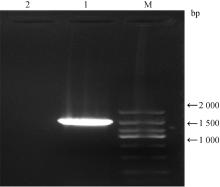
图 2
分离菌株PCR扩增结果 M. DL2000 DNA相对分子质量标准;1. 分离菌株;2. 阴性对照"

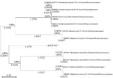
图 3
分离菌株16S rRNA系统进化树"

表 3
分离菌株药物敏感性结果"
| 抗菌药物 Antibacterial drugs | 抑菌圈直径/mm Inhibitory zone Diameter | 抑菌圈判定标准/mm | 敏感性 Sensitivity | ||
| 耐药 Resistent | 中介 Intermediate | 敏感 Sensitive | |||
| 阿米卡星 Amikacin | 20 | ≤14 | 15~18 | ≥19 | S |
| 头孢他啶 Ceftazidime | 19 | ≤14 | 14~18 | ≥18 | S |
| 诺氟沙星 Norfloxacin | 14 | ≤12 | 13~17 | ≥18 | I |
| 氟苯尼考 Florfenicol | 7 | ≤12 | 13~17 | ≥18 | R |
| 红霉素 Eythromycin | 11 | ≤15 | 16~20 | ≥21 | R |
| 四环素 Tetracycline | 16 | ≤14 | 15~18 | ≥19 | I |
| 环丙沙星 Ciprofloxacin | 12 | ≤15 | 16~20 | ≥21 | R |
| 米诺环素 Minocycline | 9 | ≤14 | 15~18 | ≥19 | R |
| 亚胺培南 Imipenem | 18 | ≤10 | 11~15 | ≥16 | S |
| 头孢哌酮 Cefoperazone | 17 | ≤15 | 16~20 | ≥21 | I |
| 庆大霉素 Gentamycin | 3 | ≤6 | 7~9 | ≥10 | R |
| 哌拉西林 Piperacillin | 23 | ≤17 | 18~20 | ≥21 | S |
| 头孢曲松 Ceftriaxone | 20 | ≤14 | 15~22 | ≥23 | I |
| 青霉素 Penicillin | 11 | ≤13 | 13~17 | ≥17 | R |
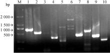
图 4
毒力基因检测结果 M. DL2000 DNA相对分子质量标准;1. WABG;2. Uge;3. Wyz-K1;4. Urea;5. Alls;6. Peg-344-1;7. ENTB;8. Fimh;9. Mrkd;10. Irp2"


图 5
试验组小鼠剖检结果 A. 试验组小鼠脾;B. 对照组小鼠脾;C. 试验组小鼠肝;D. 对照组小鼠肝"


图 6
小鼠回归试验PCR产物扩增电泳图 M. DL2000 DNA相对分子质量标准;1. 脾;2. 肝;3. 肺;4. 肾;5. 阴性对照"
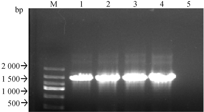

图 7
分离菌株感染小鼠试验组器官HE染色结果 A. LD50组小鼠肺(400×);B. LD50组小鼠肝(400×);C.LD50组小鼠脾(400×);D. LD50组小鼠肾(400×);E. LD50组小鼠心(400×)"


图 8
分离菌株外膜囊泡鉴定结果 A. 纳米颗粒跟踪分析(NTA);B. 透射电镜观察;C. SDS-PAGE试验(1. OMVs样品液;M. DNA相对分子质量标准)"

| 1 |
王佳宁, 张自强, 孔德婧, 等. 家兔肺炎克雷伯菌的分离鉴定[J]. 畜牧兽医学报, 2023, 54 (12): 5198- 5206.
doi: 10.11843/j.issn.0366-6964.2023.12.029 |
|
WANG J N , ZHANG Z Q , KONG D J , et al. Isolation and identification of Klebsiella pneumoniae in rabbits[J]. Acta Veterinaria et Zootechnica Sinica, 2023, 54 (12): 5198- 5206.
doi: 10.11843/j.issn.0366-6964.2023.12.029 |
|
| 2 |
AHMAD T A , EL-SAYED L H , HAROUN M , et al. Development of immunization trials against Klebsiella pneumoniae[J]. Vaccine, 2012, 30 (14): 2411- 2420.
doi: 10.1016/j.vaccine.2011.11.027 |
| 3 |
徐红云, 刘春林, 陈弟, 等. 2010—2016年耐碳青霉烯类肺炎克雷伯菌及大肠埃希菌临床分布及其耐药特征[J]. 中国感染控制杂志, 2018, 17 (8): 688- 692.
doi: 10.3969/j.issn.1671-9638.2018.08.007 |
|
XU H Y , LIU C L , CHEN D , et al. Distribution and antimicrobial resistance of carbapenem-resistant Klebsiella pneumoniae and Escherichia coli in 2010-2016[J]. Chinese Journal of Infection Control, 2018, 17 (8): 688- 692.
doi: 10.3969/j.issn.1671-9638.2018.08.007 |
|
| 4 |
MAK C Y , HO M , IU L P L , et al. Clinical features and treatment outcomes of endogenous Klebsiella endophthalmitis: a 12-year review[J]. Int J Ophthalmol, 2020, 13 (12): 1933- 1940.
doi: 10.18240/ijo.2020.12.14 |
| 5 | 马纾薏. 新疆南疆马鹿肺炎克雷伯菌分子流行病学调查及耐药特性研究[D]. 阿拉尔: 塔里木大学, 2022. |
| MA S Y. Molecular epidemiological investigation and drug resistance characteristics on Klebsiella pneumoniae of Red deer in southern Xinjiang[D]. Alaer City: Tarim University, 2022. (in Chinese) | |
| 6 |
LIU Y N , LEUNG S S Y , HUANG Y , et al. Identification of two depolymerases from phage IME205 and their antivirulent functions on K47 capsule of Klebsiella pneumoniae[J]. Front Microbiol, 2020, 11, 218.
doi: 10.3389/fmicb.2020.00218 |
| 7 |
BRINK A J . Epidemiology of carbapenem-resistant gram-negative infections globally[J]. Curr Opin Infect Dis, 2019, 32 (6): 609- 616.
doi: 10.1097/QCO.0000000000000608 |
| 8 | 陈琪, 吴敏, 白宏震, 等. 细菌外膜囊泡纳米载体的制备及其免疫调节作用[J]. 浙江大学学报: 医学版, 2017, 46 (2): 118- 124. |
| CHEN Q , WU M , BAI H Z , et al. Bacterial outer membrane vesicles as nano carriers to study immunological activities[J]. Journal of Zhejiang University: Medical Sciences, 2017, 46 (2): 118- 124. | |
| 9 | 冯文艳, 张扣兴. 革兰阴性菌外膜囊泡的研究进展[J]. 中国抗生素杂志, 2019, 44 (1): 32- 39. |
| FENG W Y , ZHANG K X . Research development of outer membrane vesicles in Gram-negative bacteria[J]. Chinese Journal of Antibiotics, 2019, 44 (1): 32- 39. | |
| 10 | 陈桥桥, 涂仕娟, 夏修文, 等. 细菌外膜囊泡发生机制的研究进展[J]. 泰山医学院学报, 2019, 40 (12): 980- 982. |
| CHEN Q Q , TU S J , XIA X W , et al. Research progress on the mechanism of bacterial outer membrane vesicle generation[J]. Journal of Taishan Medical College, 2019, 40 (12): 980- 982. | |
| 11 |
ELMI A , WATSON E , SANDU P , et al. Campylobacter jejuni outer membrane vesicles play an important role in bacterial interactions with human intestinal epithelial cells[J]. Infect Immun, 2012, 80 (12): 4089- 4098.
doi: 10.1128/IAI.00161-12 |
| 12 |
SHEN Y , TORCHIA M L G , LAWSON G W , et al. Outer membrane vesicles of a human commensal mediate immune regulation and disease protection[J]. Cell Host Microbe, 2012, 12 (4): 509- 520.
doi: 10.1016/j.chom.2012.08.004 |
| 13 | 喻胜猛. 致羊腹泻大肠杆菌噬菌体的分离鉴定及其在环境中的杀菌效果评估[D]. 咸阳: 西北农林科技大学, 2022. |
| YU S M. Isolation and identification of Escherichia coli bacteriophages causing sheep diarrhea and evaluation of their bactericidal efficacy in environment[D]. Xianyang: Northwest A&F University, 2022. (in Chinese) | |
| 14 | 东秀珠, 蔡妙英. 常见细菌系统鉴定手册[M]. 北京: 科学出版社, 2001. |
| DONG X Z , CAI M Y . Handbook of systematic identification of common bacteria[M]. Beijing: Science Press, 2001. | |
| 15 | CLSI. Vet01-A4 Performance standards for antimicrobial disk and dilution susceptibility tests for bacteria isolated from animals[S]. 4th ed. Wayne: Clinical and Laboratory Standard Institue, 2013. |
| 16 | 郭良帅. 和田地区羊源肺炎克雷伯菌的分离及生物学特征鉴定[J]. 中南农业科技, 2023, 44 (6): 74- 80. |
| GUO L S . Isolation and biological characterization of sheep-derived Klebsiella pneumoniae in Hotan[J]. South-Central Agricultural Science and Technology, 2023, 44 (6): 74- 80. | |
| 17 |
QIAN Y , WONG C C , LAI S C , et al. Klebsiella pneumoniae invasive liver abscess syndrome with purulent meningitis and septic shock: a case from mainland China[J]. World J Gastroenterol, 2016, 22 (9): 2861- 2866.
doi: 10.3748/wjg.v22.i9.2861 |
| 18 | 耿响, 刘希望, 李剑勇. 肺炎克雷伯菌耐药机制和毒力因子研究进展[J]. 中兽医医药杂志, 2024, 43 (1): 29- 38. |
| GENG X , LIU X W , LI J Y . Advances in mechanisms of resistance and virulence factors of Klebsiella pneumoniae[J]. Journal of Traditional Chinese Veterinary Medicine, 2024, 43 (1): 29- 38. | |
| 19 |
SHON A S , BAJWA R P S , RUSSO T A . Hypervirulent (hypermucoviscous) Klebsiella pneumoniae: a new and dangerous breed[J]. Virulence, 2013, 4 (2): 107- 118.
doi: 10.4161/viru.22718 |
| 20 |
PACZOSA M K , MECSAS J . Klebsiella pneumoniae: going on the offense with a strong defense[J]. Microbiol Mol Biol Rev, 2016, 80 (3): 629- 661.
doi: 10.1128/MMBR.00078-15 |
| 21 |
COMPAIN F , BABOSAN A , BRISSE S , et al. Multiplex PCR for detection of seven virulence factors and K1/K2 capsular serotypes of Klebsiella pneumoniae[J]. J Clin Microbiol, 2014, 52 (12): 4377- 4380.
doi: 10.1128/JCM.02316-14 |
| 22 | BULGER J , MACDONALD U , OLSON R , et al. Metabolite transporter PEG344 is required for full virulence of hypervirulent Klebsiella pneumoniae strain hvKP1 after pulmonary but not subcutaneous challenge[J]. Infect Immun, 2017, 85 (10): e00093- 17. |
| 23 |
KAKUTA N , NAKANO R , NAKANO A , et al. Molecular characteristics of extended-spectrum β-lactamase-producing Klebsiella pneumoniae in Japan: predominance of CTX-M-15 and emergence of hypervirulent clones[J]. Int J Infect Dis, 2020, 98, 281- 286.
doi: 10.1016/j.ijid.2020.06.083 |
| 24 |
李华明, 项维, 卢文兵, 等. 1株猪源ST-35型肺炎克雷伯菌的致病性和药物敏感性分析[J]. 畜牧兽医学报, 2022, 53 (12): 4356- 4366.
doi: 10.11843/j.issn.0366-6964.2022.12.021 |
|
LI H M , XIANG W , LU W B , et al. Pathogenicity and drug sensitivity analysis of a porcine Klebsiella pneumoniae type ST-35[J]. Acta Veterinaria et Zootechnica Sinica, 2022, 53 (12): 4356- 4366.
doi: 10.11843/j.issn.0366-6964.2022.12.021 |
|
| 25 | 田李均, 王晓丽, 肖淑珍, 等. 医院内高黏液性肺炎克雷伯菌的流行分布、毒力基因及临床特征分析[J]. 上海交通大学学报: 医学版, 2017, 37 (1): 43- 48. |
| TIAN L J , WANG X L , XIAO S Z , et al. Epidemiological distribution, virulent genes and clinical characteristics of hypermucoviscous Klebsiella pneumonia in a Hospital[J]. Journal of Shanghai Jiaotong University: Medical Science, 2017, 37 (1): 43- 48. | |
| 26 |
XU M , FU Y Q , FANG Y H , et al. High prevalence of KPC-2-producing hypervirulent Klebsiella pneumoniae causing meningitis in eastern China[J]. Infect Drug Resist, 2019, 12, 641- 653.
doi: 10.2147/IDR.S191892 |
| 27 | 姚崧源. 细菌外膜囊泡在疫苗领域的研究进展[J]. 微生物学免疫学进展, 2021, 49 (1): 78- 82. |
| YAO S Y . Advances in research on bacterial outer membrane vesicles in vaccine[J]. Progress in Microbiology and Immunology, 2021, 49 (1): 78- 82. | |
| 28 | 张靖. 高毒力肺炎克雷伯菌外膜囊泡能够诱发宿主细胞产生炎性反应[D]. 重庆: 重庆医科大学, 2021. |
| ZHANG J. Outer membrane vesicles derived from Hypervirulent Klebsiella pneumoniae stimulatr the inflammatory response[D]. Chongqing: Chongqing Medical University, 2021. (in Chinese) | |
| 29 |
WANG S J , HUANG W W , LI K , et al. Engineered outer membrane vesicle is potent to elicit HPV16E7-specific cellular immunity in a mouse model of TC-1 graft tumor[J]. Int J Nanomed, 2017, 12, 6813- 6825.
doi: 10.2147/IJN.S143264 |
| 30 | 邱晓涵, 李泳江, 吴军勇, 等. 细菌外膜囊泡: 疾病治疗的新途径[J]. 药学学报, 2021, 56 (12): 3441- 3450. |
| QIU X H , LI Y J , WU J Y , et al. Bacterial outer membrane vesicles: a new approach to diseases therapy[J]. Acta Pharmaceutica Sinica, 2021, 56 (12): 3441- 3450. | |
| 31 |
DOYLE L M , WANG M Z . Overview of extracellular vesicles, their origin, composition, purpose, and methods for exosome isolation and analysis[J]. Cells, 2019, 8 (7): 727.
doi: 10.3390/cells8070727 |
| 32 |
FURI I , MOMEN-HERAVI F , SZABO G . Extracellular vesicle isolation: present and future[J]. Ann Transl Med, 2017, 5 (12): 263.
doi: 10.21037/atm.2017.03.95 |
| 33 |
CASTILLO-ROMERO K F , SANTACRUZ A , GONZÁLEZ-VALDEZ J . Production and purification of bacterial membrane vesicles for biotechnology applications: challenges and opportunities[J]. Electrophoresis, 2023, 44 (1-2): 107- 124.
doi: 10.1002/elps.202200133 |
| 34 |
RIDER M A , HURWITZ S N , MECKES D G . ExtraPEG: a polyethylene glycol-based method for enrichment of extracellular vesicles[J]. Sci Rep, 2016, 6, 23978.
doi: 10.1038/srep23978 |
| 35 | 华雨能. 高毒力肺炎克雷伯菌通过外膜囊泡介导毒力因子转移至耐药肺炎克雷伯菌及宿主细胞的机制研究[D]. 广州: 南方医科大学, 2022. |
| HUA Y N. Outer membrane vesicles from hypervirulent Klebsiella pneumoniae mediate virulence factor transfer to antimicrobial-resistant Klebsiella pneumoniae and host cells[D]. Guangzhou: Southern Medical University, 2022. (in Chinese) |
| [1] | 刘建华, 撒瑞雪, 张嗣玉, 李银涛, 邓智超, 贾晗铎, 赵敏, 付玉, 杨一明, 冉多良, 加尔肯. 马疱疹病毒1型分离毒株对叙利亚金黄地鼠的致病性[J]. 畜牧兽医学报, 2025, 56(1): 327-334. |
| [2] | 王浩, 王世玉, 肖金龙, 沈珏, 潘天灵, 张靖松, 肖鹏, 高洪. 猪源大肠杆菌高致病性毒力岛结构基因进化分析及其对TNF/NF-κB通路的影响[J]. 畜牧兽医学报, 2025, 56(1): 392-403. |
| [3] | 贾宇航, 郭良富, 张茹楠, 赵阿勇, 刘玉芳, 储明星. miR-127调控绵羊骨骼肌细胞增殖分化及其转录因子PAX3筛选[J]. 畜牧兽医学报, 2024, 55(9): 3864-3875. |
| [4] | 龚一鸣, 贾一轩, 李佳骏, 王翔宇, 贺小云, 储明星, 狄冉. 不同FecB基因型和不同直径绵羊卵泡中BMP/SMAD通路活性及蛋白表达差异[J]. 畜牧兽医学报, 2024, 55(9): 3957-3967. |
| [5] | 张姗, 刘大虎, 刘宝京, 梁琳, 梁瑞英, 汤新明, 仇旭升, 丁铲, 丁家波, 侯绍华. 一株鸽副黏病毒Ⅰ型分离鉴定及致病性分析[J]. 畜牧兽医学报, 2024, 55(9): 4051-4060. |
| [6] | 谢碧林, 林志敏, 林彬彬, 徐以娟, 林锋强, 闫露, 伍慧妮, 李翠婷, 周海欧, 李兆龙. 鸭疫里氏杆菌LC1和CX1株的分离鉴定及致病性分析[J]. 畜牧兽医学报, 2024, 55(9): 4196-4203. |
| [7] | 王艳, 高亚东, 蒋成辉, 曾巧英. 一株鹅源禽腺病毒4型的分离及致病性[J]. 畜牧兽医学报, 2024, 55(9): 4232-4240. |
| [8] | 张帆帆, 李杰茂, 谭佳, 黄江南, 吴玲, 韦启鹏, 康昭风. 禽偏肺病毒的研究进展[J]. 畜牧兽医学报, 2024, 55(8): 3344-3353. |
| [9] | 孙雨点, 宋紫玥, 张洪亮, 秦志华, 单虎, 杨瑞梅. 鸭短喙与侏儒综合征病毒分离与鉴定[J]. 畜牧兽医学报, 2024, 55(8): 3623-3630. |
| [10] | 王奇林, 曹润来, 王威阳, 张博, 刘志杰, 王晓旭. 狐狸流产胎儿体内肺炎克雷伯菌的分离鉴定及耐药性分析[J]. 畜牧兽医学报, 2024, 55(8): 3640-3648. |
| [11] | 王婷, 张元庆, 闫益波, 上官明军, 郭宏宇, 王志武. “特藏寒羊”群体遗传结构分析与选择信号的对比分析[J]. 畜牧兽医学报, 2024, 55(7): 2913-2926. |
| [12] | 刘维哲, 罗成刚, 袁蓉, 廖艺杰, 文艺悯, 孙莹, 俞恩波, 曹三杰, 黄小波. 一株猪流行性腹泻病毒强毒株的分离与鉴定[J]. 畜牧兽医学报, 2024, 55(7): 3049-3063. |
| [13] | 班玛王清, 陈曦, 岳怡, 苏玉蓉, 岳华, 汤承. 一株牛呼吸道冠状病毒的分离鉴定及部分生物学特征[J]. 畜牧兽医学报, 2024, 55(7): 3094-3104. |
| [14] | 刘博华, 符汉宇, 王玉恒, 索朗斯珠, 牛家强, 包玉花, 李家奎, 徐业芬. 西藏那曲市牦牛源B型多杀性巴氏杆菌的分离鉴定及基因组分析[J]. 畜牧兽医学报, 2024, 55(7): 3105-3118. |
| [15] | 刘畅, 郝科兴, 陈岩, 曾维斌, 喻恒彬, 陈磊, 王静, 胡广东. 干扰PPARγ基因对绵羊滋养层细胞增殖、凋亡、迁移和脂质积累的影响[J]. 畜牧兽医学报, 2024, 55(6): 2421-2430. |
| 阅读次数 | ||||||
|
全文 |
|
|||||
|
摘要 |
|
|||||